Oncology

Beyond cancer care: Why the heart must be part of the plan

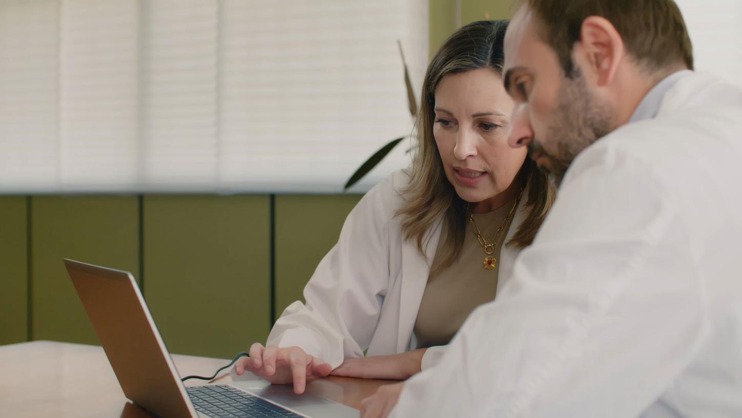
At La Paz University Hospital in Madrid, cardiology and oncology specialists work side by side to monitor one of the most serious side effects of cancer therapy: cardiotoxicity. Their experience shows that AI-supported reproducible ultrasound imaging and structured collaboration are essential to protect patients’ hearts without compromising treatment.
Sophie Gräf
Published on May 8, 2026
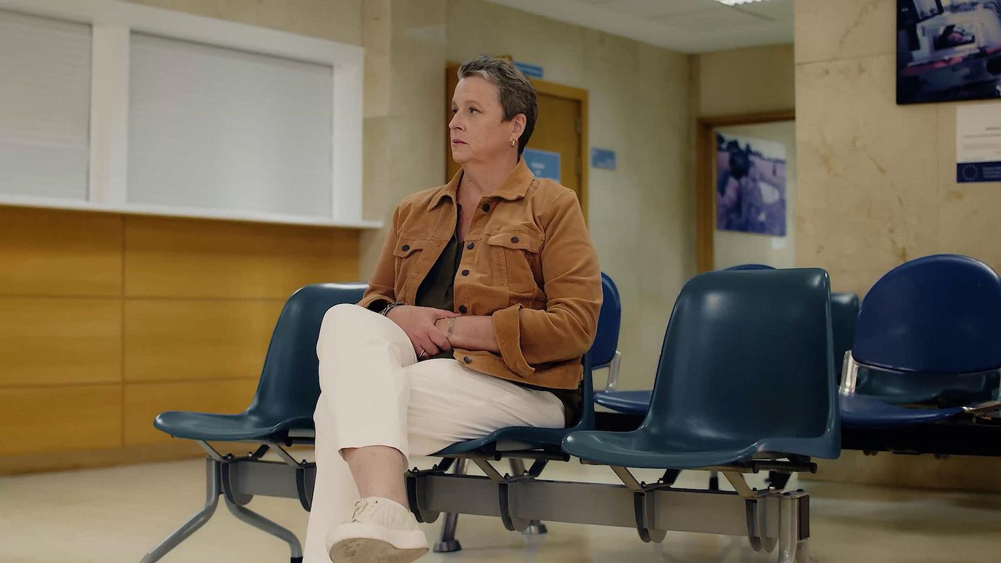
Cancer care is not only about treating cancer, it’s also about protecting the heart. For breast cancer patient Kathy S. Compton, this became reality during her treatment. At La Paz University Hospital in Madrid, cardiologists and oncologists work together as a dedicated cardio-oncology team to monitor and manage cardiotoxicity throughout therapy. Compton’s heart was not treated as a secondary concern. It was part of the plan from the very beginning.
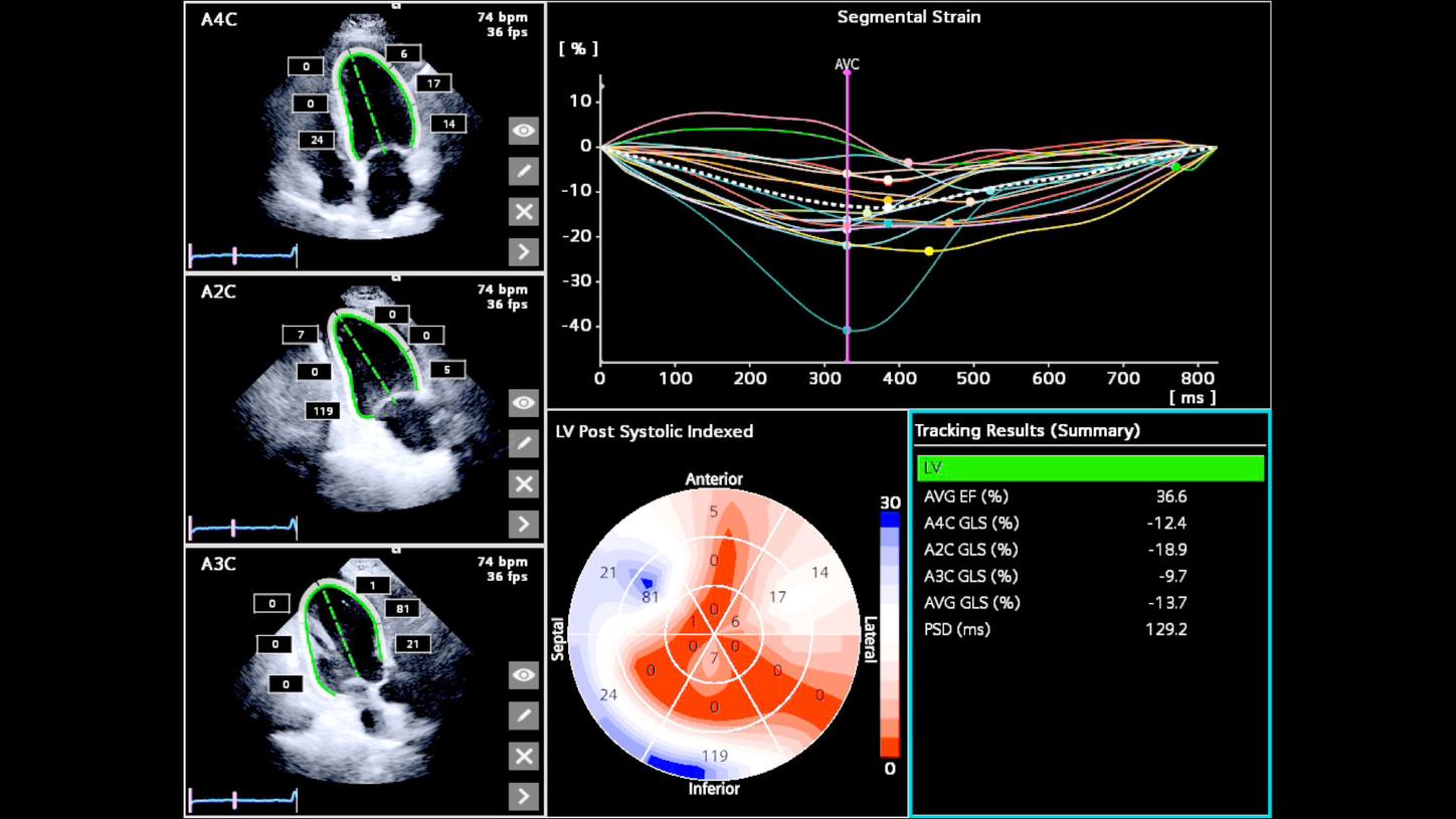
 

Breast cancer treatment brings the heart into focus in a unique way. Radiation therapy, particularly for tumors in the left breast, is delivered close to the heart and may affect surrounding cardiac tissue over time. In addition, certain systemic therapies, including anthracyclines and targeted treatments, can directly impact heart muscle cells or interfere with normal cardiac function. These effects can accumulate throughout the course of treatment, making heart health an important consideration from the very beginning.

“Cardiac imaging is central in cardio-oncology,” says Teresa Lopez-Fernandez, MD, head of cardio-oncology at La Paz University Hospital in Madrid. Certain chemotherapies and targeted therapies are known to carry a risk of cardiotoxicity. They may weaken heart muscle cells or interfere with normal contraction, gradually reducing the heart’s pumping efficiency. Cardiotoxicity often begins silently. Patients can feel entirely well while early myocardial changes are already occurring. In many cases, these subtle changes are not detectable with traditional echocardiographic measurements such as ejection fraction, which typically declines only after more significant impairment has developed.

To identify earlier signs of stress, the team at La Paz relies on strain analysis, in particular global longitudinal strain (GLS). Performed during a standard cardiac ultrasound, strain imaging measures myocardial deformation, i.e., how the heart muscle shortens and relaxes with each beat. This allows clinicians to detect subtle functional changes before symptoms even present and at a stage where preventive measures can still be taken. But early detection is only valuable if the data are reliable.

In cardio-oncology, small changes matter. “When we need to monitor patients who receive cardiotoxic therapies, we face two main challenges,” explains Lopez-Fernandez. “We must schedule our echo timely, the correct day to minimize the risk of cancer treatment delays, but we also need to be very reproducible.” Changes in GLS provide an early signal that the heart may be under stress. These findings lead to closer monitoring and joint assessment by the cardiology and oncology teams, allowing the implementation of cardioprotective strategies while maintaining cancer treatment.

Further changes in cardiac function — in particular, a significant decline in left ventricular ejection fraction (LVEF) — also guide decisions on further intervention. “When we get these numbers, we can make appropriate clinical decisions,” says Oliver Higuera Gómez, MD, oncologist at La Paz University Hospital in Madrid. Cardio-oncology teams rely on these cardiac measurements to assess risk, initiate cardioprotection or heart failure therapy when needed, and support the continuation of cancer treatment. This is where reproducibility becomes essential.

If different operators obtain significantly different cardiac measurements in the same patient, uncertainty enters the decision-making process. A patient might be taken off a potentially life-saving therapy — not because the heart has truly deteriorated, but because of measurement variability.

At La Paz, the team addresses this challenge by using AI-supported ultrasound imaging on the ACUSON Originsystem. Automated view recognition and contour placement help standardize measurements, reduce operator variability, and enable consistent follow-up — even in technically challenging examinations. “To be confident on the results, AI tools are really, really useful,” says Lopez-Fernandez.

For breast cancer patients like Compton, this consistency provides reassurance. It means that early changes can be identified and managed in good time — helping to protect the heart without unnecessary interruptions to treatment. “I went into my treatment not really knowing what that meant but the cardiologist team here is amazing and they both just guided me along, the oncologist and the cardiologist, and really worked together in helping me get better,” says Compton.

The collaboration of the cardio-oncology and oncology departments from the beginning is a success story for the team: “Prevention of cardiotoxicity starts before cancer treatment,” says Lopez-Fernandez. When cardiac monitoring is integrated into the therapeutic timeline, the need for abrupt decisions is reduced and clinicians can weigh cardiac findings in context — supported by consistent, reproducible data.


Sophie Gräf
Sophie Gräf
By Sophie Gräf
Sophie Gräf is a digital editor and multimedia content creator at Siemens Healthineers.