


Press release
Siemens Healthineers introduces industry-first seamlessly integrated end-to-end solution for CT-guided percutaneous coronary interventions
European Course on Percutaneous Cardiovascular Interventions (EuroPCR) 2026
Published on May 19, 2026
Image Gallery
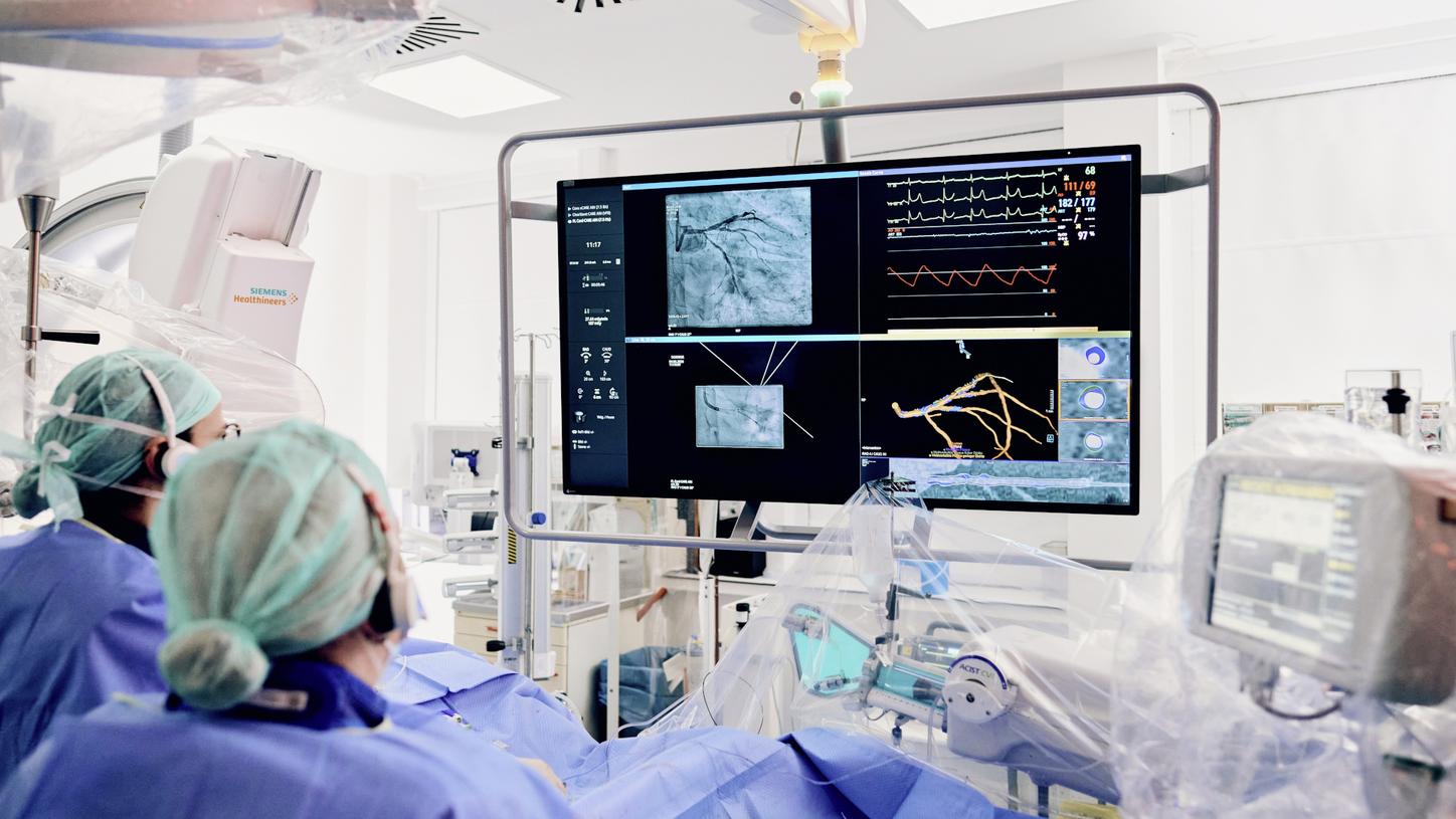
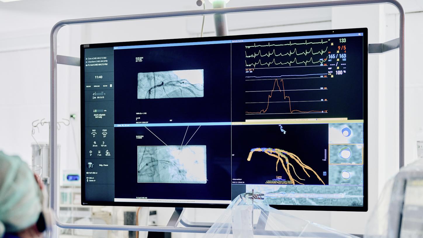
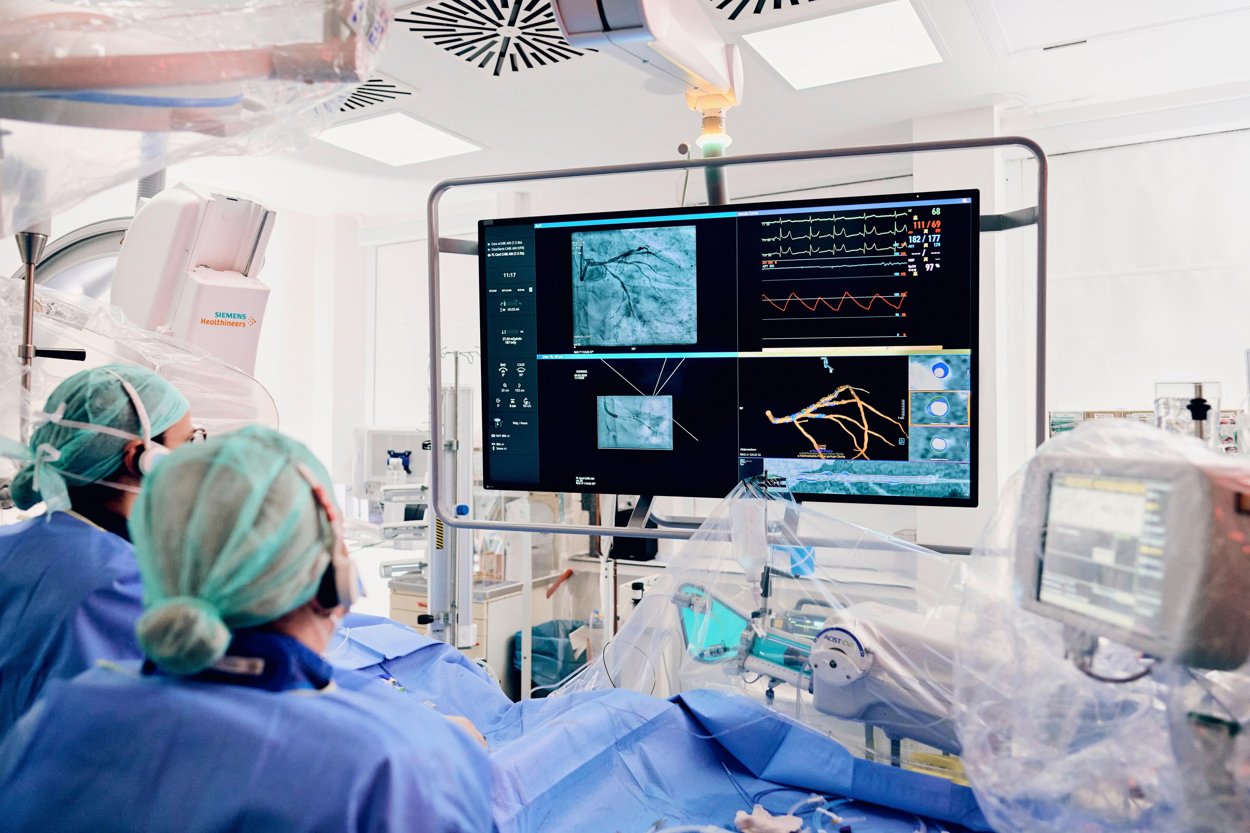
The combination of Syngo.CT Coronary Cockpit and Syngo PCI Connect gives interventional cardiologists valuable insights on morphology, plaque and lesion details and allows for strategic planning of the intervention.
Courtesy of Kerckhoff-Klinik, Bad Nauheim, Germany
Download image
Download this press release
(pdf, 287.84 KB)Contact

Kathrin Palder
Innovation, Artificial Intelligence, Advanced Therapies
The products/features mentioned herein are 510 (k) pending and not commercially available in all countries. Their future availability cannot be guaranteed. syngo.CT Coronary Cockpit is not for sale in the USA. Its future availability cannot be guaranteed.
Siemens Healthineers 2026
Siemens Healthineers pioneers breakthroughs in healthcare. For everyone. Everywhere. Sustainably. The company is a global provider of healthcare equipment, solutions and services, with activities in more than 180 countries and direct representation in more than 70. The group comprises Siemens Healthineers AG, listed as SHL in Frankfurt, Germany, and its subsidiaries. As a leading medical technology company, Siemens Healthineers is committed to improving access to healthcare for underserved communities worldwide and is striving to overcome the most threatening diseases. The company is principally active in the areas of imaging, diagnostics, cancer care and minimally invasive therapies, augmented by digital technology and artificial intelligence. In fiscal 2025, which ended on September 30, 2025, Siemens Healthineers had approximately 74,000 employees worldwide and generated revenue of around €23.4 billion. Further information is available at siemens-healthineers.com.