Imaging

“This technology is here to stay”

At the front line of stroke and neurological care, photon-counting computed tomography (CT) comes with high resolution both in-plane and cross-plane to enable physicians evaluate fine lesions and anatomical structures.
4min
Doris Pischitz
Published on November 28, 2022

Associate Professor Tobias Granberg, MD, PhD, speaks about his expectations and experiences.
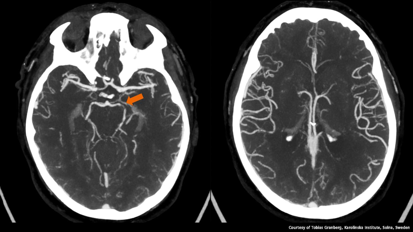
Tobias Granberg is a research group leader at the Karolinska Institute and section head at the Department of Neuroradiology, Karolinska University Hospital, Stockholm, Sweden
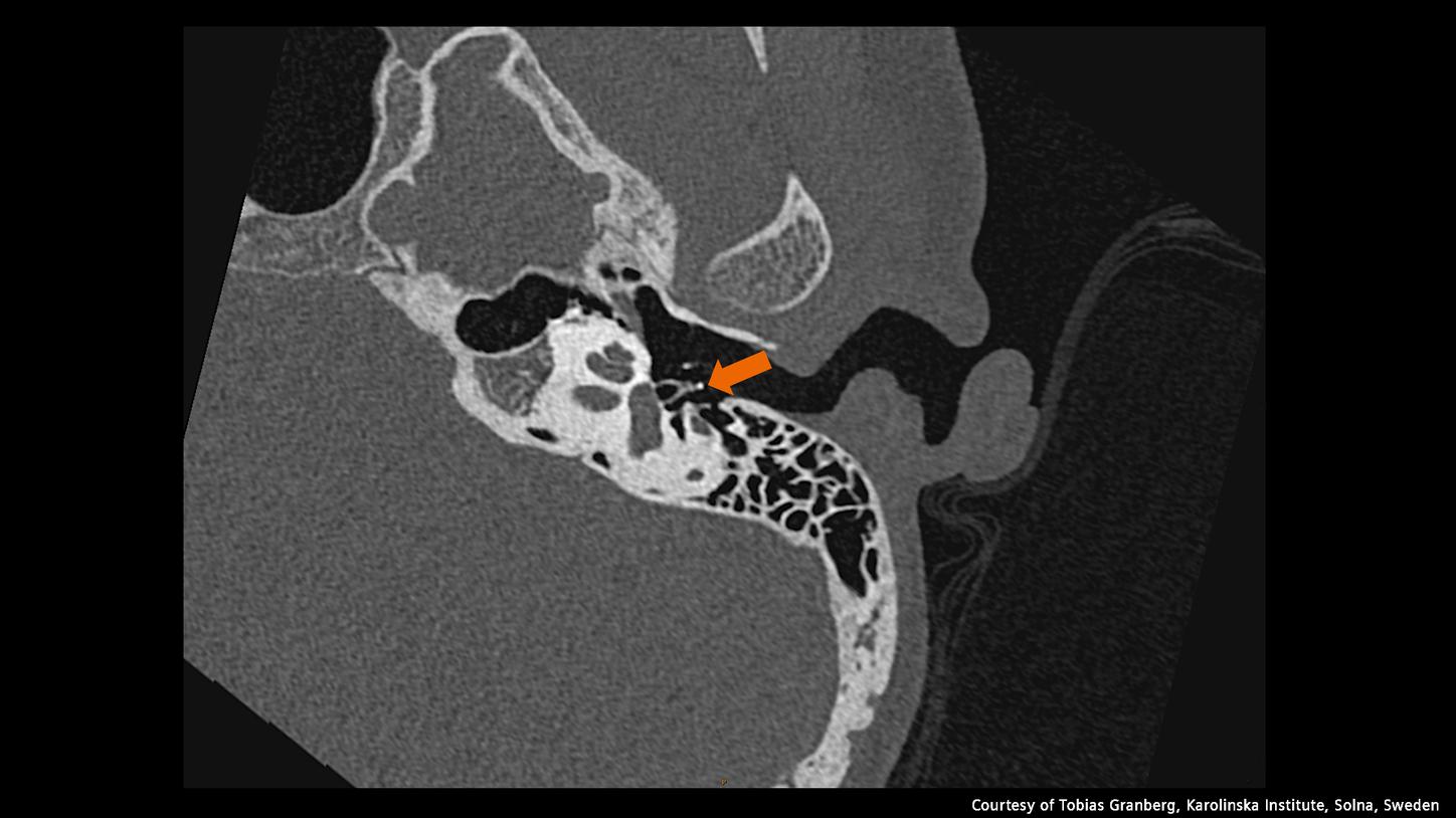
. He leads the clinical implementation and technical developments of Karolinska’s NAEOTOM Alpha1 photon-counting CT system within neurology applications.

We started using our NAEOTOM Alpha system in September 2021. We expected to be able to acquire diagnostic scans with higher spatial resolution and less noise, and to reduce both the ionizing radiation and contrast agent dose. We also envisioned using Dual Source for fast scans with multi-energy acquisitions.
The major challenge with CT applications in neurology is achieving high tissue contrast while minimizing beam hardening and cupping artifacts. In our practice, we routinely acquire dual-energy scans on our conventional scanners for optimized gray–white matter differentiation, bone removal, and iodine/virtual non-contrast maps. However, that comes with longer scan times, which can lead to motion artifacts. This means we can’t always use that strategy. Obtaining multi-energy data combined with fast scanning can solve this dilemma.

The most apparent improvement in terms of diagnostic quality has been the increased spatial resolution. We mainly use that for temporal bone scans [Figure 1], but it’s also useful for angiographies. We’ve also been able to reduce contrast agent dose by about 30 percent so far, and we expect further reductions will come. Dual or multi-energy data in fast scan modes (FLASH) has also proven valuable.


Tobias Granberg reports on photo-counting CT in neurology.

Our patients benefit from the fact that we can now use fast scanning modes to reduce the risk of motion artifacts while still obtaining dual- or multi-energy data. We’ve also been able to show that the attenuation measurements are more stable with photon-counting CT than with our traditional scanners. This opens up many interesting opportunities, not least for brain volumetrics, which is an important field in neurodegeneration that has so far mainly been dependent on high-quality 3D MRI (magnetic resonance imaging). We’re planning to explore whether we can now provide similar data for dementia investigations using CT instead of MRI.

The improved image quality gives us more confidence when reading the images. We see the biggest benefit when assessing the cochlea and the smallest bones in the middle ear [Figure 1]. Interestingly, we’re now seeing new anatomical details that don’t seem to have been documented in the literature yet. Another advantage is that we can assess pathology more confidently in the smallest cerebral blood vessels.
Photon-counting CT scan of the temporal bone

The new system benefits all patients who have a particular need for a lower ionizing radiation dose – especially children, young adults, and pregnant woman. So far, about ten percent of our scans have been pediatric patients, and it’s great to see that the dose reductions are already benefiting them. Another concrete example is when we have patients who need an angiography but can’t tolerate conventional doses of iodine contrast agents due to impaired renal function. Now we can reduce the contrast dose by at least half, making it possible to scan many of these patients. That’s particularly helpful when MRI or ultrasound aren’t an option.

We’re used to applying dual energy on our conventional systems for all neurology scans, and we now also do this with our NAEOTOM Alpha system. One way we use the iodine maps and virtual noncontrast images is to assess whether intra-axial findings of high-attenuating material are more likely to represent blood, iodine, or calcifications. The material decomposition based on multi-energy data helps us obtain more robust measurements than are possible with our dual-energy systems.
These images are less prone to certain artifacts and are more comparable across time for patients who have longitudinal follow-ups. With time, I expect that we will see more and more benefits from using monoenergetic images in our radiological assessments.
We have essentially used the system like any other clinical CT scanner. In the first year, we performed nearly 3,000 scans across all organs. About ten percent of them were emergency scans.
One example is of a patient that came in with an acute/subacute infarction in the left posterior cerebral artery territory. He had severely impaired renal function, so normally we would have been unable to perform an angiography. But an ultrasound wouldn’t have visualized the intracranial vessels, and arranging an MRI would take time. We decided to perform a multiphase angiography with half the normal contrast dose, which was within the theoretical limits of what was deemed possible for this patient. The images still had high diagnostic quality [Figure 2]. This is something that has happened multiple times, so photon-counting CT has been a problem-solver for these situations.

The most important thing with increased amounts of data is to have automated and standardized viewing protocols in the PACS (picture archiving and communication system) so that the information is always at hand where you need it. I hope that AI will make setting up these protocols easier and that algorithms will be able to further improve our contrast-to-noise ratio and spatial resolution.

I think that more stable attenuation values, virtual monoenergetic imaging, and multi-energy data might start to challenge some of the applications that we currently use other modalities for. We’ll have to think about CT data more like we currently think of MRI data, in terms of different weightings to highlight the desired tissue properties. We’re also currently using the heightened iodine sensitivity and increased spatial resolution to improve our cerebral angiographies. The idea is to reduce the need for conventional interventional digital subtraction angiographies for diagnostic purposes—for example in patients with suspected vasculitis or small vascular malformations. That would reduce the risks for the patient while still allowing us to obtain the clinically necessary information.
Photon-counting technology is here to stay. We’re currently in the early adopter phase, where there is still a lot of optimizing of protocols to make the best use of the scanner. With time, and once the technology has also been introduced in single-source CT scanners, it will come to benefit many patients worldwide.

By Doris Pischitz
Doris Pischitz is an editor in corporate communications at Siemens Healthineers. The team specializes in topics related to healthcare, medical technology, disease areas, and digitalization.