Pediatrics

Shedding light on the unseen

Visualize the smallest structures of the heart realistically without surgery? About the use of a prototype that makes this vision almost tangible. 

10min
Marc Engelhardt
Published on March 24, 2021

Cardiac surgery is one of the most difficult procedures, and it gets even more complex when it comes to operating on tiny children’s hearts. Visualization techniques like cinematic rendering provide a detailed 3-D view of the patient’s heart and surrounding anatomy. But what happens when these images are turned into a hologram that can be rotated, zoomed, and looked at from all possible perspectives? 
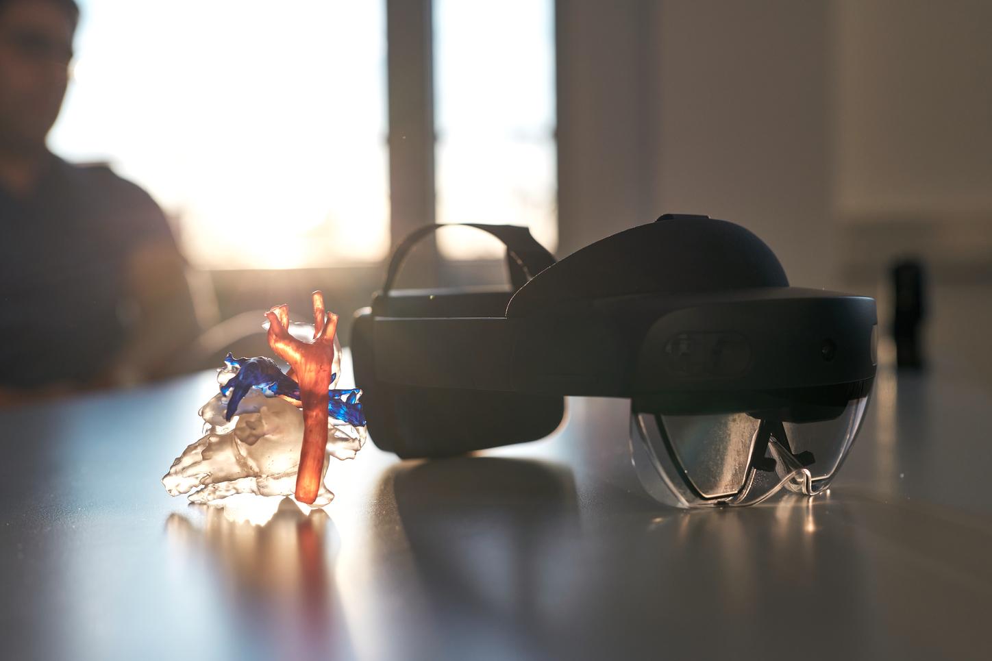
Doctors in the maternity ward of the Erlangen University Hospital were worried by the low oxygen saturation of only 85 percent in the newborn’s blood and alerted pediatric cardiologist Muhannad Alkassar less than an hour after the birth. “Within 30 minutes we had done a CT scan that confirmed our suspicion of a pulmonary vein malformation,” recalls the 40-year-old. For such emergency surgeries – Alkassar’s specialty – success depends on the best possible surgical planning.

Pulmonary vein malformation is a condition that affects blood flow between the heart and the lungs. Affected people have an abnormal connection between the pulmonary vein that carries blood from the lungs to the heart and the pulmonary artery that carries blood from the heart to the lungs.

Muhannad Alkassar, MD, holds a doctorate in biology and medicine. He has been working and researching in the Department of Pediatric Cardiology at the Erlangen University Hospital for five years. 

Portrait of MD Muhannad Alkassar
“When we look at an ultrasound image of a heart that is affected like this, we have to put the different layers together in our head to create a three-dimensional image,” explains Alkassar. “This is particularly difficult for a surgeon who doesn’t do this quite as often as we cardiologists do.” Every heart defect is unique. In the case of the newborn, the oxygen-enriched blood did not flow from the lungs into the left heart chamber, but instead through an abdominal vessel into the inferior vena cava. The surgeon and Alkassar were able to study the vessels and the heart in color and 3D – using the HoloLens 2 developed by Microsoft. This presented them with a hologram of the patient’s heart, based on the CT scan, allowing them to view the organ from all angles and down to the smallest detail. “Once you’ve tried HoloLens technology, you’ll never want to do without it,” smiles Alkassar.

Inferior vena cava is the largest vein of the human body. It carries deoxygenated blood from the lower and middle body to the heart.

The latest technology meant the surgeon could examine the newborn as if he were lying on the operating table with his chest open. And as if his walnut-sized heart were made of glass.

MD Alkassar tests the HoloLens
When Alkassar first put on the HoloLens headset more than a year ago, he admits he was skeptical. “I never imagined that the image of the object would be in such high resolution. I had seen the photorealistic image on the computer screen, but seeing a spatial representation of the heart in 3D in this quality is something else altogether.”
This is Dr. Alkassar’s reaction as he tried out the HoloLens with Cinematic Rendering for the first time. In the background on his computer screen you can see what he sees through his HoloLens, but in 3D, he is able to zoom in an out of structures, turn them around, and observe them from every possible angle.

The CT images are processed with an app using the Cinematic Rendering process, a technology that draws inspiration from Hollywood’s computer-generated images to incorporate the shadows cast by every single pixel in the image rendered.


CT images are procced with an app using Cinematic Rendering

“At first it sounds like a gimmick, but our eyes are so used to seeing the effects of light. And Cinematic Rendering means – considering the delicate vessels and structures we’re dealing with – that we can look and work the way we’re used to. That makes such a huge difference.”

MD Alkassar explores Cinematic Rendering
In the case of 26 patients, Alkassar and his team compared the procedure and quality of surgical planning using HoloLens augmented reality with using a 3D-printed model – the method of choice for preparing surgery. In the subjective assessment of the physicians, the HoloLens performed significantly better – probably because in addition to the heart, it also visualized the surrounding muscles, bones, and vessels. This explains why surgeons need less time to prepare for a heart surgery with HoloLens than they would without it. And what is more: it only takes a few minutes to process the data. “I have to wait about 24 hours for 3D printing,” says Alkassar.
When comparing the use of a 3D-printed model – the current method of choice for preparing surgery – with HoloLens augmented reality, Alkassar and his team felt HoloLens performed significantly better.


HoloLens on a table
The hologram was literally an eye-opener for Alkassar and the surgeons treating the newborn: The 3D representation enabled them to clearly see when the opening between the right ventricle and the pulmonary artery was closed by a membrane. “Only then did we see that we could insert a cardiac catheter into the pulmonary artery and avoid surgery for the time being.” Further, they clearly saw the intricate opening of a misdirected pulmonary vein, which was not visible on the ultrasound.
“The next thing we want to do is to redesign our meeting rooms so that the hologram appears in the middle of the conference table,” says Alkassar. “Then we can view the image as a team when we confer with one another, and it will simplify discussions.”


MD Alkassar explains future operation planning
The technology offers potential for training, and for briefing relatives and patients. This is especially relevant because many cardiac patients will need life-long medical care. “It definitely makes sense to use this technology in the future to provide support in the operating room, but the technology still needs to be developed further, for example in the area of artificial intelligence.” Muhannad Alkassar is in no doubt that this can be achieved. Anything seems possible to a doctor who sees into the hearts of children everyday. And luckily he was able to see into the heart of the newborn just in time.
What would Wilhelm Conrad Röntgen, born 175 years ago and discoverer of the X-ray, have made of HoloLens and cinematic 3D images from inside the body? “I think that would have been considered magic at the time,” says Alkassar, smiling whimsically.


Roentgen and MD Alkassar
“But you don’t even have to go back as far as that: even 30, 40 years ago you couldn’t have imagined seeing these kinds of images on a monitor. Now, what used to happen only in Star Trek, in the holodeck device, will become reality.” And that is not all.


By Marc Engelhardt

As a freelance journalist and author, Marc Engelhardt reports worldwide on the latest developments in business, science and politics. He has worked as a correspondent for a number of German-language media including Neue Zürcher Zeitung, ARD, and Die Zeit.