The tangible product of a radiologist’s work is the radiology report, a central part of patient management that is subject to ever-expanding requirements. Reporting modules which enable standardized structured reports expanded with multimedia can often prepare reports faster and export them in various formats for other information systems. Thereby, they open up opportunities for improved treatment, new pathways for research, and improved referrer satisfaction.
Growing requirements
The radiology report is the radiologist’s calling card. It bears witness to his or her expertise and experience, influences treatment decisions, and forms the basis for compensation.1,2
Meanwhile, reporting practices in radiology are changing: A trend toward structured reporting is gaining ground. While radiologists have traditionally drafted their findings as free text, structured reports use preset templates with defined terminology and incorporated guidelines to report specific findings and recommendations (e. g. BiRADS, PiRADS, LungRADS, etc.). The aim is to present the interpretation of an imaging study as clearly and transparently as possible for human readers, but also to make it easy for computer systems to interpret in order to fill searchable data bases.
Until now, this process was commonly used only in mammography, but it is playing an ever-greater role in other fields, such as cardiac imaging. This trend is being driven by several international initiatives – not least in order to improve the general quality of reporting and adapt it to modern medical requirements and workflows.3,4
Living up to referring doctors’ needs
Studies demonstrate the advantages of this new reporting style. For instance, computed tomography (CT) findings can often be communicated more clearly to referring physicians using structured reports.5,6 Many referring doctors also appreciate reports that include selected images and summarizing charts on important measured values, along with written information. Reports with such multimedia additions can remove the need for follow-up inquiries, speed up treatment planning, and increase clinicians’ confidence in the findings.7,8
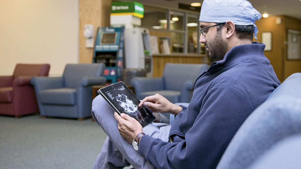
“As radiologists, we have to provide structured data in order to adequately meet the needs of today’s medical practice,” confirms Tobias Heye, MD, Department of Abdominal and Oncological Diagnostics at Basel University Hospital in Switzerland. “Structured reporting is a step in the direction of value-based radiology.”
In a survey of 400 Swiss private and hospital physicians, Heye’s team recently showed that referring doctors preferred structured and image reports over other forms of reporting that are less easy to interpret, and see them as an aid in their day-to-day work and in their communication with patients.9
Bridging reading and reporting
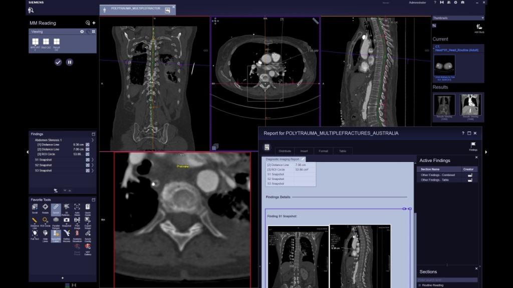
Since 2014, Heye and his colleagues have used the reporting module of multimodality software syngo.via to prepare radiology reports for all cardiac CT and cardiac magnetic resonance imaging (MRI) scans. Typical procedures include calcium scoring, CT-angiographic analysis of coronary stenoses, and MRI tests of cardiac function and volumes.
Through smart automation, syngo.via largely erases the conventional distinction between image reading and report preparation.
“The strength of the system is that the report is already being generated in the background during the diagnosis,” Heye remarks. For example, measurements which the physician takes on the monitor during the image analysis are automatically transferred to the report and marked-up images are added.
Moreover, digital templates geared to the specific test are available in which all the important parameters of analysis can be entered using drop-down lists. These templates can also be customized – for example, in order to link findings and diagnoses with specific billing codes for further data processing.
“Radiologists traditionally spend two-thirds of their working hours preparing reports. For complex cardiac tests, using syngo.via reduces this time spent by at least half.”
Faster and standardized workflows
Radiologists sometimes complain that structured reporting is more time consuming than writing up findings freely.10 However, when a smart computer system is used, the opposite can be true. “Radiologists traditionally spend two-thirds of their working hours preparing reports,” Heye estimates. “For complex cardiac tests, using syngo.via reduces this time spent by at least half.”
In addition, the software provides an opportunity to guide the image analysis itself. For instance, for the standardized measurements required in selecting a prosthetic heart valve, one could imagine giving specific measurement instructions in a guided process via the software and then preparing reports automatically with consistent quality.
“Using the same software to do all the measurements and compare them with prior scans, and then output the report as a multimedia document – this is where the future lies.”
Integrating results into information systems
syngo.via also facilitates the radiological evaluation of treatment results in oncology, where RECIST (Response Evaluation Criteria in Solid Tumors) criteria are usually applied. The principle behind them is to measure the sizes of select tumor foci multiple times in the course of treatment and incorporate the results into treatment decisions.
“With syngo.via, we can automatically compare these measurements with previous results and generate easily understandable trend charts with a few mouse clicks,” explains Dr. Philipp Lust, a radiologist at Klinikum Wels-Grieskirchen in Austria. This is possible because the software has a pattern recognition algorithm that detects identical anatomical structures on different scans.
Lust and his colleagues are using the reporting module of syngo.via to electronically transfer RECIST measurements of oncology study patients to the hospital information system through an HL7 interface. This could soon become a routine clinical practice, says Lust. “Using the same software to do all the measurements and compare them with prior scans, and then output the report as a multimedia document – this is where the future lies.”
Leveraging reports for better care and research
Generally speaking, the wide range of options for exporting data from syngo.via allows for better integration of reporting into more advanced patient management. For instance, when nodules are discovered by chance in a lung CT, the report encodes this finding so that a receiving information system can unambiguously interpret it. It could then be linked to appointment scheduling software and the patient could be invited to come in for a follow-up scan. It is a known fact that these kinds of incidental findings are otherwise frequently overlooked in everyday clinical practice.11
Radiology research would also very likely benefit from structured data preparation. Pilot projects are already underway to test whether data mining methods can be used in a pile of reports to filter out, for example, correlations between the volume of certain brain structures and clinical-neurological deficits. In this way, structured reporting goes well beyond good radiology documentation and gains a scientific value in its own right.
Individual approaches across the globe
BASEL UNIVERSITY HOSPITAL, SWITZERLAND
Radiologists in the Department of Cardiac and Thoracic Diagnostics prepare multimedia reports for all cardiac CT and MRI scans using syngo.via. For structured reporting, they use templates customized for their clinic, which are stored in the software.
CITY GENERAL HOSPITAL, STOKE-ON-TRENT, UK
CT scans of the colon are diagnosed with syngo.via. In three-quarters of the workflows, the system is then used to produce a radiology report.
CLEVELAND CLINIC, USA
Radiologists use syngo.via for standardized and structured diagnosis of cardiac scans. Results are transferred to Nuance PowerScribe 360 and integrated into electronic patient files as rich media PDFs. Benefits include the avoidance of dictation errors, output of concise and multimedia-enhanced reports, and better support for treatment decisions.
KLINIKUM WELS-GRIESKIRCHEN, AUSTRIA
The treatment result measurements for oncology study patients (according to RECIST criteria) are transferred to the hospital information system through an HL7 interface. The software automatically compares previous tests, generates precise case-progression information for each patient, and presents it in easy-to-understand charts. Electronic data export eliminates the need for paper documentation and avoids calculation and transmission errors.
ST TERESA’S HOSPITAL, HONG KONG
Radiologists use syngo.via in CT imaging of coronary arteries and for calcium scoring. In about 70 percent of cases, a structured report is generated directly from the software.