- ホーム
- 画像診断・治療装置
- X線CT装置
- SOMATOM
- SOMATOM X. プラットフォーム
- SOMATOM X.cite
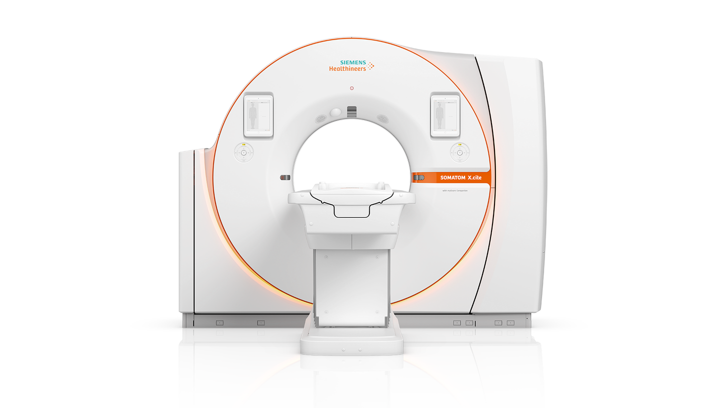
SOMATOM X.cite
with myExam Companion
Intelligent CT
with myExam Companion
AI技術を用いて開発された最新の全自動撮影システム「myExam Companion」を搭載。個々の患者の状況に合わせた検査内容を自動で作成し、技師の経験に関わらず最適な検査の実施と一貫性のある結果を提供します。また、 3D Cameraを用いた撮影ポジショニングの自動化や低線量撮影、患者の側で検査を行えるタブレット型端末など、SOMATOMシリーズのCTで培われてきた数多くの技術を統合し、患者の快適性を向上します。
特長
Intelligent imaging. Excellence empowered.
SOMATOM X.citeは、患者一人ひとりの状況やニーズに合った最適な画像を、技術者の経験レベルや環境に依存せずに、常に一貫したクオリティで提供できるインテリジェントイメージングを実現します。
 FAST 3D CameramyExam CompanionVectron® X-ray tubeVPI and Patient Observation CameraGO technologies
Mobile Workflow
Large 82 cm bore
StellarInfinity detector®Low kV imaging and Tin FilterIntelligent navigation for enhanced consistency
Cardiac imaging、Neuro imaging、Spectral imaging、3つの分野における "myExam Companion" の卓越したインテリジェントイメージングを動画でご覧いただけます。
1/3
Cardiac01
Neuro01
Spectral01
Intelligent spectral imaging in CT
Intelligent cardiac imaging in CT
Intelligent neuro imaging in CT
Intelligent spectral imaging in CT
Intelligent cardiac imaging in CT
Intelligent neuro imaging in CT
Intelligent spectral imaging in CT
1/3
82 cm large bore
さまざまな体格の患者や厳しい条件にも対応
大容量の X線管と耐荷重に優れる患者テーブル、82 cmのガントリボアサイズを実現したことで、体格の大きな患者や緊急検査に柔軟に対応することができます。
高齢で検査体位に制限のある患者や、救急検査でフレキシブルなポジショニングが求められるケース、また、インターベンション手技においても患者や術者の特徴に応じて、安全にアプローチすることが可能です。


Personalized imaging for consistent
Vectron X-ray tubeの説明動画
検査目的や患者背景に応じて個別化された
検査プロトコルの構築
X線やヨード造影剤を使用する CT検査では、検査目的や患者背景に応じて検査プロトコルを最適化することが重要です。SOMATOM X.citeは、最大1,200 mAの管電流出力を実現するVectron X-ray tubeによってハイパワーな低管電圧撮影、および、Siemens Healthineers 独自の Tin Filter technologyによる低被ばく撮影を実現します。患者と疾患の両方の特性に合わせた個別化されたCT検査を実施することができます。


Extra room to help patients feel relaxed
Mobile Workflow - Lead to Next
タブレット端末をベースとしたMobile Workflow
タブレット端末で患者選択・登録から撮影プロトコルの選択、撮影範囲の設定や画像確認を行うことが可能です。最新の自動化技術を組み合わせることで、検査開始から画像配信までの一連の検査ワークフローを効率化することができます。
クリニカル情報
1/10

Courtesy of University Zurich, Zurich, Switzerland
Inner ear assessment with Tin Filter - High spatial resolution with no dose penalty
SOMATOM X.cite
Sn 130 kV
CTDIvol: 24.25 mGy
DLP: 230 mGy cm
Rotation time: 1 sec
Scan time: 6.4 sec
- 1 mm Mpr

Courtesy of University Hospital Erlangen, Erlangen, Germany
Coronary heart disease patient after multiple bypass - Follow-up by coronary CTA spiral scan
SOMATOM X.cite
120 kV
CTDIvol: 13.8 mGy
DLP: 302.2 mGy cm
Scan length: 219 mm
Rotation time: 0.3 sec
- Curved MPR and Cinematic VRTs shown

Courtesy of Clínica Universidad de Navarra, Pamplona, Spain
Low-dose coronary CTA spiral scan
SOMATOM X.cite
70 kV
CTDIvol: 6 mGy
DLP: 91 mGy cm
Scan length: 150 mm
Rotation time: 0.3 sec
- Excellent visualization of bypass and coronaries
- Cinematic Rendering

Courtesy of Clínica Universidad de Navarra, Pamplona, Spain
Acute arterial pulmonary embolism - TwinBeam DE CT of the lung
SOMATOM X.cite
AuSN 120 kV
CTDIvol: 5.52 mGy
DLP: 188 mGy CM
Exposure time: 9.8 sec
Scan length: 356 mm
Rotation time: 0.3 sec
- Fused and perfusion surrogate iodine image demonstrating extension of perfusion deficiencies caused by embolism
- MIP at 40 keV

Courtesy of University Hosptital Erlangen, Erlangen, Germany
Trauma follow-up - Native TwinSpiralDE CT of the knee
SOMATOM X.cite
90 / Sn 150 kV
CTDIvol: 16.65 mGy
DLP: 417 mGy cm
Exposure time: 12 sec
Rotation time: 0.5 sec
- 0.8 mm MPRs
- VRT
- Bone marrow visualization

Courtesy of University Hospital Erlangen, Erlangen, Germany
Re-Staging - Single phase CME TwinBeam DE thorax to pelvis CT
SOMATOM X.cite
AuSn 120 kV
CTDIvol: 8.06 mGy
DLP: 657 mGy cm
Exposure time: 18 sec
Scan length: 689 mm
Rotation time: 0.3 sec
- Monoenergetic 70 keV
- Mixed 120 kVp equivalent
- Iodine map
- Fused Mixed / Iodine with iMAR

Courtesy of Hospital Zurich, Zurich, Switzerland
Stroke Assessment - Non-enhanced, perfusion and head CTA
SOMATOM X.cite
Native head:120 kV
CTDIvol: 40.07 mGy
DLP: 732 mGy cm
Carotid CTA:90 kV
CTDIvol: 7.07 mGy
DLP: 282 mGy cm
Rotation time: 0.3 sec
Perfusion:70 kV
CTDIvol: 158.11 mGy
DLP: 1.887 mGy cm
Rotation time: 0.3 sec

Courtesy of University Hospital Erlangen, Erlangen, Germany
Low kV imaging with high power reserves - Low kV even for bigger patients
SOMATOM X.cite
100 kV
CTDIvol: 27.7 mGy
DLP: 1450 mGy cm
Exposure time: 15.71 sec
Scan length: 533 mm
Rotation time: 0.5 sec
- Cinematic VRT
- 8 mm MIP

Courtesy of Clínica Universidad de Navarra, Pamplona, Spain
Lung evaluation with Tin Filter - Low dose even for bigger patients
SOMATOM X.cite
Sn 140 kV
CTDIvol: 1.63 mGy
DLP: 49 mGy cm
Rotation time: 0.3 sec
Tin Filter topogram
- Coronal MPR
- MIP

Courtesy of Clínica Universidad de Navarra, Pamplona, Spain
Evaluation of pancreas and abdomen - Arterial phase
SOMATOM X.cite
90 kV
CTDIvol: 17.41 mGy
DLP: 837 mGy cm
Exposure time: 6.5 sec
Rotation time: 0.5 sec
Pitch: 1
- 3 mm MPRs
- MIP and Cinematic VRT
Courtesy of University Zurich, Zurich, Switzerland
Inner ear assessment with Tin Filter - High spatial resolution with no dose penalty
SOMATOM X.cite
Sn 130 kV
CTDIvol: 24.25 mGy
DLP: 230 mGy cm
Rotation time: 1 sec
Scan time: 6.4 sec
- 1 mm Mpr
Courtesy of University Hospital Erlangen, Erlangen, Germany
Coronary heart disease patient after multiple bypass - Follow-up by coronary CTA spiral scan
SOMATOM X.cite
120 kV
CTDIvol: 13.8 mGy
DLP: 302.2 mGy cm
Scan length: 219 mm
Rotation time: 0.3 sec
- Curved MPR and Cinematic VRTs shown
Courtesy of Clínica Universidad de Navarra, Pamplona, Spain
Low-dose coronary CTA spiral scan
SOMATOM X.cite
70 kV
CTDIvol: 6 mGy
DLP: 91 mGy cm
Scan length: 150 mm
Rotation time: 0.3 sec
- Excellent visualization of bypass and coronaries
- Cinematic Rendering
Courtesy of Clínica Universidad de Navarra, Pamplona, Spain
Acute arterial pulmonary embolism - TwinBeam DE CT of the lung
SOMATOM X.cite
AuSN 120 kV
CTDIvol: 5.52 mGy
DLP: 188 mGy CM
Exposure time: 9.8 sec
Scan length: 356 mm
Rotation time: 0.3 sec
- Fused and perfusion surrogate iodine image demonstrating extension of perfusion deficiencies caused by embolism
- MIP at 40 keV
Courtesy of University Hosptital Erlangen, Erlangen, Germany
Trauma follow-up - Native TwinSpiralDE CT of the knee
SOMATOM X.cite
90 / Sn 150 kV
CTDIvol: 16.65 mGy
DLP: 417 mGy cm
Exposure time: 12 sec
Rotation time: 0.5 sec
- 0.8 mm MPRs
- VRT
- Bone marrow visualization
Courtesy of University Hospital Erlangen, Erlangen, Germany
Re-Staging - Single phase CME TwinBeam DE thorax to pelvis CT
SOMATOM X.cite
AuSn 120 kV
CTDIvol: 8.06 mGy
DLP: 657 mGy cm
Exposure time: 18 sec
Scan length: 689 mm
Rotation time: 0.3 sec
- Monoenergetic 70 keV
- Mixed 120 kVp equivalent
- Iodine map
- Fused Mixed / Iodine with iMAR
Courtesy of Hospital Zurich, Zurich, Switzerland
Stroke Assessment - Non-enhanced, perfusion and head CTA
SOMATOM X.cite
Native head:120 kV
CTDIvol: 40.07 mGy
DLP: 732 mGy cm
Carotid CTA:90 kV
CTDIvol: 7.07 mGy
DLP: 282 mGy cm
Rotation time: 0.3 sec
Perfusion:70 kV
CTDIvol: 158.11 mGy
DLP: 1.887 mGy cm
Rotation time: 0.3 sec
Courtesy of University Hospital Erlangen, Erlangen, Germany
Low kV imaging with high power reserves - Low kV even for bigger patients
SOMATOM X.cite
100 kV
CTDIvol: 27.7 mGy
DLP: 1450 mGy cm
Exposure time: 15.71 sec
Scan length: 533 mm
Rotation time: 0.5 sec
- Cinematic VRT
- 8 mm MIP
Courtesy of Clínica Universidad de Navarra, Pamplona, Spain
Lung evaluation with Tin Filter - Low dose even for bigger patients
SOMATOM X.cite
Sn 140 kV
CTDIvol: 1.63 mGy
DLP: 49 mGy cm
Rotation time: 0.3 sec
Tin Filter topogram
- Coronal MPR
- MIP
Courtesy of Clínica Universidad de Navarra, Pamplona, Spain
Evaluation of pancreas and abdomen - Arterial phase
SOMATOM X.cite
90 kV
CTDIvol: 17.41 mGy
DLP: 837 mGy cm
Exposure time: 6.5 sec
Rotation time: 0.5 sec
Pitch: 1
- 3 mm MPRs
- MIP and Cinematic VRT
Courtesy of University Zurich, Zurich, Switzerland
Inner ear assessment with Tin Filter - High spatial resolution with no dose penalty
SOMATOM X.cite
Sn 130 kV
CTDIvol: 24.25 mGy
DLP: 230 mGy cm
Rotation time: 1 sec
Scan time: 6.4 sec
- 1 mm Mpr
1/10
このページの情報はお役に立ちましたか?
ありがとうございます。
具体的なコメントがあればご記入ください . 弊社からの連絡が必要な場合はお問い合わせフォームよりをご利用ください
125 / 125
本ページの3D画像はsyngo.viaで作成したものです。
ゾマトム X
302AABZX00030000
汎用画像解析処理プログラム
syngo.via307AABZX00052000
302AABZX00030000
汎用画像解析処理プログラム
syngo.via307AABZX00052000