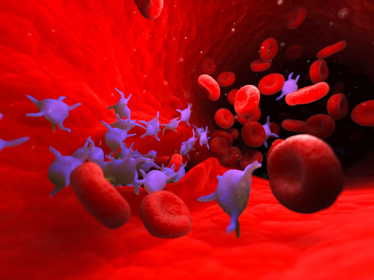
CellaVision DM9600 Digital Cell Morphology System

Offering high-volume first-pass efficiency through the next step in practical automation

The CellaVision® DM9600 digital cell morphology system is designed to automate the time-consuming, manual effort associated with traditional microscopy. The system leverages proven digital image analysis technology to locate and examine cells in blood and other body fluids, saving time, accelerating turnaround, and increasing technologists' productivity throughout high-volume labs. In addition, the CellaVision DM9600 system easily adapts to any hematology workflow within hospital IT environments.

  • Reduces review time for differentials to make more efficient use of experienced morphologists' skills
  • Standardizes differential results by automating cell location and pre-classification
  • Enables the utilization of skills across the entire organization through real-time collaboration and centrally stored images
  • Supports skill expansion via a comprehensive reference library and historical patient images and comments
  • Improves ergonomics by reducing the neck, wrist, and eye fatigue associated with traditional microscopy

Features & Benefits

CellaVision DM96

With the practical automation capabilities afforded by the CellaVision DM9600 Digital Cell Morphology System, you can eliminate the majority of manual steps commonly performed in blood cell differentials for maximum speed and productivity. The CellaVision DM9600 system enables this by automatically locating cells on a stained peripheral blood or body fluid smear, then pre-classifying, storing, and presenting the data for confirmation by a technologist. 


  • Automatic differential count on peripheral blood or body fluid smears
  • Pre-classification and grouping of similar white blood cells into 18 distinct cell classes
  • Pre-characterization of red blood cells for polychromasia, hypochromasia, aniso-, micro-, and macrocytosis, and poikilocytosis
  • Platelet estimation
  • High-quality images to detect cell inclusions
  • View of all cells within a specific cell class
  • Comparison of different cell classes on a single screen
  • Magnification of individual cells for detailed analysis
  • Software that enables the comparison of individual cells with user-defined reference cells
  • Continuous feed of slides, allowing for an escalating workload without increasing labor
  • Digital slide creation for future education and assessment
  • Improved ergonomics versus traditional microscopes
  • Bidirectional LIS support
  • Ability to share a database with other CellaVision analyzers
  • Real-time, remote access to slide data from anywhere within the network

The CellaVision DM9600 system also integrates seamlessly with your existing Siemens Healthineers hematology investments, including:

  • ADVIA® 2120i Hematology System
  • ADVIA Autoslide Slide Maker
  • Atellica® Data Manager
  • Scan body fluids, including cerebrospinal, peritoneal, and pleural fluids
  • Perform a preliminary differential of nucleated cells, allowing the user to scan the entire sample area
  • Pre-classify up to seven WBC classes with two magnification options
  • Tag Regions of Interest (ROI), add comments, and store and share images from anywhere in your network
  • Run body fluid slides concurrently with WBC and RBC differentials on stained peripheral blood smears
  • Analyze, reclassify, and sign slides from a remote location
  • Request second opinions and collaborate within and across hospitals
  • Compare new cell images with patient’s historical images in real time
  • Develop a program for education and competency testing of manual blood cell differentials
  • Standardize differential results, and assess, track, and document proficiency
  • Benchmark to a morphology expert

Technical Specifications

System components 

  • Slide scanning unit
  • CellaVision DM software
  • System Computer with Windows


Slide preparation methods (wedged)

  • Automated slide maker and stainers
  • Hemaprep® Automated Blood Smearing Instruments
  • Manual smears
  • Standard cytocentrifuge preparation


Slide handling

  • Requires barcoded-labeled slides with clipped/round corners
  • Slides are loaded into magazines with a capacity of 12 slides each, fits 8 magazines
  • Analyzes slides with blood smears and body fluids either batched in dedicated magazines or mixed when interfaced to a LIS


Stains

  • Romanowsky stains (May Grünwald Giemsa, Wright Giemsa, Wright)


Throughput

  • Peripheral blood 

        Up to 30 slides/hour for complete differential (100 WBC + RBC + PLT)

  • Digital slides

       Up to 20 slides/hour for 10 x 10 mm in 10X
       Up to 1.5 slides/hour for 10 x 10 mm in 10X + 50X

  • Body fluids (based on 6 mm sample area)

        Up to 15 slides/hour for differential (100 WBCs + 10X)
        Up to 3 slides/hour for differential (100 WBCs + 10X + 50X)


Storage capacity

  • Primary storage: On local hard drive up to 4000 slides (20 GB)
  • Secondary storage: Unlimited when transferred to external storage media


Weight and dimensions

  • Weight: 93 kg (205 lb)
  • Height: 790 mm (33.1 in.)
  • Width: 580 mm (22.8 in.)
  • Depth: 560 mm (22 in.)


Optional software/applications

  • CellaVision Advanced RBC Application
  • CellaVision Body Fluid Application
  • CellaVision Remote Review Software
  • CellaVision Server Software
  • CellaVision Proficiency Software


Accessories 

  • CellaVision Immersion Oil Pack
  • Barcode-labeled slide magazines
  • QC barcode labels
  • Label printer kit


Clearance

  • CE, 510(k)