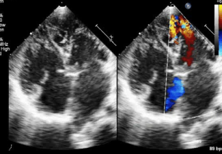
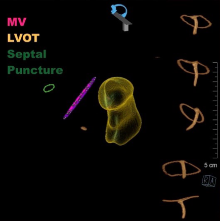
Typically, the left ventricle contains two groups of papillary muscles (PMs): the anterolateral and posteromedial PMs. These muscles give rise to chordae tendineae, which in turn anchor to the leaflets of the mitral valve.
In the developing heart, the PMs are initially seen as compacting columns emerging from the left ventricular (LV) wall, directed toward the mitral valve leaflets. These columns form a muscular ridge that later evolves into two distinct anterior and posterior papillary muscles through a process known as delamination. Abnormalities in this delamination process led to congenital anomalies of the papillary muscles, such as parachute mitral valve (PMV).