Discussion
This case involves a patient with a history of uterine cancer and a family history of breast cancer, raising the likelihood of an underlying hereditary cancer syndrome. Given her personal and family history, there is a potential risk for hereditary breast and ovarian cancer (HBOC) syndromes, particularly linked to BRCA1 and BRCA2 mutations. These mutations significantly increase the risk of developing breast, ovarian, and other cancers. Genetic counseling and, potentially, genetic testing are recommended to assess her risk profile further. This information would be crucial in tailoring her treatment and guiding future management strategies.


Clinical Case Study 1
Clinical History and Findings
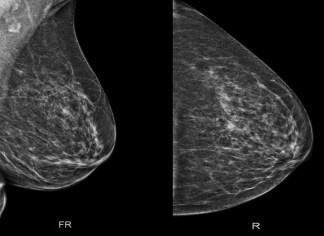
The 63-year-old patient was diagnosed with breast cancer based on the following imaging modalities: mammography, ultrasound, and subsequent ultrasound-guided biopsy results.
Key Findings
Mammogram and Ultrasound Findings
• Left breast tissue: Heterogeneously dense.
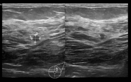
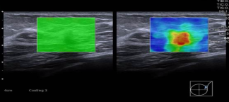
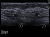
• Mass 1 (12-1 o’clock): Round, partially obscured mass located in the mid-upper/slightly outer quadrant of the left breast. Correlated with a hypoechoic mass on ultrasound.
• Mass 2 (2 o’clock): Irregular mass located in the deep upper outer quadrant of the left breast. Correlated with an angular margin and hypoechoic appearance on ultrasound.
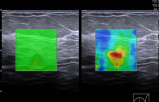
• Elastography: Demonstrated increased stiffness and hardness in both masses, suggesting a higher likelihood of malignancy.
Biopsy Results
Both Mass 1 (12-1 o’clock) and Mass 2 (2 o’clock) were biopsied under ultrasound guidance and confirmed to be malignant.
Impression
• The imaging and elastography findings, combined with the biopsy results, confirm the diagnosis of breast cancer, with malignancies identified in both masses.
• The initial BI-RADS 4 category, indicating a “suspicious abnormality” with a low to moderate suspicion of malignancy, justified the decision to pursue a biopsy, which confirmed the cancer diagnosis.
Discussion
This case involves a patient with a history of uterine cancer and a family history of breast cancer, raising the likelihood of an underlying hereditary cancer syndrome. Given her personal and family history, there is a potential risk for hereditary breast and ovarian cancer (HBOC) syndromes, particularly linked to BRCA1 and BRCA2 mutations. These mutations significantly increase the risk of developing breast, ovarian, and other cancers. Genetic counseling and, potentially, genetic testing are recommended to assess her risk profile further. This information would be crucial in tailoring her treatment and guiding future management strategies.


Clinical Case Study 1
Clinical History and Findings
The 63-year-old patient was diagnosed with breast cancer based on the following imaging modalities: mammography, ultrasound, and subsequent ultrasound-guided biopsy results.
Key Findings
Mammogram and Ultrasound Findings
• Left breast tissue: Heterogeneously dense.
• Mass 1 (12-1 o’clock): Round, partially obscured mass located in the mid-upper/slightly outer quadrant of the left breast. Correlated with a hypoechoic mass on ultrasound.
• Mass 2 (2 o’clock): Irregular mass located in the deep upper outer quadrant of the left breast. Correlated with an angular margin and hypoechoic appearance on ultrasound.
• Elastography: Demonstrated increased stiffness and hardness in both masses, suggesting a higher likelihood of malignancy.
Biopsy Results
Both Mass 1 (12-1 o’clock) and Mass 2 (2 o’clock) were biopsied under ultrasound guidance and confirmed to be malignant.
Impression
• The imaging and elastography findings, combined with the biopsy results, confirm the diagnosis of breast cancer, with malignancies identified in both masses.
• The initial BI-RADS 4 category, indicating a “suspicious abnormality” with a low to moderate suspicion of malignancy, justified the decision to pursue a biopsy, which confirmed the cancer diagnosis.

Discussion
This case involves a patient with a history of uterine cancer and a family history of breast cancer, raising the likelihood of an underlying hereditary cancer syndrome. Given her personal and family history, there is a potential risk for hereditary breast and ovarian cancer (HBOC) syndromes, particularly linked to BRCA1 and BRCA2 mutations. These mutations significantly increase the risk of developing breast, ovarian, and other cancers. Genetic counseling and, potentially, genetic testing are recommended to assess her risk profile further. This information would be crucial in tailoring her treatment and guiding future management strategies.
Conclusion
This case highlights the importance of vigilant follow-up in patients with new findings on imaging, even in the context of routine cancer screening. The discovery of metastatic adenocarcinoma during a breast cancer workup demonstrates the complexity of cancer detection and the role of advanced imaging and biopsy in guiding diagnosis and treatment.
Early detection of primary lung cancer might have altered the patient’s prognosis; however, at this stage, the extensive metastasis significantly limits treatment options. Further management will likely focus on palliative care and symptom control.

Clinical Case Study 2
Advanced Metastatic Adenocarcinoma presenting in a 55-Year-Old Female
Clinical History
A 55-year-old female with a positive family history of breast cancer (sister diagnosed in her 40s and a paternal aunt with breast cancer). The patient underwent mammogram screening in 2020, which was negative and categorized as BI-RADS 1. Four years later, she presented for a follow-up mammogram.
Mammogram Findings
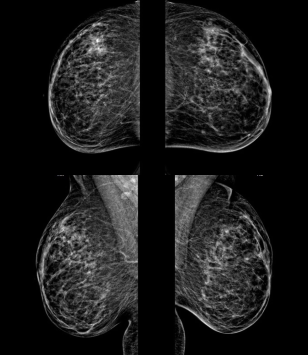
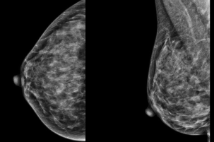
• Views Obtained: CC (Craniocaudal) and MLO (Mediolateral Oblique) views of both breasts.
• Breast Density: Heterogeneously dense breast tissue.
• Masses: Newly identified round isodense masses in the upper outer and lower inner quadrants of the left breast, as well as a round mass in the lower inner quadrant of the right breast.
• Microcalcifications: No suspicious clustered microcalcifications observed.
Ultrasound Findings
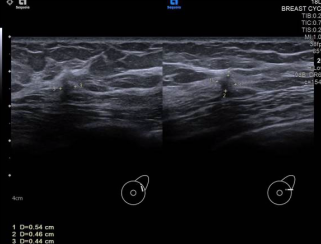
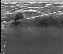
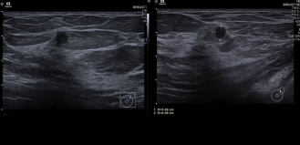
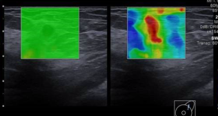
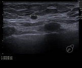
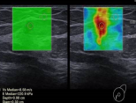
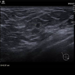
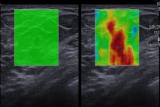
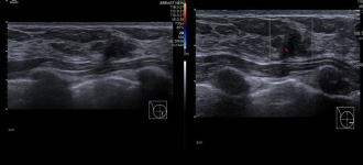
• Upper Outer Left Breast: A round mass with microlobulated margins was noted in the upper outer quadrant, measuring 0.5 x 0.6 x 0.6 cm. The mass appeared hard on elastography.
• Lower Inner Left Breast: Multiple round hypoechoic masses were identified.
• Lower Inner Right Breast: A similar round mass was identified, measuring 0.5 x 0.3 x 0.3 cm.
• Axillary Lymph Nodes: No axillary lymphadenopathy.


Impression
Several newly identified round hypoechoic masses were observed in the left and right breasts. The most prominent lesion, located in the upper outer quadrant of the left breast, displayed irregular margins, raising suspicion for malignancy.
This mass appeared to be more concerning due to its size, shape, and the presence of irregular borders, which are often indicative of malignant lesions. Further imaging studies and biopsy were recommended to better characterize the lesion and determine the appropriate course of action for diagnosis and management.
• BI-RADS Category 4: Suspicious abnormality with low to moderate suspicion of malignancy. A biopsy was recommended.
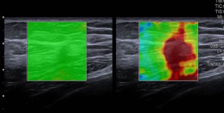
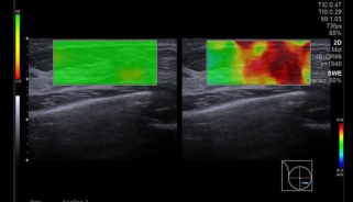
• Ultrasound 2D-SWE: Shows high shear wave velocities and hard tissue characteristics, suggesting a high level of suspicion for malignancy (as shown in the images).
Biopsy and Diagnosis
• Ultrasound-guided Core Biopsy: A core biopsy was performed on the mass in the upper outer left breast. Three clips were placed at the biopsy site, and a post-procedure mammogram confirmed correct clip placement.
• Diagnosis: The biopsy confirmed metastatic adenocarcinoma, consistent with a pulmonary origin.
• Recommendations: Further clinical evaluation and imaging were advised to identify the suspected primary lung cancer.
Subsequent Imaging and Findings
CT Scan
Multiple mass lesions of varying sizes were detected in the left lung, liver, spleen, left adrenal gland, and left ovary. Extensive sclerotic metastatic lesions were observed in the ribs, dorsal spine, lumbosacral spine, and pelvic bones.
Brain MRI
Numerous supra- and infratentorial metastatic brain lesions, some measuring up to 1 cm, were identified. Several lesions exhibited hemorrhagic components but did not show perifocal edema.
Discussion
This case presents an unusual sequence of events. The patient, who was undergoing routine breast cancer screening due to a family history of breast cancer, was ultimately diagnosed with metastatic adenocarcinoma of pulmonary origin.
The initial mammogram revealed new, suspicious masses, prompting an ultrasound and biopsy. The biopsy confirmed that these masses were metastatic, consistent with adenocarcinoma from the lung.
Although breast cancer was the primary concern due to her family history, this case underscores the importance of comprehensive assessment when new findings emerge during routine screenings.
Metastatic Adenocarcinoma of Pulmonary Origin
The patient’s widespread metastases, confirmed through CT and MRI, involved multiple organs, including the lung, liver, spleen, adrenal gland, ovary, as well as bones, and extensive brain involvement. The diagnosis of metastatic adenocarcinoma of pulmonary origin suggests that the patient had an undiagnosed primary lung malignancy that had already metastasized by the time of her breast cancer screening.
Conclusion
This case highlights the importance of vigilant follow-up in patients with new findings on imaging, even in the context of routine cancer screening. The discovery of metastatic adenocarcinoma during a breast cancer workup demonstrates the complexity of cancer detection and the role of advanced imaging and biopsy in guiding diagnosis and treatment.
Early detection of primary lung cancer might have altered the patient’s prognosis; however, at this stage, the extensive metastasis significantly limits treatment options. Further management will likely focus on palliative care and symptom control.

Clinical Case Study 2
Advanced Metastatic Adenocarcinoma presenting in a 55-Year-Old Female
Clinical History
A 55-year-old female with a positive family history of breast cancer (sister diagnosed in her 40s and a paternal aunt with breast cancer). The patient underwent mammogram screening in 2020, which was negative and categorized as BI-RADS 1. Four years later, she presented for a follow-up mammogram.
Mammogram Findings
• Views Obtained: CC (Craniocaudal) and MLO (Mediolateral Oblique) views of both breasts.
• Breast Density: Heterogeneously dense breast tissue.
• Masses: Newly identified round isodense masses in the upper outer and lower inner quadrants of the left breast, as well as a round mass in the lower inner quadrant of the right breast.
• Microcalcifications: No suspicious clustered microcalcifications observed.
Ultrasound Findings
• Upper Outer Left Breast: A round mass with microlobulated margins was noted in the upper outer quadrant, measuring 0.5 x 0.6 x 0.6 cm. The mass appeared hard on elastography.
• Lower Inner Left Breast: Multiple round hypoechoic masses were identified.
• Lower Inner Right Breast: A similar round mass was identified, measuring 0.5 x 0.3 x 0.3 cm.
• Axillary Lymph Nodes: No axillary lymphadenopathy.


Impression
Several newly identified round hypoechoic masses were observed in the left and right breasts. The most prominent lesion, located in the upper outer quadrant of the left breast, displayed irregular margins, raising suspicion for malignancy.
This mass appeared to be more concerning due to its size, shape, and the presence of irregular borders, which are often indicative of malignant lesions. Further imaging studies and biopsy were recommended to better characterize the lesion and determine the appropriate course of action for diagnosis and management.
• BI-RADS Category 4: Suspicious abnormality with low to moderate suspicion of malignancy. A biopsy was recommended.
• Ultrasound 2D-SWE: Shows high shear wave velocities and hard tissue characteristics, suggesting a high level of suspicion for malignancy (as shown in the images).
Biopsy and Diagnosis
• Ultrasound-guided Core Biopsy: A core biopsy was performed on the mass in the upper outer left breast. Three clips were placed at the biopsy site, and a post-procedure mammogram confirmed correct clip placement.
• Diagnosis: The biopsy confirmed metastatic adenocarcinoma, consistent with a pulmonary origin.
• Recommendations: Further clinical evaluation and imaging were advised to identify the suspected primary lung cancer.
Subsequent Imaging and Findings
CT Scan
Multiple mass lesions of varying sizes were detected in the left lung, liver, spleen, left adrenal gland, and left ovary. Extensive sclerotic metastatic lesions were observed in the ribs, dorsal spine, lumbosacral spine, and pelvic bones.
Brain MRI
Numerous supra- and infratentorial metastatic brain lesions, some measuring up to 1 cm, were identified. Several lesions exhibited hemorrhagic components but did not show perifocal edema.
Discussion
This case presents an unusual sequence of events. The patient, who was undergoing routine breast cancer screening due to a family history of breast cancer, was ultimately diagnosed with metastatic adenocarcinoma of pulmonary origin.
The initial mammogram revealed new, suspicious masses, prompting an ultrasound and biopsy. The biopsy confirmed that these masses were metastatic, consistent with adenocarcinoma from the lung.
Although breast cancer was the primary concern due to her family history, this case underscores the importance of comprehensive assessment when new findings emerge during routine screenings.
Metastatic Adenocarcinoma of Pulmonary Origin
The patient’s widespread metastases, confirmed through CT and MRI, involved multiple organs, including the lung, liver, spleen, adrenal gland, ovary, as well as bones, and extensive brain involvement. The diagnosis of metastatic adenocarcinoma of pulmonary origin suggests that the patient had an undiagnosed primary lung malignancy that had already metastasized by the time of her breast cancer screening.
Conclusion
This case highlights the importance of vigilant follow-up in patients with new findings on imaging, even in the context of routine cancer screening. The discovery of metastatic adenocarcinoma during a breast cancer workup demonstrates the complexity of cancer detection and the role of advanced imaging and biopsy in guiding diagnosis and treatment.
Early detection of primary lung cancer might have altered the patient’s prognosis; however, at this stage, the extensive metastasis significantly limits treatment options. Further management will likely focus on palliative care and symptom control.


Clinical Case Study 3
Breast Cancer detection in a 49-year-old female during initial mammogram screening
Clinical History
A 49-year-old female patient presented for her first screening mammogram. She had no prior history of breast cancer screening or imaging.
Mammogram Findings
• Views Obtained: Craniocaudal (CC) and mediolateral oblique (MLO) views of both breasts, bilateral true lateral views, and a right magnification view.
• Breast Density: The breast tissue was noted to be heterogeneously dense, which may obscure the detection of small masses.
Findings
• No evidence of suspicious dominant masses.
• Scattered benign-appearing microcalcifications.
• No evidence of suspicious clustered microcalcifications.
Ultrasound Findings
• Right Breast (Lower Inner Quadrant): An irregular hypoechoic mass with angular margins was detected, measuring 1.2 x 0.6 x 1 cm. The mass demonstrated increased internal vascularity.
• Elastography: The mass appeared stiffer than the surrounding tissue, with high shear wave velocities, raising suspicion for malignancy.
• Additional Findings: A few small cysts were noted in both breasts, which were considered benign.
Impression
• The irregular hypoechoic mass in the lower inner quadrant of the right breast, along with angular margins, increased vascularity, and marked stiffness on elastography, was suspicious for malignancy.
• Recommendation: Biopsy of the mass was recommended to confirm its nature.
Biopsy and Pathology Results:
• Procedure: An ultrasound-guided core biopsy of the mass in the lower inner right breast was performed.
• Pathology Results: The biopsy confirmed the presence of malignancy.
Impression and Conclusion
The imaging findings, particularly on ultrasound and elastography, indicated a suspicious lesion, which was confirmed as malignant after biopsy. Early detection of this malignancy during the patient’s first screening mammogram underscores the importance of breast cancer screening, even in the absence of symptoms. Further management will likely involve an oncological consultation for staging, treatment planning, and potential surgical intervention.
Key Takeaways
• Breast Density: Heterogeneously dense breast tissue can obscure mammographic findings, highlighting the critical role of ultrasound in detecting subtle lesions.
• Ultrasound and Elastography: The irregular hypoechoic mass with increased vascularity and stiffness raised suspicion for malignancy, prompting the biopsy.
• Early Detection: Despite the initial screening, this case demonstrates the value of combining mammography and ultrasound for comprehensive breast cancer detection, especially in patients with dense breast tissue.
The next steps for the patient should involve a multidisciplinary team to plan for definitive treatment based on the stage and extent of the disease.
"Early detection of this malignancy during the patient’s first screening mammogram underscores the importance of breast cancer screening, even in the absence of symptoms."

King Hussein Cancer Center, Jordan