
Radiotherapy and Systemic Therapy
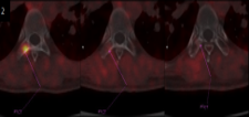
The patient with osseous metastasis to T8 initially underwent stereotactic body radiotherapy (SBRT) delivering 30 Gy in 5 fractions, resulting in resolution of bone pain ref fig2. Subsequent FAPI PET-CT revealed disease progression, prompting initiation of induction therapy with Gemcitabine, Cisplatin, and Durvalumab.
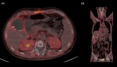
Three months later, follow-up FAPI PET-CT demonstrated significant metabolic regression of retrocrural and upper abdominal lymphadenopathies refer fig 4a & 4b; however, most hepatic nodular metastases showed increased uptake intensity and metabolic volume, indicating progression. Consequently, second-line treatment with Nalirifox was commenced, with consideration for selective Yttrium-90 internal radiation therapy (SIRT) based on post- treatment restaging.


Clinical Case Study
Advanced Gallbladder Adenocarcinoma with Osseous Metastasis Evaluated by FAPI PET-CT Patient Profile:
Patient Profile
A 31-year-old male presented with a diagnosis of poorly differentiated biliary-type adenocarcinoma of the gallbladder (Grade 3), initially identified following laparoscopic cholecystectomy. Histopathology revealed a 3 cm tumor invading the liver (pT3), with positive hepatic resection margin (R1), lymphovascular and perineural invasion, and involvement of one cystic duct lymph node (pN1).
Initial Staging and Imaging
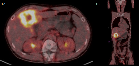
The patient underwent FAPI PET-CT on the advance PET-CT platform with FlowMotion technology which enables continuous bed movement during PET acquisition, replacing the conventional step-and-shoot approach. This method allows tailored scanning speeds for different anatomical regions, improving image quality and reducing radiation exposure. This process minimizes motion artifacts & therefore consistent quantitative accuracy across the entire scan. Due to demonstrated hypermetabolic activity in hepatic segments IV/V and a suspicious focus in the right transverse process of T8. MRI of the thoracic spine confirmed metastatic involvement at T8.

Management and Surgical Intervention
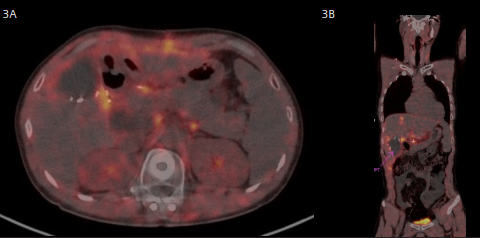
The patient underwent a second hepatobiliary resection (segments IV/V) with re-excision of the positive liver bed margin and regional lymph node sampling (Refer to figure 3A & 3B) Pathology confirmed residual moderately-to-poorly differentiated adenocarcinoma with two metastatic nodules in segment IV and additional positive lymph nodes.

Radiotherapy and Systemic Therapy
The patient with osseous metastasis to T8 initially underwent stereotactic body radiotherapy (SBRT) delivering 30 Gy in 5 fractions, resulting in resolution of bone pain ref fig2. Subsequent FAPI PET-CT revealed disease progression, prompting initiation of induction therapy with Gemcitabine, Cisplatin, and Durvalumab.
Three months later, follow-up FAPI PET-CT demonstrated significant metabolic regression of retrocrural and upper abdominal lymphadenopathies refer fig 4a & 4b; however, most hepatic nodular metastases showed increased uptake intensity and metabolic volume, indicating progression. Consequently, second-line treatment with Nalirifox was commenced, with consideration for selective Yttrium-90 internal radiation therapy (SIRT) based on post- treatment restaging.


Clinical Case Study
Advanced Gallbladder Adenocarcinoma with Osseous Metastasis Evaluated by FAPI PET-CT Patient Profile:
Patient Profile
A 31-year-old male presented with a diagnosis of poorly differentiated biliary-type adenocarcinoma of the gallbladder (Grade 3), initially identified following laparoscopic cholecystectomy. Histopathology revealed a 3 cm tumor invading the liver (pT3), with positive hepatic resection margin (R1), lymphovascular and perineural invasion, and involvement of one cystic duct lymph node (pN1).
Initial Staging and Imaging
The patient underwent FAPI PET-CT on the advance PET-CT platform with FlowMotion technology which enables continuous bed movement during PET acquisition, replacing the conventional step-and-shoot approach. This method allows tailored scanning speeds for different anatomical regions, improving image quality and reducing radiation exposure. This process minimizes motion artifacts & therefore consistent quantitative accuracy across the entire scan. Due to demonstrated hypermetabolic activity in hepatic segments IV/V and a suspicious focus in the right transverse process of T8. MRI of the thoracic spine confirmed metastatic involvement at T8.

Management and Surgical Intervention
The patient underwent a second hepatobiliary resection (segments IV/V) with re-excision of the positive liver bed margin and regional lymph node sampling (Refer to figure 3A & 3B) Pathology confirmed residual moderately-to-poorly differentiated adenocarcinoma with two metastatic nodules in segment IV and additional positive lymph nodes.

Radiotherapy and Systemic Therapy
The patient with osseous metastasis to T8 initially underwent stereotactic body radiotherapy (SBRT) delivering 30 Gy in 5 fractions, resulting in resolution of bone pain ref fig2. Subsequent FAPI PET-CT revealed disease progression, prompting initiation of induction therapy with Gemcitabine, Cisplatin, and Durvalumab.
Three months later, follow-up FAPI PET-CT demonstrated significant metabolic regression of retrocrural and upper abdominal lymphadenopathies refer fig 4a & 4b; however, most hepatic nodular metastases showed increased uptake intensity and metabolic volume, indicating progression. Consequently, second-line treatment with Nalirifox was commenced, with consideration for selective Yttrium-90 internal radiation therapy (SIRT) based on post- treatment restaging.

Teaching Point
Ga-68 FAPI PET imaging stands out as a transformative tracer in oncology due to its ability to target fibroblast activation protein (FAP), which is highly expressed in cancer-associated fibroblasts within the tumor microenvironment. Unlike conventional tracers such as FDG, Ga-68 FAPI offers exceptionally high tumor-to-background contrast, minimal physiological uptake in most normal tissues, and rapid clearance, enabling clearer visualization of lesions even in challenging regions like the abdomen and pelvis. Its unique mechanism provides superior sensitivity for detecting primary tumors, metastases, and stromal involvement, while reducing false positives linked to inflammation or glucose metabolism. These advantages position Ga-68 FAPI as a next-generation imaging agent with broad applicability and potential theranostic benefits in precision.
"Ga-68 FAPI PET imaging stands out as a transformative tracer in oncology due to its ability to target fibroblast activation protein (FAP), which is highly expressed in cancer-associated fibroblasts within the tumor microenvironment."

Medicine Consultant
Al Zahra Hospital, Dubai, UAE
