Clinical Case Study 2
Diagnostic Mammography Case
Patient History
The patient is a 55-year-old woman undergoing routine screening mammography. She has no personal or family history of breast cancer or any other type of cancer. No prior imaging studies, including those from outside facilities, are available for comparison. This routine screening is conducted to facilitate early detection and ongoing monitoring of breast health in the absence of previous findings or known risk factors.
Mammography Findings
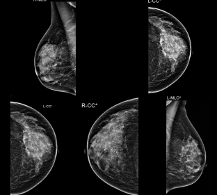
The mammography examination included bilateral digital craniocaudal (CC) and mediolateral oblique (MLO) views, revealing heterogeneously dense breast tissue (ACR C), which may obscure the detection of small masses. Findings included grouped, suspicious pleomorphic, and coarse heterogeneous calcifications located in the anterior third of the left breast, at the 3 o’clock position in the para-areolar region, with corresponding ultrasound evaluation. Additional scattered grouped calcifications, some exhibiting a popcorn-like appearance consistent with involuted fibroadenomata, were identified in both breasts. These were located in the upper outer quadrant of the right breast (middle third) and the posterior third of the left breast (lower outer quadrant). No architectural distortion or definite masses were detected. The ultrasound findings correlated well with the mammographic observations.
Image Impression and Clinical Management
The mammography findings indicate a BIRADS Assessment Category 4: Suspicious. In the left breast, there is a non-palpable cluster of morphologically suspicious calcifications in the para-areolar region. This finding warrants further investigation, either through ultrasound-guided biopsy and pathology or wire localization with surgical excision. Additionally, bilateral dynamic contrast-enhanced (DCE) MRI of the breasts with contrast is recommended to evaluate the extent of the disease and to rule out bilaterality.
Partially calcified involuted fibroadenomata were identified in both breasts, each demonstrating morphologically benign features on ultrasound. These findings are recommended for follow-up in six months to monitor stability.The primary priority is to investigate the suspicious calcifications in the left breast to ensure timely diagnosis and appropriate management.

Clinical Case Study 1
Diagnostic Mammography Case
Patient History
The patient is a 70-year-old woman who presented for diagnostic mammography due to a palpable lump in the left breast. She has no personal or family history of breast or other cancers. No prior imaging studies, including external facility reports, were available for comparison. Mammography was performed to evaluate the findings and provide further diagnostic insights.
Mammography Findings
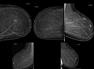
Bilateral digital craniocaudal (CC) and mediolateral oblique (MLO) views were obtained, showing breasts with predominantly fatty composition (ACR A). Two areas of benign-appearing focal asymmetries were identified in the anterior and middle thirds of the left breast, at approximately the 12 o’clock and 1 o’clock positions, located 5 cm and 9 cm from the nipple, respectively.
The asymmetry closer to the nipple corresponds to the clinically palpable lump, with no associated microcalcifications, spiculations, or architectural distortion observed. Diffuse, bilateral, benign-appearing round calcifications were noted throughout both breasts. No suspicious masses, microcalcifications, or distortions were identified in the left breast.
Ultrasound Findings
Targeted ultrasound of the left breast revealed an oval-shaped, homogenous, hyperechoic lesion measuring 1.6 x 1.0 cm at the site of clinical concern. The lesion exhibited no abnormal vascularity. An additional asymmetry 5 cm above the primary lesion was identified, likely representing an intramammary lymph node.
No solid lesions, cysts, retroareolar ductal dilation, or abnormalities in the nipple-areolar complex were noted in the right breast.
This case highlights the importance of integrating mammography and ultrasound findings for comprehensive evaluation of breast abnormalities, particularly in patients with fatty breast composition.
Image Impression and Clinical Management
The mammography findings indicate a BI-RADS Assessment Category 2: Benign. The primary finding is a solitary, palpable, non-calcified lesion in the left breast, most likely a hematoma. A follow-up examination is recommended in four weeks to monitor for any changes in size or characteristics.
Additional findings include bilateral coarse benign calcific foci, with no evidence of malignancy detected in either breast. For ongoing surveillance and patient management, a routine follow-up mammogram is advised in one year.

Clinical Case Study 2
Diagnostic Mammography Case
Patient History
The patient is a 55-year-old woman undergoing routine screening mammography. She has no personal or family history of breast cancer or any other type of cancer. No prior imaging studies, including those from outside facilities, are available for comparison. This routine screening is conducted to facilitate early detection and ongoing monitoring of breast health in the absence of previous findings or known risk factors.
Mammography Findings
The mammography examination included bilateral digital craniocaudal (CC) and mediolateral oblique (MLO) views, revealing heterogeneously dense breast tissue (ACR C), which may obscure the detection of small masses. Findings included grouped, suspicious pleomorphic, and coarse heterogeneous calcifications located in the anterior third of the left breast, at the 3 o’clock position in the para-areolar region, with corresponding ultrasound evaluation. Additional scattered grouped calcifications, some exhibiting a popcorn-like appearance consistent with involuted fibroadenomata, were identified in both breasts. These were located in the upper outer quadrant of the right breast (middle third) and the posterior third of the left breast (lower outer quadrant). No architectural distortion or definite masses were detected. The ultrasound findings correlated well with the mammographic observations.
Image Impression and Clinical Management
The mammography findings indicate a BIRADS Assessment Category 4: Suspicious. In the left breast, there is a non-palpable cluster of morphologically suspicious calcifications in the para-areolar region. This finding warrants further investigation, either through ultrasound-guided biopsy and pathology or wire localization with surgical excision. Additionally, bilateral dynamic contrast-enhanced (DCE) MRI of the breasts with contrast is recommended to evaluate the extent of the disease and to rule out bilaterality.
Partially calcified involuted fibroadenomata were identified in both breasts, each demonstrating morphologically benign features on ultrasound. These findings are recommended for follow-up in six months to monitor stability.The primary priority is to investigate the suspicious calcifications in the left breast to ensure timely diagnosis and appropriate management.

Clinical Case Study 1
Diagnostic Mammography Case
Patient History
The patient is a 70-year-old woman who presented for diagnostic mammography due to a palpable lump in the left breast. She has no personal or family history of breast or other cancers. No prior imaging studies, including external facility reports, were available for comparison. Mammography was performed to evaluate the findings and provide further diagnostic insights.
Mammography Findings
Bilateral digital craniocaudal (CC) and mediolateral oblique (MLO) views were obtained, showing breasts with predominantly fatty composition (ACR A). Two areas of benign-appearing focal asymmetries were identified in the anterior and middle thirds of the left breast, at approximately the 12 o’clock and 1 o’clock positions, located 5 cm and 9 cm from the nipple, respectively.
The asymmetry closer to the nipple corresponds to the clinically palpable lump, with no associated microcalcifications, spiculations, or architectural distortion observed. Diffuse, bilateral, benign-appearing round calcifications were noted throughout both breasts. No suspicious masses, microcalcifications, or distortions were identified in the left breast.
Ultrasound Findings
Targeted ultrasound of the left breast revealed an oval-shaped, homogenous, hyperechoic lesion measuring 1.6 x 1.0 cm at the site of clinical concern. The lesion exhibited no abnormal vascularity. An additional asymmetry 5 cm above the primary lesion was identified, likely representing an intramammary lymph node.
No solid lesions, cysts, retroareolar ductal dilation, or abnormalities in the nipple-areolar complex were noted in the right breast.
This case highlights the importance of integrating mammography and ultrasound findings for comprehensive evaluation of breast abnormalities, particularly in patients with fatty breast composition.
Image Impression and Clinical Management
The mammography findings indicate a BI-RADS Assessment Category 2: Benign. The primary finding is a solitary, palpable, non-calcified lesion in the left breast, most likely a hematoma. A follow-up examination is recommended in four weeks to monitor for any changes in size or characteristics.
Additional findings include bilateral coarse benign calcific foci, with no evidence of malignancy detected in either breast. For ongoing surveillance and patient management, a routine follow-up mammogram is advised in one year.

Clinical Case Study 2
Diagnostic Mammography Case
Patient History
The patient is a 55-year-old woman undergoing routine screening mammography. She has no personal or family history of breast cancer or any other type of cancer. No prior imaging studies, including those from outside facilities, are available for comparison. This routine screening is conducted to facilitate early detection and ongoing monitoring of breast health in the absence of previous findings or known risk factors.
Mammography Findings
The mammography examination included bilateral digital craniocaudal (CC) and mediolateral oblique (MLO) views, revealing heterogeneously dense breast tissue (ACR C), which may obscure the detection of small masses. Findings included grouped, suspicious pleomorphic, and coarse heterogeneous calcifications located in the anterior third of the left breast, at the 3 o’clock position in the para-areolar region, with corresponding ultrasound evaluation. Additional scattered grouped calcifications, some exhibiting a popcorn-like appearance consistent with involuted fibroadenomata, were identified in both breasts. These were located in the upper outer quadrant of the right breast (middle third) and the posterior third of the left breast (lower outer quadrant). No architectural distortion or definite masses were detected. The ultrasound findings correlated well with the mammographic observations.
Image Impression and Clinical Management
The mammography findings indicate a BIRADS Assessment Category 4: Suspicious. In the left breast, there is a non-palpable cluster of morphologically suspicious calcifications in the para-areolar region. This finding warrants further investigation, either through ultrasound-guided biopsy and pathology or wire localization with surgical excision. Additionally, bilateral dynamic contrast-enhanced (DCE) MRI of the breasts with contrast is recommended to evaluate the extent of the disease and to rule out bilaterality.
Partially calcified involuted fibroadenomata were identified in both breasts, each demonstrating morphologically benign features on ultrasound. These findings are recommended for follow-up in six months to monitor stability.The primary priority is to investigate the suspicious calcifications in the left breast to ensure timely diagnosis and appropriate management.
Conclusion
Siemens Healthineers’ 3D Tomosynthesis Digital Mammography technology offers advanced imaging capabilities for the precise evaluation of breast conditions. Both case studies highlight the clinical efficacy and reliability of this cutting-edge imaging system in routine screening and diagnostic settings.
Continued adoption and utilization of 3D Tomosynthesis Digital Mammography have the potential to enhance early detection and improve patient outcomes in breast health management.
"Siemens Healthineers’ 3D Tomosynthesis Digital Mammography technology offers advanced imaging capabilities for the precise evaluation of breast conditions."

Consultant of Diagnostic Breast Imaging
Women and Fetal Imaging Center (WAFI)
Cairo, Egypt
"Continued adoption and utilization of 3D Tomosynthesis Digital Mammography have the potential to enhance early detection and improve patient outcomes in breast health management."

Women and Fetal Imaging Center (WAFI)
Cairo, Egypt