Clinical Impact of Deep Resolve in MSK Imaging
Existing MSK imaging protocols were adapted to incorporate Deep Resolve reconstruction. Given that MSK MRI relies heavily on turbo spin echo (TSE) based acquisitions, the majority of protocols were successfully implemented using the deep learning reconstruction framework. These adaptations allowed improvements in both spatial resolution and acquisition efficiency without altering the underlying contrast mechanisms.
Workflow Enhancement
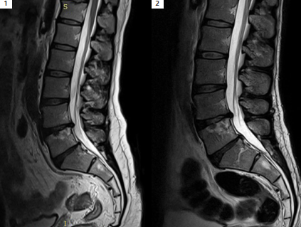
AI-based image reconstruction significantly reduces MRI acquisition times, directly improving patient throughput and departmental workflow efficiency. Shorter scan durations decrease overall examination time, enabling higher system utilization and greater scheduling flexibility. While increased throughput may require complementary operational adjustments, such as enhanced patient preparation and staffing support, the net effect is improved access to MRI services.
For selected examinations, particularly large-joint studies such as knee MRI, AI-driven acceleration enables a shift toward more flexible workflows, including walk-in imaging. Reduced scan times and simplified positioning allow examinations to be performed promptly upon patient arrival, enhancing patient convenience and reducing waiting times without compromising diagnostic image quality.
Resolution Enhancement
A primary focus of protocol optimization was achieving higher spatial resolution, particularly for imaging small joints such as the wrist, elbow, fingers, and toes. This was accomplished through matrix size enhancement enabled by the Deep Resolve DL algorithm, which reconstructs images at twice the acquired in-plane resolution. For example, an image acquired with a 256 × 256 matrix is reconstructed to an effective resolution of 512 × 512 using the AI-based reconstruction network, resulting in improved delineation of fine anatomical details.
Further gains in spatial resolution were achieved by reducing slice thickness. In many finger and small-joint examinations, slice thickness was successfully reduced from the conventional 3 mm to 2 mm, allowing improved visualization of small structures while maintaining acceptable signal-to-noise characteristics.
Acceleration and Workflow Improvement
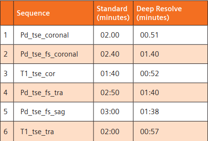
In a high-capacity MSK imaging environment, reducing scan duration while preserving diagnostic image quality is a key operational objective. Minimizing the time required for each patient examination cycle directly enhances MRI accessibility and throughput, enabling a greater number of patients to be scanned within the same operational timeframe.
The acceleration benefits provided by Deep Resolve are particularly valuable in clinical scenarios where rapid image acquisition is essential, including patients who are claustrophobic, experiencing severe pain, pediatric patients, elderly individuals, or those unable to remain still for extended periods. Additional benefits are observed in high-risk patients, such as those requiring sedation, intensive care unit (ICU) patients, or individuals with implanted devices that necessitate reduced specific absorption rate (SAR) exposure, including neurostimulators.
By enabling shorter scan times and enhanced image quality simultaneously, Deep Resolve supports more efficient MSK imaging workflows while maintaining diagnostic reliability across a wide range of patient populations.
Workflow Enhancement
AI-based image reconstruction significantly reduces MRI acquisition times, directly improving patient throughput and departmental workflow efficiency. Shorter scan durations decrease overall examination time, enabling higher system utilization and greater scheduling flexibility. While increased throughput may require complementary operational adjustments, such as enhanced patient preparation and staffing support, the net effect is improved access to MRI services.
For selected examinations, particularly large-joint studies such as knee MRI, AI-driven acceleration enables a shift toward more flexible workflows, including walk-in imaging. Reduced scan times and simplified positioning allow examinations to be performed promptly upon patient arrival, enhancing patient convenience and reducing waiting times without compromising diagnostic image quality.
Resolution Enhancement
A primary focus of protocol optimization was achieving higher spatial resolution, particularly for imaging small joints such as the wrist, elbow, fingers, and toes. This was accomplished through matrix size enhancement enabled by the Deep Resolve DL algorithm, which reconstructs images at twice the acquired in-plane resolution. For example, an image acquired with a 256 × 256 matrix is reconstructed to an effective resolution of 512 × 512 using the AI-based reconstruction network, resulting in improved delineation of fine anatomical details.
Further gains in spatial resolution were achieved by reducing slice thickness. In many finger and small-joint examinations, slice thickness was successfully reduced from the conventional 3 mm to 2 mm, allowing improved visualization of small structures while maintaining acceptable signal-to-noise characteristics.
Acceleration and Workflow Improvement
In a high-capacity MSK imaging environment, reducing scan duration while preserving diagnostic image quality is a key operational objective. Minimizing the time required for each patient examination cycle directly enhances MRI accessibility and throughput, enabling a greater number of patients to be scanned within the same operational timeframe.
The acceleration benefits provided by Deep Resolve are particularly valuable in clinical scenarios where rapid image acquisition is essential, including patients who are claustrophobic, experiencing severe pain, pediatric patients, elderly individuals, or those unable to remain still for extended periods. Additional benefits are observed in high-risk patients, such as those requiring sedation, intensive care unit (ICU) patients, or individuals with implanted devices that necessitate reduced specific absorption rate (SAR) exposure, including neurostimulators.
By enabling shorter scan times and enhanced image quality simultaneously, Deep Resolve supports more efficient MSK imaging workflows while maintaining diagnostic reliability across a wide range of patient populations.
Workflow Enhancement
AI-based image reconstruction significantly reduces MRI acquisition times, directly improving patient throughput and departmental workflow efficiency. Shorter scan durations decrease overall examination time, enabling higher system utilization and greater scheduling flexibility. While increased throughput may require complementary operational adjustments, such as enhanced patient preparation and staffing support, the net effect is improved access to MRI services.
For selected examinations, particularly large-joint studies such as knee MRI, AI-driven acceleration enables a shift toward more flexible workflows, including walk-in imaging. Reduced scan times and simplified positioning allow examinations to be performed promptly upon patient arrival, enhancing patient convenience and reducing waiting times without compromising diagnostic image quality.
Clinical Cases
Findings: Thumb
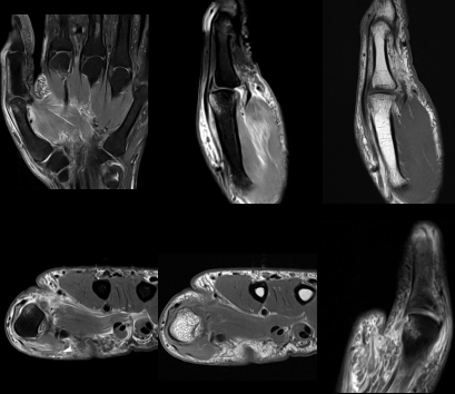
MRI demonstrates complete avulsion of the ulnar collateral ligament from the head of the first metacarpal. A small associated avulsion fracture fragment is present, with subtle proton density (PD) hyperintense marrow edema at the fracture site. These findings are consistent with an acute ulnar collateral ligament injury of the thumb.
Findings: Shoulder
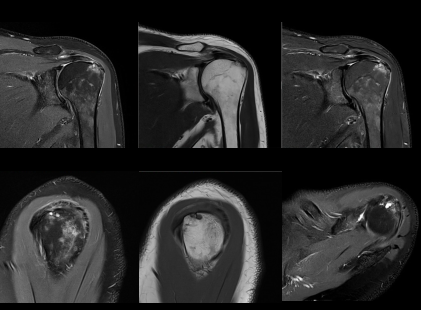
Magnetic resonance images demonstrate supraspinatus tendinosis with a high-grade partial-thickness tear involving the articular surface of the anterior fibers. Small T2-weighted hyperintense subcortical cystic changes are noted within the greater tuberosity at the supraspinatus tendon insertion, accompanied by surrounding marrow edema. These findings are consistent with chronic rotator cuff pathology with associated insertional changes.
Findings: Wrist (TFCC)
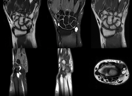
MRI of the wrist reveals T2 intermediate signal synovial thickening in the prestyloid region along the ulnar aspect, consistent with prestyloid synovitis. Additionally, a T2 hyperintense volar ganglion cyst is identified on the ulnar side of the wrist. No definitive full-thickness tear of the triangular fibrocartilage complex is visualized.
Findings: Thumb
MRI demonstrates complete avulsion of the ulnar collateral ligament from the head of the first metacarpal. A small associated avulsion fracture fragment is present, with subtle proton density (PD) hyperintense marrow edema at the fracture site. These findings are consistent with an acute ulnar collateral ligament injury of the thumb.
Findings: Shoulder
Magnetic resonance images demonstrate supraspinatus tendinosis with a high-grade partial-thickness tear involving the articular surface of the anterior fibers. Small T2-weighted hyperintense subcortical cystic changes are noted within the greater tuberosity at the supraspinatus tendon insertion, accompanied by surrounding marrow edema. These findings are consistent with chronic rotator cuff pathology with associated insertional changes.
Findings: Wrist (TFCC)
MRI of the wrist reveals T2 intermediate signal synovial thickening in the prestyloid region along the ulnar aspect, consistent with prestyloid synovitis. Additionally, a T2 hyperintense volar ganglion cyst is identified on the ulnar side of the wrist. No definitive full-thickness tear of the triangular fibrocartilage complex is visualized.
Findings: Thumb
MRI demonstrates complete avulsion of the ulnar collateral ligament from the head of the first metacarpal. A small associated avulsion fracture fragment is present, with subtle proton density (PD) hyperintense marrow edema at the fracture site. These findings are consistent with an acute ulnar collateral ligament injury of the thumb.
Authors

Medcare Orthopaedics and Spine Hospital Dubai, UAE

Medcare Orthopaedics and Spine Hospital Dubai, UAE