Discussion
Reports of papillary muscle (PM) aberrations include parachute mitral valve, anomalous insertion of papillary muscle, and hypertrophic cardiomyopathy-associated PM abnormalities. These aberrations often affect mitral valve function, leading to mitral stenosis or incompetence, and may contribute to the development of mid-cavitary or left ventricular outflow tract (LVOT) obstruction. Identifying these aberrations can significantly impact surgical management.
The octopus papillary muscle is a congenital abnormality that affects papillary muscle development and typically leads to significant mid-cavitary obstruction. Shah et al. reported an octopus papillary muscle causing symptomatic mid-left ventricular cavity obstruction. Patel MS reported a second case of octopus papillary muscle associated with a left lateral accessory pathway.4,5
The diagnosis of octopus papillary muscle is quite unfamiliar to most cardiologists, and to our knowledge, this is the third case report worldwide. In our case, the patient was initially referred to Aswan Heart Center with a diagnosis of obstructive hypertrophic cardiomyopathy. However, the absence of left ventricular hypertrophy made this diagnosis unlikely.
Echocardiography clearly showed significant mid-cavitary obstruction with a peak gradient of 120 mmHg, along with an anomalous papillary muscle morphology. CMR clearly identified a solitary, apically displaced, centrally localized papillary muscle resembling the body of an octopus. The “head” sent accessory muscular bands to all LV walls, mimicking the arms of an octopus, leading to significant mid-cavitary obstruction. This abnormality was identified through short-axis and long-axis views. The patient was symptomatic and subsequently referred for surgical management.
Conclusion
Octopus papillary muscle is a rare, unfamiliar etiology of mid-cavitary obstruction and should be interpreted using multimodal imaging for better anatomical delineation and to guide surgical management.


Case Report
A 6-year-old girl presented to Aswan Heart Center with chest pain and palpitations triggered by strenuous exercise, with an initial diagnosis of hypertrophic cardiomyopathy. She denied syncope and any relevant family history. On examination, a grade 4/6 systolic ejection murmur was audible throughout the precordium. The peripheral pulses were normal, and the rest of her examination was unremarkable. The electrocardiogram showed normal sinus rhythm with frequent atrial ectopic beats and no signs of left ventricular (LV) hypertrophy.
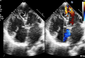
Echocardiography revealed normal left ventricular size, function, and wall thickness, but it identified an abnormally hypertrophied large papillary muscle causing significant mid-cavitary obstruction, with a peak gradient of 120 mmHg (Fig. 1).
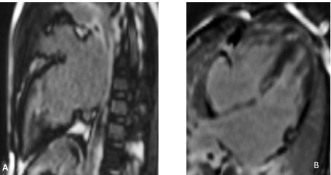
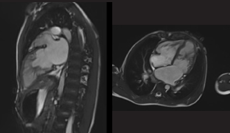
The patient was referred for cardiac magnetic resonance (CMR) imaging for better delineation of the papillary muscle hypertrophy and to confirm or exclude accompanying LV hypertrophy. CMR confirmed the absence of LV hypertrophy and clearly illustrated the abnormal papillary muscle. The primary band of the anomalous papillary muscle was 2 cm thick and extended radially from the central muscle mass, resulting in multiple smaller muscle elements that split the LV cavity into apical and subaortic chambers, leading to significant mid-cavitary obstruction (Fig. 2). The abnormal papillary muscle exhibited isointense signals in both T1- and T2-weighted images with no evidence of fibrosis on late gadolinium enhancement sequences. No myocardial fibrosis was detected (Fig. 3).
Despite the presence of the anomaly, the systolic function of the left ventricle remained normal. The mitral valve appeared structurally and functionally intact, apart from trivial mitral incompetence. The patient was significantly symptomatic and was referred for further surgical management.
Discussion
Reports of papillary muscle (PM) aberrations include parachute mitral valve, anomalous insertion of papillary muscle, and hypertrophic cardiomyopathy-associated PM abnormalities. These aberrations often affect mitral valve function, leading to mitral stenosis or incompetence, and may contribute to the development of mid-cavitary or left ventricular outflow tract (LVOT) obstruction. Identifying these aberrations can significantly impact surgical management.
The octopus papillary muscle is a congenital abnormality that affects papillary muscle development and typically leads to significant mid-cavitary obstruction. Shah et al. reported an octopus papillary muscle causing symptomatic mid-left ventricular cavity obstruction. Patel MS reported a second case of octopus papillary muscle associated with a left lateral accessory pathway.4,5
The diagnosis of octopus papillary muscle is quite unfamiliar to most cardiologists, and to our knowledge, this is the third case report worldwide. In our case, the patient was initially referred to Aswan Heart Center with a diagnosis of obstructive hypertrophic cardiomyopathy. However, the absence of left ventricular hypertrophy made this diagnosis unlikely.
Echocardiography clearly showed significant mid-cavitary obstruction with a peak gradient of 120 mmHg, along with an anomalous papillary muscle morphology. CMR clearly identified a solitary, apically displaced, centrally localized papillary muscle resembling the body of an octopus. The “head” sent accessory muscular bands to all LV walls, mimicking the arms of an octopus, leading to significant mid-cavitary obstruction. This abnormality was identified through short-axis and long-axis views. The patient was symptomatic and subsequently referred for surgical management.
Conclusion
Octopus papillary muscle is a rare, unfamiliar etiology of mid-cavitary obstruction and should be interpreted using multimodal imaging for better anatomical delineation and to guide surgical management.


Case Report
A 6-year-old girl presented to Aswan Heart Center with chest pain and palpitations triggered by strenuous exercise, with an initial diagnosis of hypertrophic cardiomyopathy. She denied syncope and any relevant family history. On examination, a grade 4/6 systolic ejection murmur was audible throughout the precordium. The peripheral pulses were normal, and the rest of her examination was unremarkable. The electrocardiogram showed normal sinus rhythm with frequent atrial ectopic beats and no signs of left ventricular (LV) hypertrophy.
Echocardiography revealed normal left ventricular size, function, and wall thickness, but it identified an abnormally hypertrophied large papillary muscle causing significant mid-cavitary obstruction, with a peak gradient of 120 mmHg (Fig. 1).
The patient was referred for cardiac magnetic resonance (CMR) imaging for better delineation of the papillary muscle hypertrophy and to confirm or exclude accompanying LV hypertrophy. CMR confirmed the absence of LV hypertrophy and clearly illustrated the abnormal papillary muscle. The primary band of the anomalous papillary muscle was 2 cm thick and extended radially from the central muscle mass, resulting in multiple smaller muscle elements that split the LV cavity into apical and subaortic chambers, leading to significant mid-cavitary obstruction (Fig. 2). The abnormal papillary muscle exhibited isointense signals in both T1- and T2-weighted images with no evidence of fibrosis on late gadolinium enhancement sequences. No myocardial fibrosis was detected (Fig. 3).
Despite the presence of the anomaly, the systolic function of the left ventricle remained normal. The mitral valve appeared structurally and functionally intact, apart from trivial mitral incompetence. The patient was significantly symptomatic and was referred for further surgical management.
Discussion
Reports of papillary muscle (PM) aberrations include parachute mitral valve, anomalous insertion of papillary muscle, and hypertrophic cardiomyopathy-associated PM abnormalities. These aberrations often affect mitral valve function, leading to mitral stenosis or incompetence, and may contribute to the development of mid-cavitary or left ventricular outflow tract (LVOT) obstruction. Identifying these aberrations can significantly impact surgical management.
The octopus papillary muscle is a congenital abnormality that affects papillary muscle development and typically leads to significant mid-cavitary obstruction. Shah et al. reported an octopus papillary muscle causing symptomatic mid-left ventricular cavity obstruction. Patel MS reported a second case of octopus papillary muscle associated with a left lateral accessory pathway.4,5
The diagnosis of octopus papillary muscle is quite unfamiliar to most cardiologists, and to our knowledge, this is the third case report worldwide. In our case, the patient was initially referred to Aswan Heart Center with a diagnosis of obstructive hypertrophic cardiomyopathy. However, the absence of left ventricular hypertrophy made this diagnosis unlikely.
Echocardiography clearly showed significant mid-cavitary obstruction with a peak gradient of 120 mmHg, along with an anomalous papillary muscle morphology. CMR clearly identified a solitary, apically displaced, centrally localized papillary muscle resembling the body of an octopus. The “head” sent accessory muscular bands to all LV walls, mimicking the arms of an octopus, leading to significant mid-cavitary obstruction. This abnormality was identified through short-axis and long-axis views. The patient was symptomatic and subsequently referred for surgical management.
Conclusion
Octopus papillary muscle is a rare, unfamiliar etiology of mid-cavitary obstruction and should be interpreted using multimodal imaging for better anatomical delineation and to guide surgical management.
"Octopus papillary muscle is a rare, unfamiliar etiology of mid-cavitary obstruction and should be interpreted using multimodal imaging for better anatomical delineation and to guide surgical management."

Director of the Advanced Cardiac Imaging Unit
Aswan Heart Centre, Cardiac Imaging unit, Aswan, Egypt
Associate Professor of Cardiology, Tanta University