Clinical Case Study 1
A 47-year-old woman presented with a palpable mass in the left breast. Histopathology confirmed invasive ductal carcinoma (IDC), grade II. Contrast-enhanced spectral mammography (CESM) was performed on the Siemens Healthineers.
MAMMOMAT B.Brilliant Mammography system to assess the local extent of disease and screen the contralateral breast.
Conclusion
In this case, the MAMMOMAT B.Brilliant Mammography system with CESM enabled comprehensive assessment of both breasts in a single examination. The system not only characterized the known IDC in the left breast but also revealed multiple additional enhancing foci suggestive of multifocal disease and detected a suspicious enhancing focus in the contralateral breast.
By improving visualization of tumor extent and uncovering additional lesions that may be occult on conventional imaging, MAMMOMAT B.Brilliant CESM can support more accurate staging and inform surgical and systemic treatment planning, ultimately contributing to optimized patient management.

CESM Findings – Left Breast
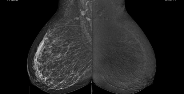
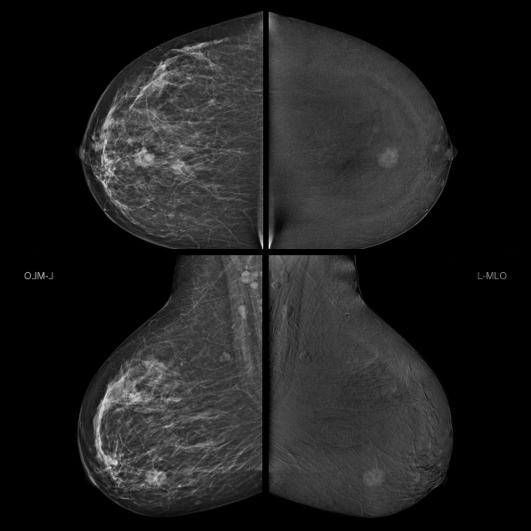
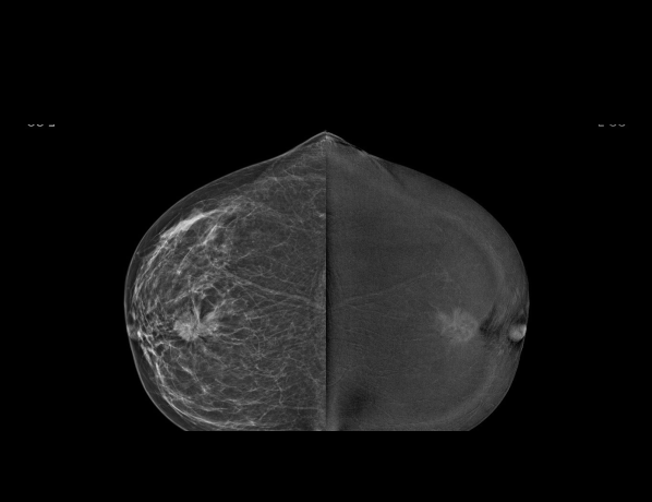
CESM on the MAMMOMAT B.Brilliant demonstrated a moderately enhancing, heterogeneous mass in the lower outer quadrant of the left breast, in a rather central location, measuring approximately 1.9 × 2.1 cm.
CESM Findings – Right Breast
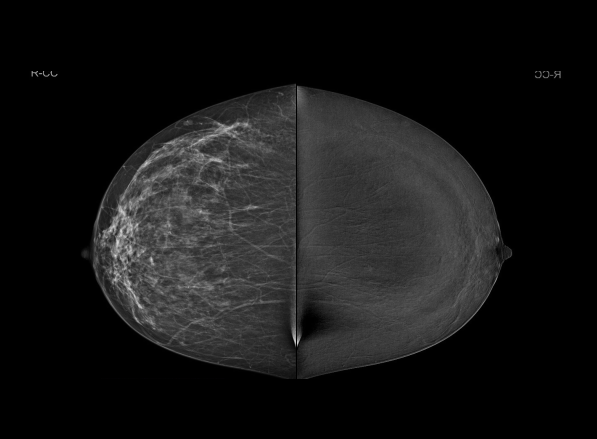
In the right breast, MAMMOMAT B.Brilliant CESM revealed an additional enhancing focus in the lower inner quadrant, posterior third. This lesion measured approximately 0.4 × 0.6 cm and was located about 10 cm from the nipple, raising suspicion for a contralateral lesion and prompting further diagnostic work-up.

Conclusion
In this case, the MAMMOMAT B.Brilliant Mammography system with CESM enabled comprehensive assessment of both breasts in a single examination. The system not only characterized the known IDC in the left breast but also revealed multiple additional enhancing foci suggestive of multifocal disease and detected a suspicious enhancing focus in the contralateral breast.
By improving visualization of tumor extent and uncovering additional lesions that may be occult on conventional imaging, MAMMOMAT B.Brilliant CESM can support more accurate staging and inform surgical and systemic treatment planning, ultimately contributing to optimized patient management.

CESM Findings – Left Breast
CESM on the MAMMOMAT B.Brilliant demonstrated a moderately enhancing, heterogeneous mass in the lower outer quadrant of the left breast, in a rather central location, measuring approximately 1.9 × 2.1 cm.

CESM Findings – Right Breast
In the right breast, MAMMOMAT B.Brilliant CESM revealed an additional enhancing focus in the lower inner quadrant, posterior third. This lesion measured approximately 0.4 × 0.6 cm and was located about 10 cm from the nipple, raising suspicion for a contralateral lesion and prompting further diagnostic work-up.

Conclusion
In this case, the MAMMOMAT B.Brilliant Mammography system with CESM enabled comprehensive assessment of both breasts in a single examination. The system not only characterized the known IDC in the left breast but also revealed multiple additional enhancing foci suggestive of multifocal disease and detected a suspicious enhancing focus in the contralateral breast.
By improving visualization of tumor extent and uncovering additional lesions that may be occult on conventional imaging, MAMMOMAT B.Brilliant CESM can support more accurate staging and inform surgical and systemic treatment planning, ultimately contributing to optimized patient management.
Clinical Case Study 2
A 56-year-old woman presented with a left breast mass. Histopathology confirmed invasive ductal carcinoma (IDC) in the left breast. Contrast-enhanced spectral mammography (CESM) was performed on the MAMMOMAT B.Brilliant Mammography system to evaluate the extent of disease and to assess the contralateral breast.
Histopathology Conclusion
Biopsy of the right breast lesion revealed mucinous carcinoma, establishing synchronous bilateral breast cancer: invasive ductal carcinoma in the left breast and mucinous carcinoma in the right breast.
This case illustrates how MAMMOMAT B.Brilliant Mammography with CESM can:
• Characterize the index malignancy and accurately depict its size and location.
• Detect a subtle, contralateral enhancing mass that might otherwise be underestimated or overlooked.
• Promptly biopsy of a BI-RADS 4 lesion, leading to the diagnosis of synchronous contralateral breast cancer.
By enabling sensitive bilateral assessment in a single examination, MAMMOMAT B.Brilliant CESM supports more accurate staging and treatment planning in patients with newly diagnosed breast cancer.

CESM Findings – Left Breast
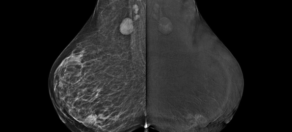
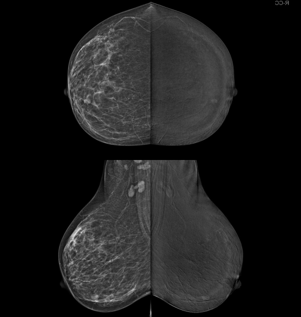
MAMMOMAT B.Brilliant CESM revealed a single, moderately enhancing heterogeneous mass in the lower outer quadrant of the left breast. The lesion measured approximately 4.0 × 3.5 cm and was located about 2 cm from the nipple. The enhancement pattern and morphologic features were consistent with a known malignancy. Based on imaging and prior pathology, this lesion was categorized as BI-RADS 6 (known biopsy-proven malignancy).
CESM Findings – Right Breast
In the right breast, CESM demonstrated a single mass in the lower inner quadrant, anterior third. The lesion showed homogeneous enhancement and measured approximately 12.5 × 11 mm, located about 2.5 cm from the nipple.
Despite its relatively small size and homogeneous enhancement, the lesion was considered suspicious on CESM and classified as BI-RADS 4 (suspicious abnormality), warranting tissue diagnosis.

Histopathology Conclusion
Biopsy of the right breast lesion revealed mucinous carcinoma, establishing synchronous bilateral breast cancer: invasive ductal carcinoma in the left breast and mucinous carcinoma in the right breast.
This case illustrates how MAMMOMAT B.Brilliant Mammography with CESM can:
• Characterize the index malignancy and accurately depict its size and location.
• Detect a subtle, contralateral enhancing mass that might otherwise be underestimated or overlooked.
• Promptly biopsy of a BI-RADS 4 lesion, leading to the diagnosis of synchronous contralateral breast cancer.
By enabling sensitive bilateral assessment in a single examination, MAMMOMAT B.Brilliant CESM supports more accurate staging and treatment planning in patients with newly diagnosed breast cancer.

CESM Findings – Left Breast
MAMMOMAT B.Brilliant CESM revealed a single, moderately enhancing heterogeneous mass in the lower outer quadrant of the left breast. The lesion measured approximately 4.0 × 3.5 cm and was located about 2 cm from the nipple. The enhancement pattern and morphologic features were consistent with a known malignancy. Based on imaging and prior pathology, this lesion was categorized as BI-RADS 6 (known biopsy-proven malignancy).

CESM Findings – Right Breast
In the right breast, CESM demonstrated a single mass in the lower inner quadrant, anterior third. The lesion showed homogeneous enhancement and measured approximately 12.5 × 11 mm, located about 2.5 cm from the nipple.
Despite its relatively small size and homogeneous enhancement, the lesion was considered suspicious on CESM and classified as BI-RADS 4 (suspicious abnormality), warranting tissue diagnosis.

Histopathology Conclusion
Biopsy of the right breast lesion revealed mucinous carcinoma, establishing synchronous bilateral breast cancer: invasive ductal carcinoma in the left breast and mucinous carcinoma in the right breast.
This case illustrates how MAMMOMAT B.Brilliant Mammography with CESM can:
• Characterize the index malignancy and accurately depict its size and location.
• Detect a subtle, contralateral enhancing mass that might otherwise be underestimated or overlooked.
• Promptly biopsy of a BI-RADS 4 lesion, leading to the diagnosis of synchronous contralateral breast cancer.
By enabling sensitive bilateral assessment in a single examination, MAMMOMAT B.Brilliant CESM supports more accurate staging and treatment planning in patients with newly diagnosed breast cancer.
Clinical Case Study 3
A 49-year-old woman presented with a recently noticed palpable lump in the right breast. Full-field digital mammography (FFDM) and 3D tomosynthesis were performed on the MAMMOMAT B.Brilliant Mammography system to evaluate the palpable finding and to assess both breasts comprehensively.
Opinion
MAMMOMAT B.Brilliant Mammography with 3D tomosynthesis identified:
• A right UOQ irregular mass with subtle distortion, corresponding to the palpable lump and considered a suspicious lesion.
• A left UOQ focal asymmetry with distortion, non-palpable but clearly suspicious on tomosynthesis.
Both lesions would be appropriately categorized as BI-RADS 4 (suspicious abnormalities), warranting further evaluation with targeted ultrasound and image-guided biopsies for histopathological confirmation.
Clinical Message
In patients presenting with a unilateral palpable lump, bilateral imaging with 3D tomosynthesis on MAMMOMAT B.Brilliant can reveal additional, clinically occult contralateral lesions.
Subtle architectural distortion and focal asymmetries may be better defined on tomosynthesis than on 2D mammography, supporting accurate BI-RADS assessment and guiding biopsy of synchronous lesions in both breasts.

Right Breast – MAMMOMAT B.Brilliant FFDM and 3D Tomosynthesis
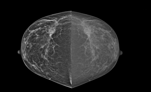
Imaging of the right breast demonstrated a small, irregular, equally dense mass lesion in the upper outer quadrant (UOQ), associated with subtle architectural distortion. The lesion measured approximately 15 × 11 × 14 mm and was located about 8 cm from the nipple.
While the finding was already suspected on 2D mammography, MAMMOMAT B.Brilliant 3D tomosynthesis clearly confirmed the presence and morphology of the mass and better delineated the surrounding distortion, increasing confidence that this represented a suspicious lesion corresponding to the palpable abnormality.

Left Breast – MAMMOMAT B.Brilliant FFDM and 3D Tomosynthesis
In the left breast, FFDM revealed a focal asymmetry with architectural distortion in the upper outer quadrant (UOQ), located approximately 76 mm from the nipple. This finding did not correspond to any known clinical symptoms.
On 3D tomosynthesis, the focal asymmetry and distortion were confirmed and became more conspicuous, indicating a second suspicious lesion in the contralateral breast. Tomosynthesis helped separate the lesion from overlapping parenchymal structures, which is particularly important in subtle bilateral findings.

Opinion
MAMMOMAT B.Brilliant Mammography with 3D tomosynthesis identified:
• A right UOQ irregular mass with subtle distortion, corresponding to the palpable lump and considered a suspicious lesion.
• A left UOQ focal asymmetry with distortion, non-palpable but clearly suspicious on tomosynthesis.
Both lesions would be appropriately categorized as BI-RADS 4 (suspicious abnormalities), warranting further evaluation with targeted ultrasound and image-guided biopsies for histopathological confirmation.
Clinical Message
In patients presenting with a unilateral palpable lump, bilateral imaging with 3D tomosynthesis on MAMMOMAT B.Brilliant can reveal additional, clinically occult contralateral lesions.
Subtle architectural distortion and focal asymmetries may be better defined on tomosynthesis than on 2D mammography, supporting accurate BI-RADS assessment and guiding biopsy of synchronous lesions in both breasts.

Right Breast – MAMMOMAT B.Brilliant FFDM and 3D Tomosynthesis
Imaging of the right breast demonstrated a small, irregular, equally dense mass lesion in the upper outer quadrant (UOQ), associated with subtle architectural distortion. The lesion measured approximately 15 × 11 × 14 mm and was located about 8 cm from the nipple.
While the finding was already suspected on 2D mammography, MAMMOMAT B.Brilliant 3D tomosynthesis clearly confirmed the presence and morphology of the mass and better delineated the surrounding distortion, increasing confidence that this represented a suspicious lesion corresponding to the palpable abnormality.

Left Breast – MAMMOMAT B.Brilliant FFDM and 3D Tomosynthesis
In the left breast, FFDM revealed a focal asymmetry with architectural distortion in the upper outer quadrant (UOQ), located approximately 76 mm from the nipple. This finding did not correspond to any known clinical symptoms.
On 3D tomosynthesis, the focal asymmetry and distortion were confirmed and became more conspicuous, indicating a second suspicious lesion in the contralateral breast. Tomosynthesis helped separate the lesion from overlapping parenchymal structures, which is particularly important in subtle bilateral findings.

Opinion
MAMMOMAT B.Brilliant Mammography with 3D tomosynthesis identified:
• A right UOQ irregular mass with subtle distortion, corresponding to the palpable lump and considered a suspicious lesion.
• A left UOQ focal asymmetry with distortion, non-palpable but clearly suspicious on tomosynthesis.
Both lesions would be appropriately categorized as BI-RADS 4 (suspicious abnormalities), warranting further evaluation with targeted ultrasound and image-guided biopsies for histopathological confirmation.
Clinical Message
In patients presenting with a unilateral palpable lump, bilateral imaging with 3D tomosynthesis on MAMMOMAT B.Brilliant can reveal additional, clinically occult contralateral lesions.
Subtle architectural distortion and focal asymmetries may be better defined on tomosynthesis than on 2D mammography, supporting accurate BI-RADS assessment and guiding biopsy of synchronous lesions in both breasts.
Clinical Case Study 4
A 39-year-old woman was recalled from routine screening after findings in the left breast on full-field digital mammography (FFDM) with the MAMMOMAT B.Brilliant Mammography system. The patient had a positive family history of breast cancer (aunt diagnosed at 65 years). Additional imaging with MAMMOMAT B.Brilliant FFDM, complementary ultrasound, and 3D tomosynthesis was performed. Clinical–radiological correlation confirmed that the palpable lump corresponded to a benign cyst, while an additional, non-palpable suspicious lesion was detected in the left breast.
Opinion
Imaging demonstrated a non-palpable, malignant looking mass in the left breast at 12 o’clock, distinct from the palpable cyst.
Based on the architectural distortion seen on MAMMOMAT B.Brilliant FFDM, the irregular vascular mass on ultrasound, and confirmatory 3D tomosynthesis findings, the lesion was classified as BI-RADS 4 (suspicious abnormality). Biopsy and bilateral contrast-enhanced mammography were recommended for further characterization and staging.
Recommendation
Referral to the breast clinic was advised for image-guided biopsy of the left 12 o’clock lesion and subsequent management planning.

Mammography Findings (MAMMOMAT B.Brilliant FFDM)
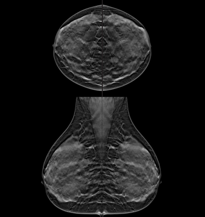
Bilateral FFDM on MAMMOMAT B.Brilliant showed heterogeneously dense breast parenchyma (ACR category C), a known challenge for lesion detection and characterization due to tissue overlap.
Bilateral scattered microcalcifications were present. In the left upper quadrant, an area of architectural distortion with an overlying tiny calcific focus was identified, separate from the area of the clinically palpable lump. Skin thickness and contour were normal, and the nipple–areolar complexes appeared unremarkable. Bilateral nonspecific axillary lymph nodes were seen.

Ultrasound Findings
Targeted ultrasound of the area corresponding to the palpable lump in the left breast demonstrated a benign-appearing cyst, explaining the palpable finding.
Additional targeted ultrasound of the left upper quadrant at 12 o’clock revealed a separate, irregular solid mass measuring approximately 2.5 × 1.1 cm, with an overlying calcific focus and mild internal vascularity on color Doppler. This lesion was located about 7.5 cm from the nipple and 0.5 cm from the skin. Both breasts showed heterogeneous mottled parenchyma with bilateral scattered cysts, consistent with fibrocystic changes.
3D Tomosynthesis (MAMMOMAT B.Brilliant)
3D tomosynthesis on the MAMMOMAT B.Brilliant Mammography system was performed to further evaluate the subtle architectural distortion seen on 2D imaging. Tomosynthesis significantly improved lesion conspicuity within the dense parenchyma, confirming an underlying mass in the left upper quadrant that corresponded to the irregular solid lesion on ultrasound.
Importantly, tomosynthesis helped clearly distinguish this suspicious, non-palpable mass from the superficial palpable cyst, overcoming the limitations of tissue overlap in dense breasts.

Opinion
Imaging demonstrated a non-palpable, malignant looking mass in the left breast at 12 o’clock, distinct from the palpable cyst.
Based on the architectural distortion seen on MAMMOMAT B.Brilliant FFDM, the irregular vascular mass on ultrasound, and confirmatory 3D tomosynthesis findings, the lesion was classified as BI-RADS 4 (suspicious abnormality). Biopsy and bilateral contrast-enhanced mammography were recommended for further characterization and staging.
Recommendation
Referral to the breast clinic was advised for image-guided biopsy of the left 12 o’clock lesion and subsequent management planning.

Mammography Findings (MAMMOMAT B.Brilliant FFDM)
Bilateral FFDM on MAMMOMAT B.Brilliant showed heterogeneously dense breast parenchyma (ACR category C), a known challenge for lesion detection and characterization due to tissue overlap.
Bilateral scattered microcalcifications were present. In the left upper quadrant, an area of architectural distortion with an overlying tiny calcific focus was identified, separate from the area of the clinically palpable lump. Skin thickness and contour were normal, and the nipple–areolar complexes appeared unremarkable. Bilateral nonspecific axillary lymph nodes were seen.

Ultrasound Findings
Targeted ultrasound of the area corresponding to the palpable lump in the left breast demonstrated a benign-appearing cyst, explaining the palpable finding.
Additional targeted ultrasound of the left upper quadrant at 12 o’clock revealed a separate, irregular solid mass measuring approximately 2.5 × 1.1 cm, with an overlying calcific focus and mild internal vascularity on color Doppler. This lesion was located about 7.5 cm from the nipple and 0.5 cm from the skin. Both breasts showed heterogeneous mottled parenchyma with bilateral scattered cysts, consistent with fibrocystic changes.
3D Tomosynthesis (MAMMOMAT B.Brilliant)
3D tomosynthesis on the MAMMOMAT B.Brilliant Mammography system was performed to further evaluate the subtle architectural distortion seen on 2D imaging. Tomosynthesis significantly improved lesion conspicuity within the dense parenchyma, confirming an underlying mass in the left upper quadrant that corresponded to the irregular solid lesion on ultrasound.
Importantly, tomosynthesis helped clearly distinguish this suspicious, non-palpable mass from the superficial palpable cyst, overcoming the limitations of tissue overlap in dense breasts.

Opinion
Imaging demonstrated a non-palpable, malignant looking mass in the left breast at 12 o’clock, distinct from the palpable cyst.
Based on the architectural distortion seen on MAMMOMAT B.Brilliant FFDM, the irregular vascular mass on ultrasound, and confirmatory 3D tomosynthesis findings, the lesion was classified as BI-RADS 4 (suspicious abnormality). Biopsy and bilateral contrast-enhanced mammography were recommended for further characterization and staging.
Recommendation
Referral to the breast clinic was advised for image-guided biopsy of the left 12 o’clock lesion and subsequent management planning.
Authors