Follow-Up & Discussion
The patient demonstrated marked clinical improvement following the procedure, with complete resolution of heart failure symptoms and was discharged in stable condition. At one-month follow-up, liver enzyme levels showed progressive normalization, coagulopathy resolved, and tricuspid regurgitation regressed significantly. The patient remained clinically stable with excellent functional recovery. This case highlights the critical role of advanced fusion imaging and 3D segmentation in enabling safe and precise transcatheter mitral valve-in-valve implantation in patients with extremely high-risk anatomy. The ability to generate accurate 3D anatomical models and integrate them seamlessly with live fluoroscopy allows contrast- and radiation-sparing workflows while delivering sub-millimetric precision in complex structural heart procedures.
Clinical Case Study
Patient History
A 63-year-old female presented with recurrent hospital admissions for decompensated heart failure and pulmonary edema. She had undergone surgical mitral valve replacement with a 29-mm Mosaic bioprosthetic valve 13 years earlier. Transthoracic and transesophageal echocardiography demonstrated:
• Severe prosthetic mitral regurgitation
• Severe tricuspid regurgitation
• Hyperdynamic left ventricular systolic function (LVEF >70%)
The patient had significant comorbidities, most notably liver cirrhosis with associated coagulopathy, rendering her a prohibitive candidate for redo open-heart surgery.
Following multidisciplinary Heart Team discussion, percutaneous transcatheter mitral valve-in-valve implantation with an LVOT obstruction prevention strategy was recommended.
Material & Method
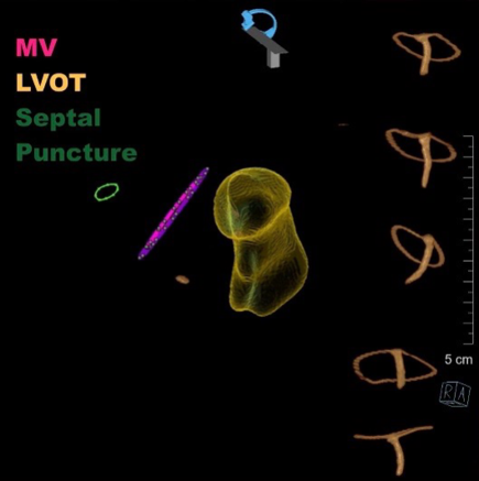
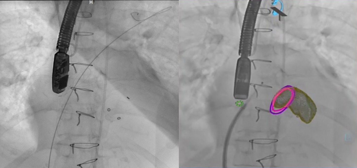
Pre-procedural cardiac computed tomography angiography (CTA) was performed to exclude significant coronary artery disease and to allow detailed anatomical assessment of the degenerated mitral prosthesis and surrounding cardiac structures. The predicted neo-LVOT area was calculated to be 144 mm², indicating a very high risk of LVOT obstruction with standard ViV implantation. Advanced imaging capabilities of the ARTIS icono angiography system were therefore utilized to enhance procedural planning and execution. Stroke-based segmentation tools were used to rapidly generate an accurate 3D model of the mitral prosthesis, LVOT, and region of interest. Using the syngo Fusion package, the CT-derived 3D model was co-registered with live fluoroscopy, enabling real-time anatomical overlay that dynamically followed all C-arm movements and allowed automatic navigation to optimal coplanar deployment angles without the need for additional contrast injections. CT-derived transseptal puncture planning was used to identify the optimal puncture location and angiographic projection, which were overlaid on live fluoroscopy to facilitate precise transseptal access.
Procedural Technique
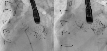
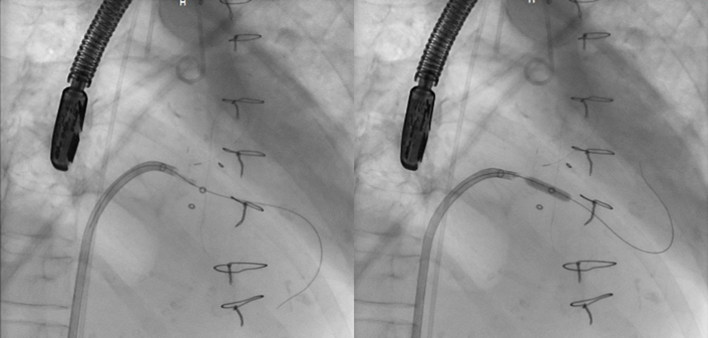
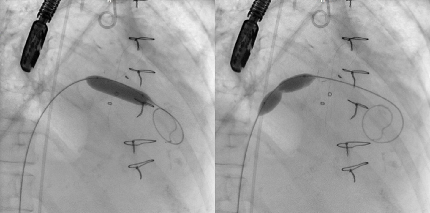
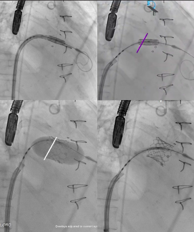
Following successful transseptal puncture at the pre-planned site, the Balloon-Assisted Translocation of the Anterior Mitral leaflet to Prevent Neo-LVOT Obstruction (BATMAN) technique was performed to mitigate the high risk of LVOT obstruction. A 26-mm Edwards SAPIEN 3 Ultra transcatheter heart valve was then deployed successfully within the degenerated Mosaic bioprosthetic valve under CT-fluoroscopy fusion guidance, using CT-derived anatomical landmarks for accurate positioning. Due to the presence of a persistent right-to-left shunt following transseptal access, the interatrial septum was closed using an atrial septal defect (ASD) closure device, achieving an excellent angiographic and fluoroscopic result. The procedure was completed without complications, with minimal contrast usage and low radiation exposure.
Imaging Guidance and Workflow
Pre-procedural CT planning allowed precise localization of the transseptal puncture site, which was overlaid onto live fluoroscopy using syngo Fusion to ensure accurate access. Fusion imaging facilitated real-time guidance during valve positioning and deployment, with continuous visualization of the prosthesis relative to surrounding anatomical structures. Final angiographic and fluoroscopic imaging confirmed optimal positioning of the SAPIEN 3 Ultra valve within the surgical bioprosthesis, as well as successful closure of the transseptal puncture site with the ASD device.
Follow-Up & Discussion
The patient demonstrated marked clinical improvement following the procedure, with complete resolution of heart failure symptoms and was discharged in stable condition. At one-month follow-up, liver enzyme levels showed progressive normalization, coagulopathy resolved, and tricuspid regurgitation regressed significantly. The patient remained clinically stable with excellent functional recovery. This case highlights the critical role of advanced fusion imaging and 3D segmentation in enabling safe and precise transcatheter mitral valve-in-valve implantation in patients with extremely high-risk anatomy. The ability to generate accurate 3D anatomical models and integrate them seamlessly with live fluoroscopy allows contrast- and radiation-sparing workflows while delivering sub-millimetric precision in complex structural heart procedures.
Clinical Case Study
Patient History
A 63-year-old female presented with recurrent hospital admissions for decompensated heart failure and pulmonary edema. She had undergone surgical mitral valve replacement with a 29-mm Mosaic bioprosthetic valve 13 years earlier. Transthoracic and transesophageal echocardiography demonstrated:
• Severe prosthetic mitral regurgitation
• Severe tricuspid regurgitation
• Hyperdynamic left ventricular systolic function (LVEF >70%)
The patient had significant comorbidities, most notably liver cirrhosis with associated coagulopathy, rendering her a prohibitive candidate for redo open-heart surgery.
Following multidisciplinary Heart Team discussion, percutaneous transcatheter mitral valve-in-valve implantation with an LVOT obstruction prevention strategy was recommended.
Material & Method
Pre-procedural cardiac computed tomography angiography (CTA) was performed to exclude significant coronary artery disease and to allow detailed anatomical assessment of the degenerated mitral prosthesis and surrounding cardiac structures. The predicted neo-LVOT area was calculated to be 144 mm², indicating a very high risk of LVOT obstruction with standard ViV implantation. Advanced imaging capabilities of the ARTIS icono angiography system were therefore utilized to enhance procedural planning and execution. Stroke-based segmentation tools were used to rapidly generate an accurate 3D model of the mitral prosthesis, LVOT, and region of interest. Using the syngo Fusion package, the CT-derived 3D model was co-registered with live fluoroscopy, enabling real-time anatomical overlay that dynamically followed all C-arm movements and allowed automatic navigation to optimal coplanar deployment angles without the need for additional contrast injections. CT-derived transseptal puncture planning was used to identify the optimal puncture location and angiographic projection, which were overlaid on live fluoroscopy to facilitate precise transseptal access.
Procedural Technique
Following successful transseptal puncture at the pre-planned site, the Balloon-Assisted Translocation of the Anterior Mitral leaflet to Prevent Neo-LVOT Obstruction (BATMAN) technique was performed to mitigate the high risk of LVOT obstruction. A 26-mm Edwards SAPIEN 3 Ultra transcatheter heart valve was then deployed successfully within the degenerated Mosaic bioprosthetic valve under CT-fluoroscopy fusion guidance, using CT-derived anatomical landmarks for accurate positioning. Due to the presence of a persistent right-to-left shunt following transseptal access, the interatrial septum was closed using an atrial septal defect (ASD) closure device, achieving an excellent angiographic and fluoroscopic result. The procedure was completed without complications, with minimal contrast usage and low radiation exposure.
Imaging Guidance and Workflow
Pre-procedural CT planning allowed precise localization of the transseptal puncture site, which was overlaid onto live fluoroscopy using syngo Fusion to ensure accurate access. Fusion imaging facilitated real-time guidance during valve positioning and deployment, with continuous visualization of the prosthesis relative to surrounding anatomical structures. Final angiographic and fluoroscopic imaging confirmed optimal positioning of the SAPIEN 3 Ultra valve within the surgical bioprosthesis, as well as successful closure of the transseptal puncture site with the ASD device.
Follow-Up & Discussion
The patient demonstrated marked clinical improvement following the procedure, with complete resolution of heart failure symptoms and was discharged in stable condition. At one-month follow-up, liver enzyme levels showed progressive normalization, coagulopathy resolved, and tricuspid regurgitation regressed significantly. The patient remained clinically stable with excellent functional recovery. This case highlights the critical role of advanced fusion imaging and 3D segmentation in enabling safe and precise transcatheter mitral valve-in-valve implantation in patients with extremely high-risk anatomy. The ability to generate accurate 3D anatomical models and integrate them seamlessly with live fluoroscopy allows contrast- and radiation-sparing workflows while delivering sub-millimetric precision in complex structural heart procedures.
Conclusion
Advanced fusion imaging and 3D segmentation using the ARTIS icono platform represent a significant advancement in the field of structural heart interventions. In high-risk mitral valve-in-valve procedures, particularly those with a predicted risk of LVOT obstruction, these technologies enable precise, safe, and efficient valve deployment while minimizing contrast load and radiation exposure. This approach has the potential to transform outcomes in frail patients with complex anatomy and significant comorbidities.
"The ability to generate accurate 3D anatomical models and integrate them seamlessly with live fluoroscopy allows contrast- and radiation-sparing workflows while delivering sub-millimetric precision in complex structural heart procedures."
Chief of Cardiac Cath Lab
King Fahad Hospital of University
Al Khobar, Kingdome of Saudi Arabia