Conclusion
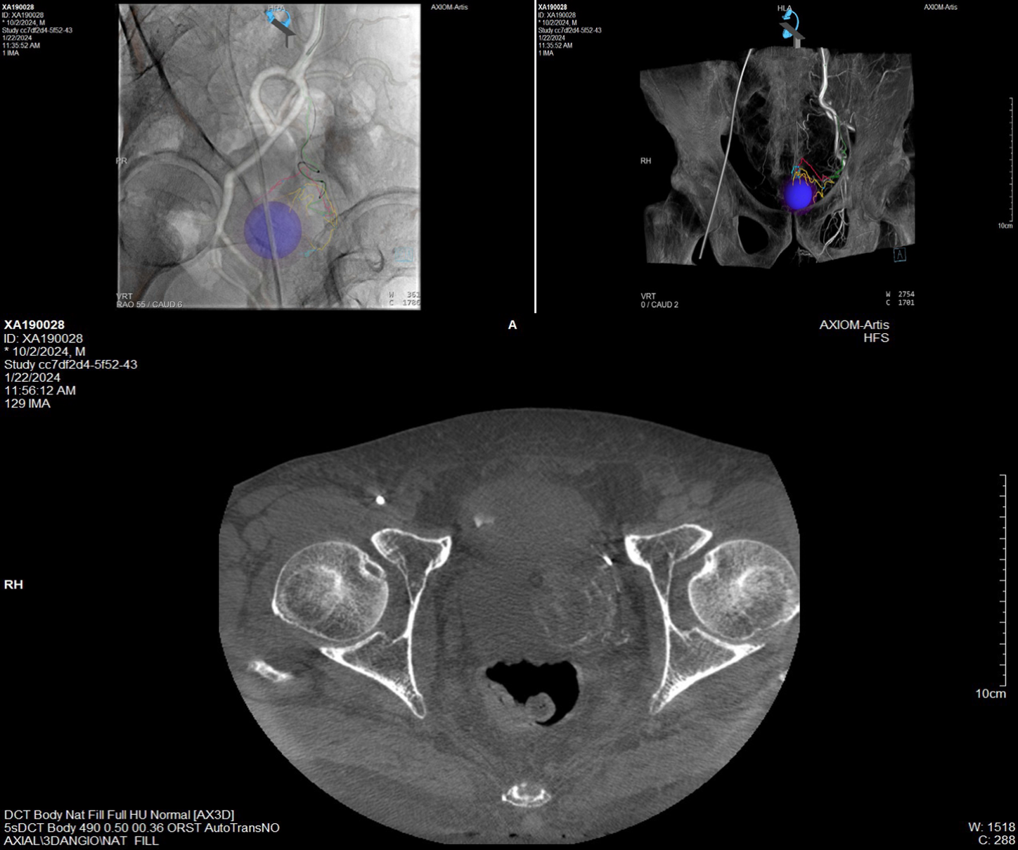
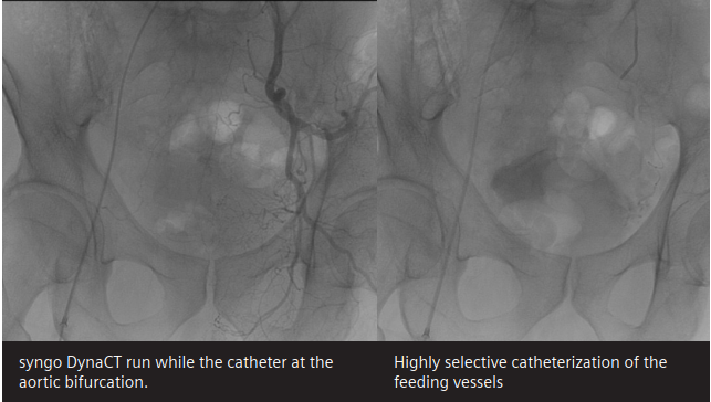
The use of syngo DynaCT and syngo Embolization Guidance software improves the planning of prostate artery embolization procedures and enhances procedural guidance, making embolizations faster. This approach offers increased treatment confidence and allows for reduced radiation dose.
Conclusion
The use of syngo DynaCT and syngo Embolization Guidance software improves the planning of prostate artery embolization procedures and enhances procedural guidance, making embolizations faster. This approach offers increased treatment confidence and allows for reduced radiation dose.
Conclusion
The use of syngo DynaCT and syngo Embolization Guidance software improves the planning of prostate artery embolization procedures and enhances procedural guidance, making embolizations faster. This approach offers increased treatment confidence and allows for reduced radiation dose.
"The use of syngo DynaCT and syngo Embolization Guidance software improves the planning of prostate artery embolization procedures and enhances procedural guidance, making embolizations faster. This approach offers increased treatment confidence and allows for reduced radiation dose."

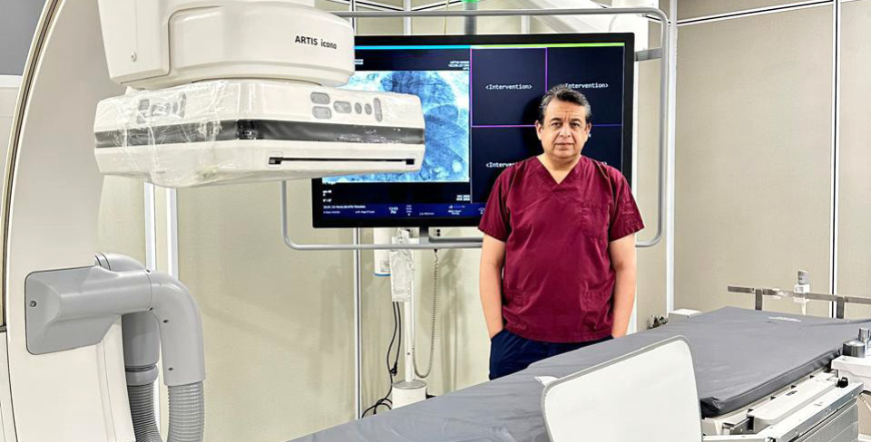
Dr. Sameh Fathy Center, Alexandria, Egypt
"The use of syngo DynaCT and syngo Embolization Guidance software improves the planning of prostate artery embolization procedures and enhances procedural guidance, making embolizations faster. This approach offers increased treatment confidence and allows for reduced radiation dose."