Computed Tomography for CardiologyYou think ahead. We innovate ahead.
Cardiovascular diseases are responsible for almost one-third of deaths worldwide.1 Since the burden coronary artery disease (CAD) and other heart conditions place on healthcare systems will continue to grow, driving innovations in cardiac imaging is crucial.
CT imaging solutions from Siemens Healthineers have long served as true game changers for cardiology, covering the broad spectrum from early detection and diagnostic support to personalization of therapy decision and follow-up. Paired with technologies that harness the power of AI, our state-of-the-art systems help reveal hidden information in your CT scans quickly and accurately. This allows you to take patient evaluation and treatment decision-making to the next level.
As pioneers in cardiac CT solutions, we invite you to witness firsthand how our state-of-the-art CT systems combined with AI-powered technologies are shaping the future of cardiology.
Cardiac CT made easy with innovative technologies
Customer voices
Clinical cases

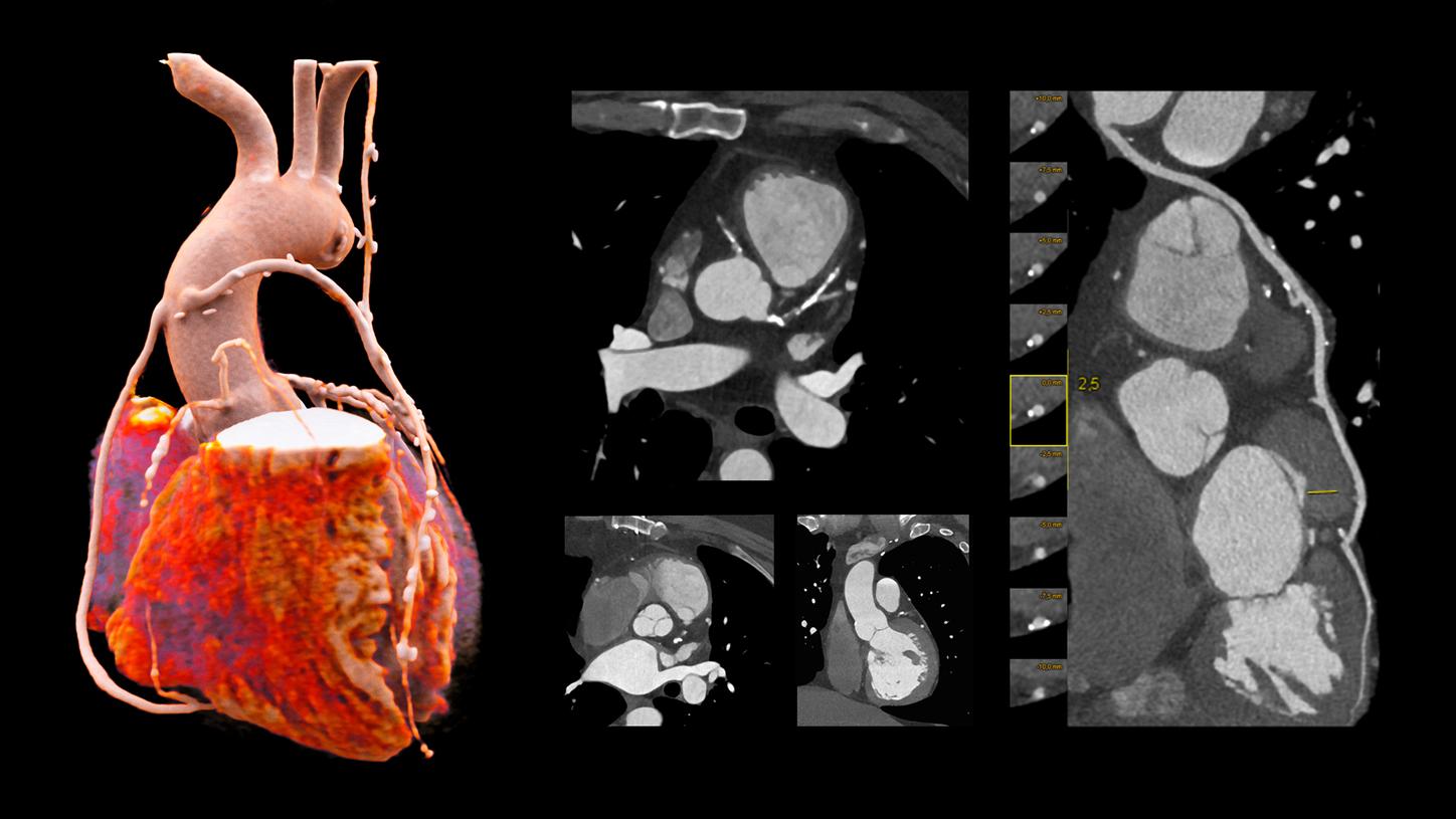
Courtesy of University Medical Center of Freiburg, Freiburg, Germany
NAEOTOM Alpha
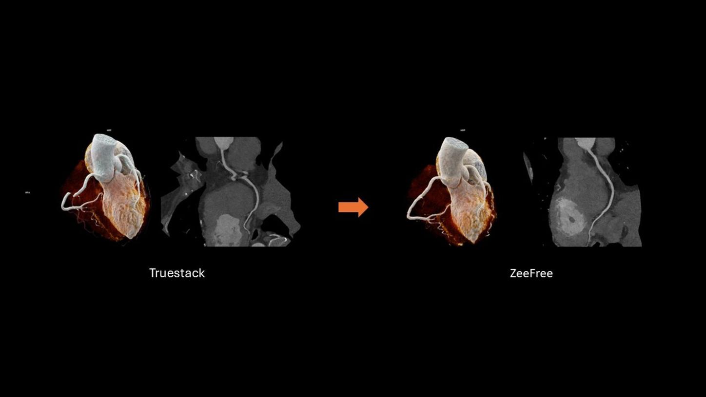
Clinical example for challenging conditions – Visualization in the presence of stents, vessel occlusions, and multiple calcifications