Biograph Vision™1 is the next-generation of PET/CT scanners that empowers you to see a whole new world of precision. It goes beyond digital to reveal the bigger picture, maximize efficiency and help you to better understand disease progression.

Biograph VisionSee a whole new world of precision.
Características e Benefícios
Features & Benefits

Accuracy to reveal the bigger picture
Biograph Vision is specifically designed to break through the limits of spatial and temporal resolution. With 3.2-mm crystals, Biograph Vision delivers high spatial resolution to reduce the impact of partial volume effect (PVE). Along with higher spatial resolution, a faster time of-flight also makes it easier to see small lesions2. This helps you quantify more accurately and more confidently understand disease progression.
“…by improving the spatial resolution…you have less partial volume effect, so you get sharper images and more accurate quantification.”


Performance to maximize efficiency
Biograph Vision can help you optimize your clinical operations with quality images and efficient workflow. With the market’s highest effective sensitivity at 100 cps/kBq3 and the fastest time-of flight in the industry4 Biograph Vision can not only reduce scan time and injected dose to boost productivity, it can also improve image quality. Combined with FlowMotion, it is designed to reduce unnecessary exposure to CT radiation, provide greater patient comfort, and decrease examination times.2
“We have now already, as compared to the older system, reduced the activity we inject… Now it's probably 30% faster with about 30% less dose which is something very acceptable.”


Reproducibility to understand disease progression
Biograph Vision can help reduce unwarranted variations to maximize patient care. Its zero-differential-deflection patient bed provides perfect registration between the CT and PET fields of view, ensuring accurate attenuation correction for more precise quantification. QualityGuard5 automates daily and weekly quality control without a radioactive source to help produce consistent and accurate results. FlowMotion Multiparametric PET Suite makes it easier and faster to perform parametric imaging in daily clinical routine. It is completely automated and integrated into the PET/CT workflow for more reproducible images.
“Being more quantitative, our reproducibility can be that much better, and it may matter when we’re trying to do a repeat scan early on in a therapy and decide what to do.”

Detalhes técnicos
Technical Details
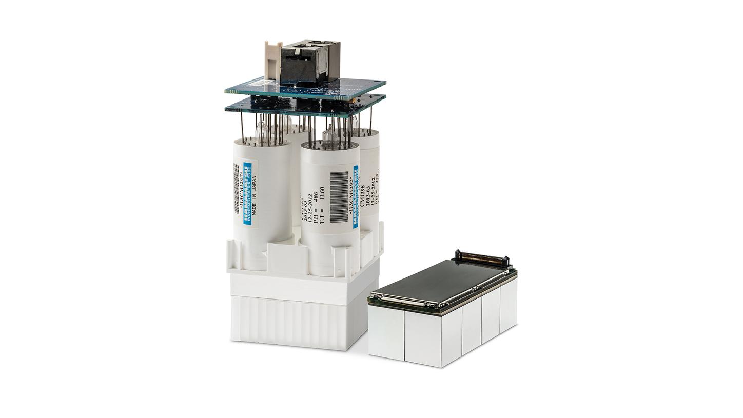
Transcend digital with the Optiso UDR detector
Optiso UDR’s proprietary 3.2 mm LSO crystals move silicon photomultiplier (SiPM) technology beyond digital to a new level of precision to help you detect small lesions, devise accurate treatment strategies and achieve optimal performance in a wide range of count rates.
See a whole new world of precision
/


/
Especificações Técnicas
Technical Specifications
Essa informação foi útil?
1
Biograph Vision is not commercially available in all countries. Due to regulatory reasons, its future availability cannot be guaranteed. Please contact your local Siemens organization for further details.
2
Compared to current state-of-the-art technologies. Data on file.
3
Based on internal measurements available at time of publication. Data on file.
4
Based on competitive literature available at time of publication. Data on file.
5
QualityGuard is currently under development on Biograph Vision and is not available for sale in the U.S. or any other country. Future availability cannot be guaranteed.
6
Optional.
The statements by Siemens Healthineers customers described herein are based on results that were achieved in the customer’s unique setting. Since there is no “typical” hospital and many variables exist (e.g., hospital size, case mix, level of IT adoption) there can be no guarantee that other customers will achieve the same results.