Taking biopsies from small lung nodules to diagnose cancer at an early stage poses an opportunity and a challenge at the same time. The smaller the nodule, the more difficult the biopsy, but the higher the survival rate of the patient. Angiographic imaging in 2D and 3D can now help the physician biopsy the patient quickly and precisely. Find clinical workflow descriptions, movies, images and articles here.

Improving the accuracy in diagnosis of bronchial carcinomas
According to the recent National Lung Screening Trial (NCI U.S.) patients who underwent screening for the early detection of lung cancer with a low-dose CT are 20% less likely to die of lung cancer than those receiving just a conventional X-ray.
Many solitary pulmonary nodules (SPN) are discovered purely by chance when the patient is undergoing thoracic imaging. The relative risk of an incidental SPN on unbiased thoracic imaging to become lung varies according to size1:
- 1% in nodules smaller than 4mm,
- 10-20% for those between 8 and 20mm, and
- 50% for nodules larger than 20mm, making clarification essential.
However, the accuracy of tissue samples taken from small lesions is relatively low: 33 percent accuracy in the case of lesions under 20 mm as opposed to 62 percent for lesions over 20 millimetres2. And this fact poses a problem. Although small SPN tend to be benign rather than malignant, they require more invasive treatment because conventional bronchoscopies on lesions of this size result in a low accuracy rate and tissue samples can be false-negative.
The German S3 guideline proposes the surgical clarification of solitary pulmonary nodules below three centimetres via transthoracic needle aspiration or wedge resection. This means that patients with very small lesions are subjected to potentially useless interventions. The alternative - watchful waiting - find many individuals too difficult to endure. Besides, small-celled bronchial carcinomas can grow rapidly in the meantime, which can worsen the prognosis substantially. Studies prove that the life expectancy of stage I or II cancer patients lies at 88% at 10 years, as opposed to around 6 months for stage III or IV patients3.
Angiographic imaging in 2D and 3D with syngo DynaCT provides an enhanced solution for diagnostic clarification of small, peripherally located lung lesions.
syngo DynaCT - Guided biopsies of suspicious lung nodules
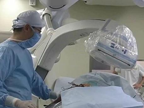
With the patient being sedated and under jet ventilation a syngo DynaCT run is performed in order to acquire CT-like images. Using only air as the contrast medium - the tumour is clearly visible with syngo DynaCT. (Image1)

The tumour can then be marked on the workstation, along with the path for the biopsy tool through the bronchie. (Image 2)

This 3D image with the markers is then being overlayed with live fluoroscopy while the physician advances the biopsy tool towards the tumour. This way he clearly sees the marked tumour (usually invisible in standard fluoroscopy alone) based on the overlayed 3D image, as well as the movement of the biopsy forceps through the bronchie. (Image 3)
With both 2D and 3D images being acquired intra-procedurally, with the patient in the same position and the diaphragm still, navigation is highly accurate and false-negative results can be greatly reduced. This differentiates this workflow from procedures in which biopsy planning is done based on pre-procedurally acquired CT-data and electromagnetic navigation of the biopsy forceps. Differences in the actual anatomy of up to 2cm especially in peripheral areas of the lungs are not rare in these cases.

High accuracy even in small and peripheral lesions
syngo DynaCT-guided bronchoscopies improve minimally-invasive diagnosis of even small and peripherally located lesions. Dr. Hohenforst-Schmidt from Hospital Coburg diagnosed the first 25 patients with this method – and with impressive results:
In lesions with an average diameter of 24x23x23mm, the hit rate was 78%. Even in very small nodules with an average diameter of 13x12x14mm the hit rate was 50% - and this during the proof-of-concept period alone.

Medical Knowledge Corner
Explore our collection of case studies, clinical workflows, videos, brochures and scientific publications for thoracic surgery / endobronchial procedures.
Brochures
- Image-guided thoracic surgery in the hybrid OR - Illustrated Workflows in Hybrid Operating Rooms, No.12 (pdf) 4.37 MB
- Electromagnetic navigation bronchoscopy with intra-operative 3D imaging - Illustrated Workflows in Hybrid Operating Rooms, No.14 (pdf) 3.42 MB
- Navigation in the Lung - Case study (pdf) 3.71 MB
Videos & Images
Multiply your Senses - Thoracic
Multiply your Senses - Thorax
Presentation Hohenforst/Schmidt - Thoracic
Multiply your Senses - Thoracic Case
Workflow for one-stop image guided surgery of small pulmonary nodules, SHG Clinic Völklingen
3D image-guidance for video-assisted thoracoscopic surgery (VATS) at Saga University Hosital, Japan
5 seconds syngo DynaCT run to acquire CT-like 3D images – syngo DynaCT*
Overlay of marked path / tumour with live fluoroscopy – syngo iPilot*
Overlay of syngo DynaCT 3D volume with live fluoroscopy to see both tumor and movement of biopsy forceps – syngo 3D Roadmap*
Video Virtual fly through the bronchie towards the tumour – syngo FlyThrough*

Manual marking of path for biopsy forceps and tumour out of different angulations in the 3D volume – syngo iGuide Toolbox*
* Image and video courtesy W. Hohenforst-Schmidt, J. Brachmann; Klinikum Coburg GmbH, Ketschendorfer Str. 33, 96450 Coburg, Germany
Webcasts
Clinical webcast on syngo DynaCT in interventional chest medicine
by Dr. Hohenforst-Schmidt, Coburg Hospital, Germany