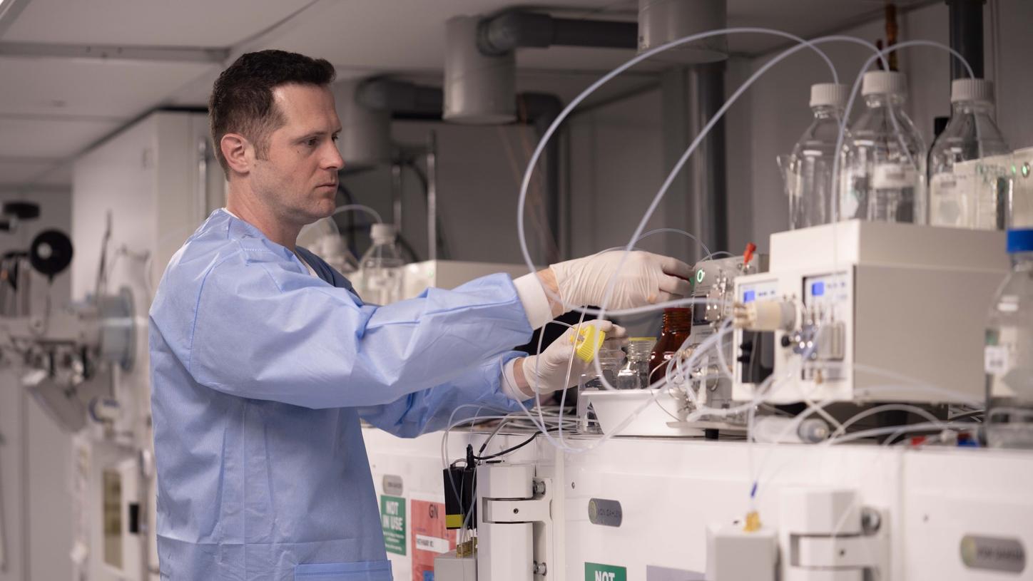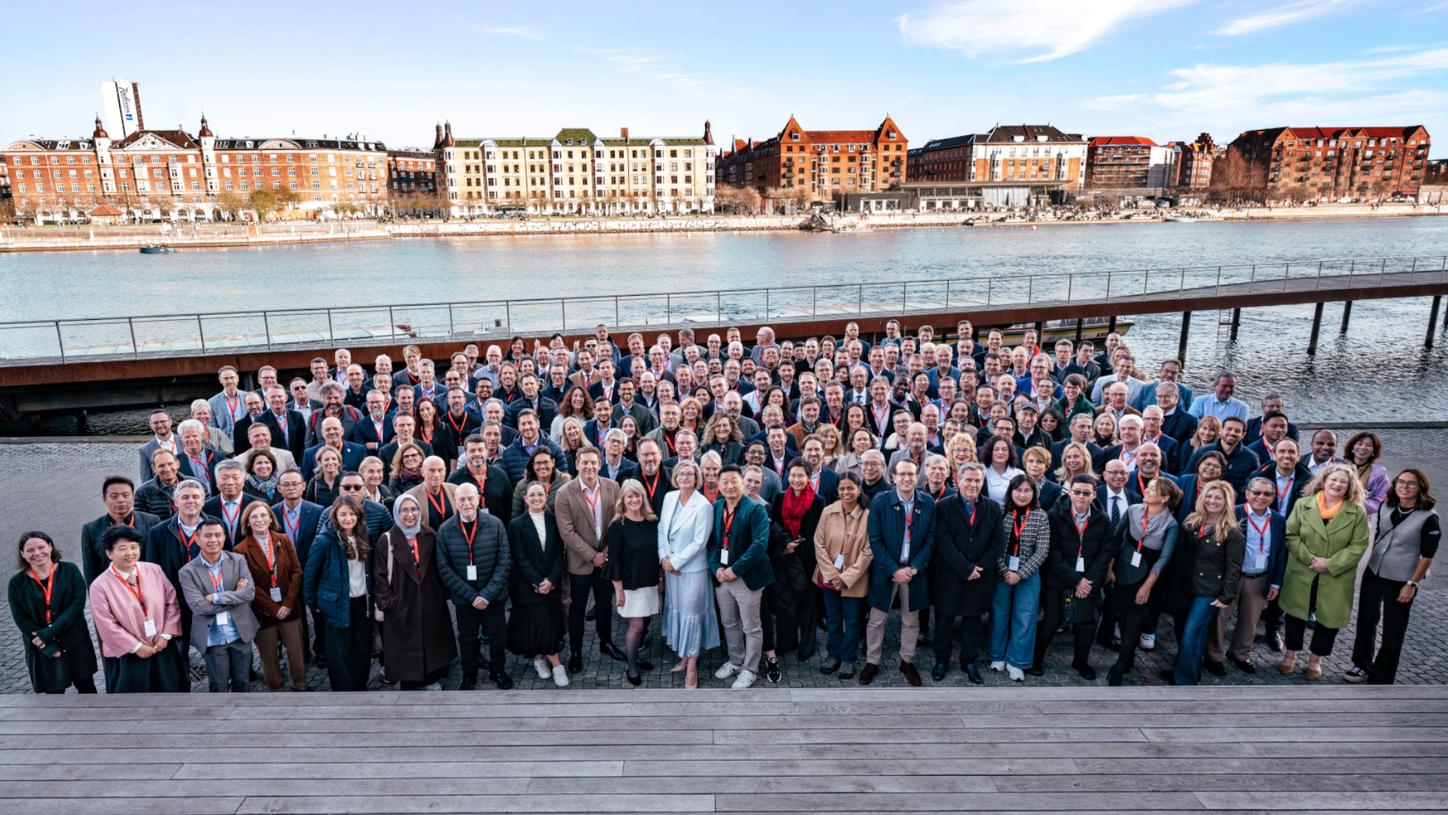
Almost 100 years ago, an interdisciplinary team of scientists laid the foundations of molecular imaging in Copenhagen, Denmark. This past April, the Danish capital welcomed molecular imaging and nuclear medicine experts from around the world for kinetic collaboration at the Molecular Imaging World Summit 2026.
Photography by Annika Daria Neubauer and Simon Lau
"We have so many more questions to answer,” affirmed Dr. James Williams, chief executive officer of Siemens Healthineers Molecular Imaging (MI) Business Line, to the global audience at the MI World Summit 2026. The event explored the latest breakthroughs in diagnostic precision, theranostic advancements, clinical research, artificial intelligence (AI), reporting methods, and successful synergies across institutions. The MI World Summit 2026, hosted by Siemens Healthineers and the fourth event of its kind, welcomed experts from 39 countries in Copenhagen, Denmark, this past April.
Copenhagen: an incubator for nuclear medicine
Reflections on Copenhagen’s contributions to nuclear medicine permeated the Summit’s opening keynote address. The keynote speaker, a nuclear medicine physician and professor from Denmark, explained how the city will be forever famous for the Copenhagen interpretation of quantum mechanics, outlined by Nobel laureates Niels Bohr and Werner Heisenberg in 1927. She continued to say that Copenhagen also holds a special place in the history of nuclear medicine.
As the speaker explained during her keynote, the name to know is George de Hevesy, a chemist, who spent time in Copenhagen during the 1920 and 1930s and discovered the indicator principle. Within molecular imaging, the indicator principle is essential to quantifying dynamic biological processes by tracking labeled indicators as they metabolize in tissues. Hevesy received the Nobel Prize in chemistry for his work on the use of isotopes as tracers in 1943.
Reflecting on the history set the stage for what was to become a vibrant and compelling scientific summit focused on today’s global molecular imaging efforts and the future direction it may take.
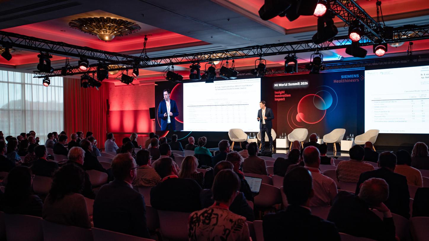
More than 30 presentations across two days from global leaders captivated the audience on a wide range of topics during the MI World Summit.
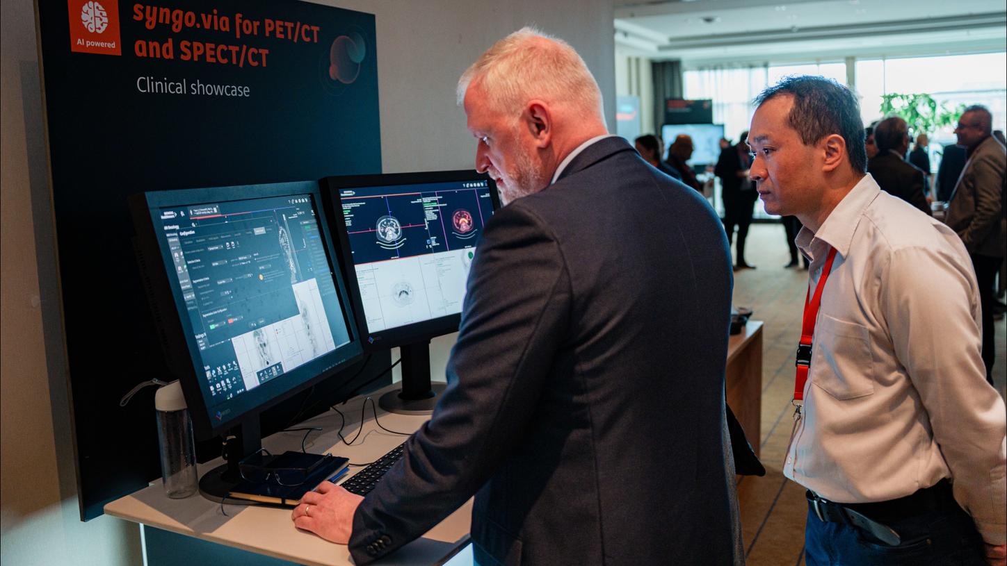
During breaks, attendees explored AI-assisted reporting and a virtual-reality simulation to experience a modern theranostics center.
Exploring advances shaping molecular imaging
Over the course of two days, the MI World Summit 2026 conducted five themed sessions featuring more than 30 presentations and panel discussions by global experts in their fields, as well as an exhibition area that allowed for deep dives into inspiring innovations. These included imaging solutions for care pathways in Alzheimer’s disease, various showcases on AI-assisted reporting, and a virtual-reality simulation to experience a modern theranostics center. In addition to the talks, there were smaller conversational “Ask the developers” sessions that covered automation in reading and reporting, fast time of flight, and the future of quantitative SPECT/CT.
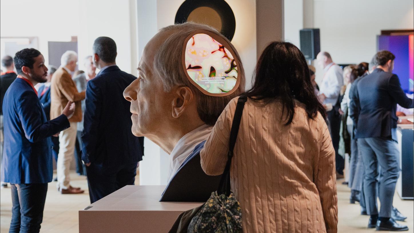
Alzheimer’s disease was also highlighted in the exhibition space.
Diagnostic precision for personalized care
On Friday, April 17, the sessions began with the latest advancements in PET/CT, quantitative SPECT/CT, and tracers focused on diagnostic precision for cancer, cardiovascular, and neurodegenerative diseases.
Presenters discussed how high-sensitivity PET/CT scans can help to diagnose cancers and cancer recurrences without delay. Imaging with targeted radionuclides, including immunoPET, can help to understand individual tumors better, which might lead to more personalized cancer therapies in the longer run. The goal would be to not only target tumor antigens, but also immune cells to get a more complete picture of what is going on in the tissue. Dr. Erik Aarntzen, nuclear medicine physician at University Medical Center Groningen, stated, “Long axial field-of-view (LAFOV) scanners unlock unprecedented opportunities for immunoPET.”
LAFOV scanners indeed loomed large in many presentations. A key feature mentioned in several presentations was the ability to perform whole-body perfusion imaging. This makes it possible to evaluate disease not only with relation to one organ, but systemically, which can provide new insights on various types of diseases. One area is cardiovascular diseases: LAFOV scanning can be used in this context to not only image cardiac perfusion, but simultaneously show how, for example, heart disease affects other areas of the body like the brain.
Brain neuropsychiatric imaging was the third clinical area discussed in this session. Dr. Maria Rosana Ponisio of the Mallinckrodt Institute of Radiology at Washington University in St. Louis, Missouri, U.S., explained that molecular brain imaging extends well beyond PET/CT. “The way we approach neuromolecular imaging is fundamentally shifting,” she said, noting that PET/MRI enables simultaneous acquisition of molecular and multiparametric structural data — something PET/CT alone cannot achieve. With key technical barriers now resolved and radiation exposure significantly reduced, she added, “As it becomes more widely available, PET/MRI will be the platform of choice.”
Faster way to reporting
The day’s second session featured presentations largely focused on artificial intelligence, data-driven patient adaptation, and AI-assisted reading and reporting. Dr. Trishul Siddarthan from the University of Miami, Florida, USA, spoke about how ventilation and perfusion from SPECT/CT can be used to explore respiratory health while Michel Koole, PhD, from KU Leuven, Belgium, contributed the why, how, and clinical impact of dosimetry.
Expanded clinical reach
To round out the day, a captivated audience listened to presentations dedicated to expanding clinical reach. Prof. Dr. Marcus Hacker, director of the Division of Nuclear Medicine at Medical University Vienna, Austria, noted, “In healthcare, there is a clear paradigm shift towards screening and disease prevention. And thanks to the LAFOV technology, PET/CT can play a role because it enables ultra-low-dose scanning.” 1, 2

The Friday evening event provided a comfortable atmosphere for attendees to reconnect with global molecular imaging experts and exchange ideas.
A consultant of pediatric nuclear medicine from Denmark pointed out that the benefits of modern PET/CT scanners in pediatrics reach beyond low radiation dose. LAFOV scanners in children are also about speed, since the short scanning times make examining younger children much easier for everyone involved.
The presenters also emphasized the impact of efficient infrastructure relevant to extending clinical reach. Prof. Dr. Pek-Lan Khong from the Clinical Imaging Research Centre (CIRC), Yong Loo Lin School of Medicine, National University of Singapore, reported about a very recent project in which they replaced two older PET/CTs with a total-body PET/CT. “Our goal was to make clinical services more efficient and to optimize our clinical research infrastructure.” Khong stated that modernizing PET/CT is about more than scanning infrastructure. “Success is very ecosystem-dependent. You need the workforce. You need a proper radiopharmaceutical supply. And you also need an IT backbone with sufficient data storage and high-performance computing capabilities.”
Making ends meet with high-end PET/CT scanners financially and logistically is easier in the industrialized world, however Summit attendees learned that staying up-to-date with technology can be possible even in more financially challenged locations. Dr. Adelina Sanches de Melo is head of the Department of Nuclear Medicine at Santa Casa de Misericórdia da Bahia in Salvador, Brazil. With the help of innovative financing models and strategic partnerships, she acquired a PET/CT for molecular diagnostics and a SPECT/CT for theranostics. “This was not the inexpensive choice, but the smart choice, an investment that makes sense for the next ten years. We more than doubled our numbers in 2025. We are on another level of expertise now. The market looks at us with different eyes, and we are also saving a lot of money.”
Advancing theranostics
Saturday, April 18, commenced with a dynamic session on theranostics. Presenters acknowledged that access to theranostics has improved worldwide and will further improve thanks to a broad spectrum of molecular imaging solutions that allow tailoring equipment to individual needs. Changes in our understanding of tumor biology are leading to new theranostic care concepts that take into account the individual immune response.
According to Dr. Ishita B. Sen of the Fortis Memorial Research Institute in Gurgaon, India, a theranostics program does not have to be complex. Sen presented several case stories that demonstrated the progressive nature at Fortis to help deliver theranostics to patients.
Pertinent to this session were two active debates on theranostics from a panel of international luminaries. The first panel discussion, moderated by Dr. Carl von Gall, of Siemens Healthineers, discussed radiopharmaceuticals and their role in defining the future of therapy. Dr. Rene Fernandez, from PositronMed, Santiago, Chile, moderated the panel focused on theranostics delivery models, from inpatient to outpatient pathways to decentralized networks and mobile approaches.
Synergies for success
Since progress does not happen spontaneously, the last session explored successful synergies. Since nuclear medicine, molecular biology, and personalized medicine are complex fields, progress will most likely be reached in interdisciplinary, highly collaborative environments. Prof. Dr. Akash Sharma, head of nuclear medicine at the Mayo Clinic in Jacksonville, Florida, put it this way: “If you want to go fast, go alone. If you want to get far, go together.”
Attendees discovered that to foster collaboration, a Siemens Healthineers Biograph Vision Quadra Consortium has been set up in North America to share learnings and develop standards around LAFOV PET/CT parametric imaging. Even more ambitious is the European PREFERENCE project that was introduced by Dr. Ronald Boellaard, professor for PET methodology at Amsterdam University Medical Center, Netherlands. PREFERENCE, funded by the European Union, brings together twelve nuclear medicine centers from all over Europe with the explicit goal to explore the benefits of LAFOV PET/CT imaging.
This will be done in seven methodological and eight clinical research projects, including projects that investigate the effect of ischemic heart disease on multi-organ perfusion, assess total-body dynamics of brown adipose tissue, aim at detecting early-stage infection and inflammation, and study the impact of multi-tracer PET imaging on holistic tumor characterization. None of this can be done alone. But, as Copenhagen’s history demonstrated, collaboration is deeply embedded in the genes of nuclear medicine.
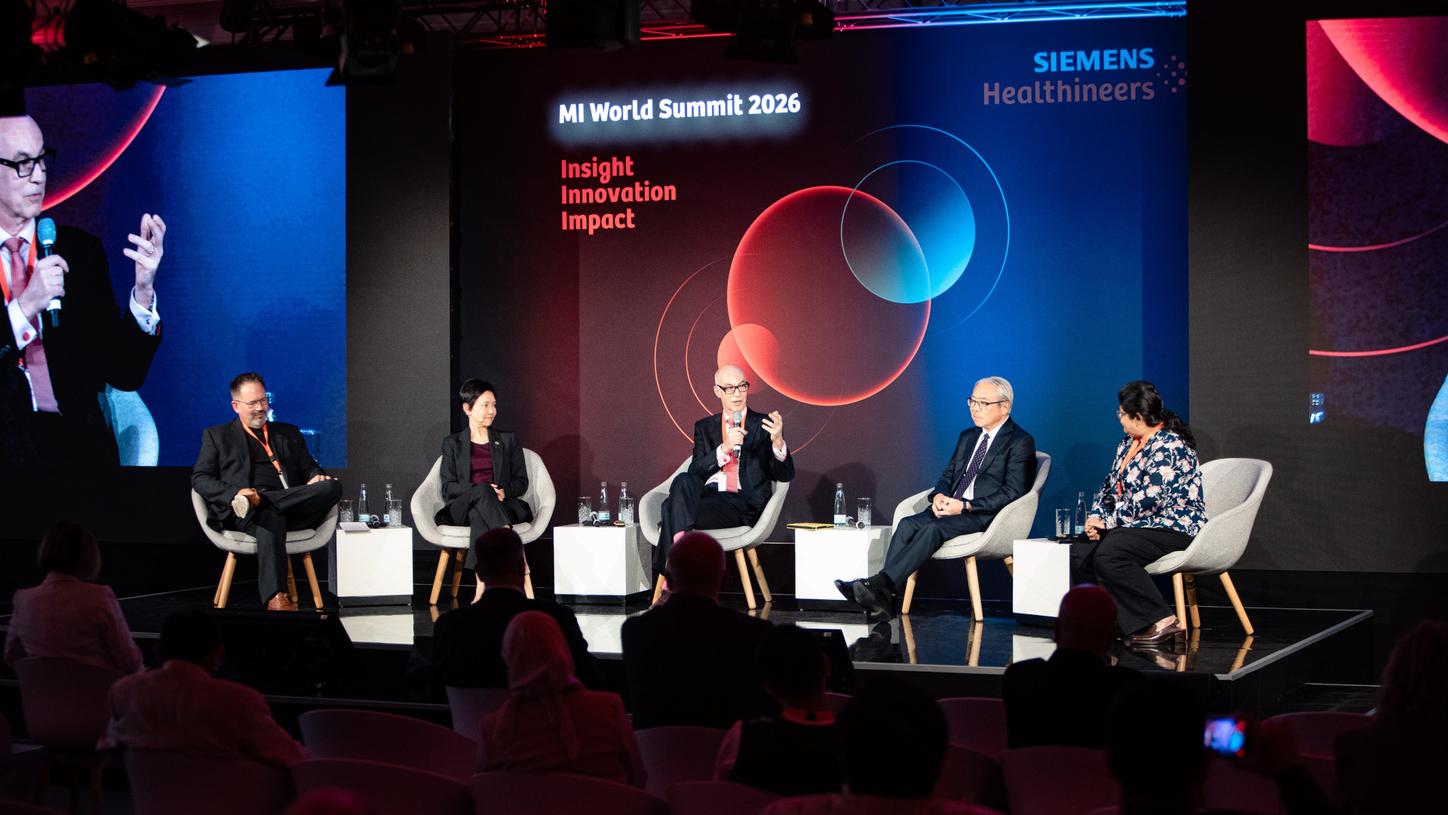
Williams hosted the closing panel discussion entitled “What does molecular imaging look like in 2036?”
Reflections from the closing panel discussion:
What will molecular imaging look like in 2036?
When leaders meet again at future MI World Summits, it will be enlightening to see what milestones are achieved and what remains on the horizon.
- More precise diagnostics: PET/CT moves more upfront in the diagnostic cascade, rather than being an adjunct method that it often is today.
- Better prevention: Ultra-low dose PET/CT imaging combined with AI to integrate digital patient data from other sources might help to intercept more diseases before becoming clinically apparent.1, 2
- Simulation first, treatment second: Building digital twins of individual patients based on imaging, laboratory diagnostics, and other data might allow to simulate therapies in-silico before applying them.
- Reduced workload: AI-powered image analysis and reporting may lead to automation at a scale unseen before. This will free human capacities to deal with more complex patients and allow room for closer patient interaction.
- More knowledge: LAFOV imaging may substantially contribute to the understanding of the complex interactions between mind and body.
- Less-invasive cancer care: New tracers and new target may extend the scope of theranostics way beyond its current areas of application.
- More personalized care: AI algorithms for ventilation-perfusion SPECT/CT and whole-body perfusion imaging might result in a more holistic and more patient-centric approach to cardio-pulmonary diseases, among others.
— Panelists and audience remarks, MI World Summit 2026
As the Summit concluded, Dr. James Williams remarked, “You cannot protect yourselves against the future. Expectations might be destroyed. Theories might break down. Paradigms might change.” The MI World Summit 2026 gathered dedicated experts who are open to that change and are energized about the future.