
Welcome to the MI Clinical Corner, your resource for expert insights and clinical perspectives across a wide range of molecular imaging applications. Explore real-world experiences, emerging trends, and best practices designed to support confident decision-making and elevate patient care.
Highlights
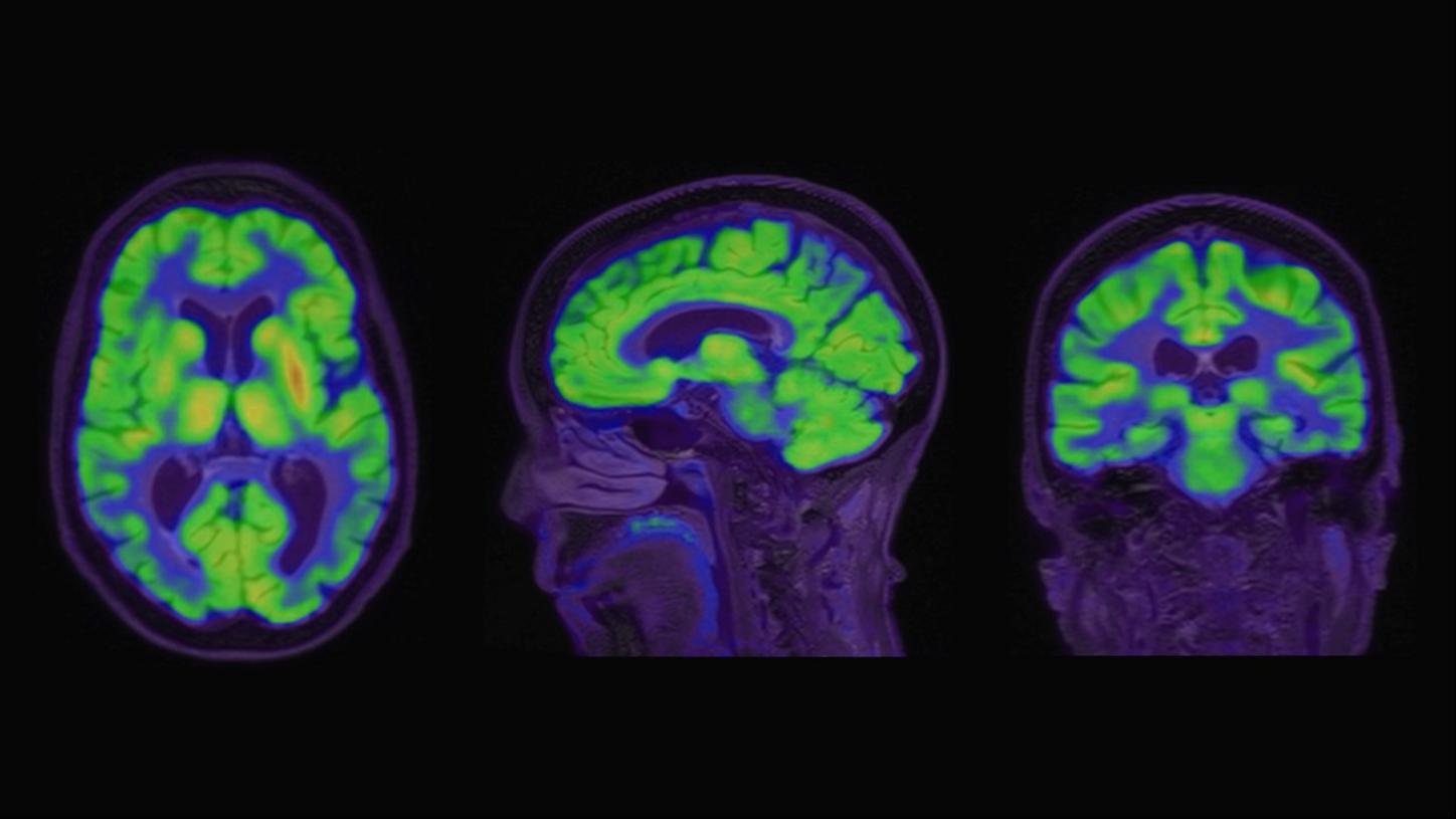
Case study:
Beta-amyloid PET/CT imaging in the assessment for anti-amyloid monoclonal therapy

MI World Summit 2026
The MI World Summit 2026 brought together global leaders in molecular imaging in Copenhagen, Denmark, for two days of scientific exchange, collaboration, and forward-looking discussion. Relive key moments, expert perspectives, and curated content that reflect the depth and energy of the global molecular imaging community on our MI World Summit 2026 webpage.
Scientific presentation:
The role of imaging in theranostics | SNMMI Symposium 2026
Dr. Munir Ghesani, MD, FACNM, FACR, FSNMMI Chief Medical Officer at United Theranostics and System Chief of Nuclear Medicine at the Mount Sinai Health System in New York
Learn how integrated molecular imaging solutions help enable diagnostic precision and advance theranostics, streamlining clinical workflows while addressing patient and imaging needs.
Scientific presentation:
Molecular neuroimaging: Differential diagnosis of Alzheimer's disease | Symposium at ECR Alzheimer’s Day 2026
Ana M. Franceschi, MD, PhD, Associate Professor of Radiology, Director of Dementia Imaging, and Director of Molecular Neuroimaging, Donald and Barbara Zucker School of Medicine at Hofstra/Northwell, Lenox Hill Hospital.
This case-based session explores how PET helps distinguish Alzheimer’s disease from other neurodegenerative disorders by using different tracers and the identification of characteristic metabolic patterns.
White paper:
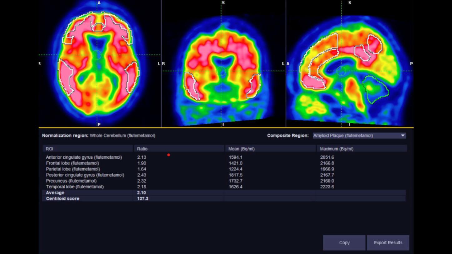
syngo.MI Neurology Cortical Analysis: Quantification software for amyloid and tau PET imaging
This white paper describes quantification methods used for amyloid and tau PET in syngo.MI Neuro Cortical Analysis. For over a decade, syngo.MI Neuro has had an analysis workflow dedicated to automated SUVr-based quantification of amyloid PET imaging. This workflow, referred to as Cortical Analysis, provides the reading physician with a user-friendly software tool that enables qualitative and quantitative assessments of amyloid PET brain studies with all three approved radiotracers.
Scientific presentation:
Leveraging long-axial field of view PET in molecular imaging and theranostics | Symposium at RSNA 2025
Philipose Mulugeta, MD, Associate Professor, University Pennsylvania School of Medicine
Dr. Philipose Mulugeta speaks about his experience with (LAFOV) PET/CT scanners to optimize the imaging of neuroendocrine tumors and discuss some potential upcoming uses of Zr-89 based PET imaging.
Case study:
Beta-amyloid PET/CT imaging in the assessment for anti-amyloid monoclonal therapy
MI World Summit 2026
The MI World Summit 2026 brought together global leaders in molecular imaging in Copenhagen, Denmark, for two days of scientific exchange, collaboration, and forward-looking discussion. Relive key moments, expert perspectives, and curated content that reflect the depth and energy of the global molecular imaging community on our MI World Summit 2026 webpage.
Scientific presentation:
The role of imaging in theranostics | SNMMI Symposium 2026
Dr. Munir Ghesani, MD, FACNM, FACR, FSNMMI Chief Medical Officer at United Theranostics and System Chief of Nuclear Medicine at the Mount Sinai Health System in New York
Learn how integrated molecular imaging solutions help enable diagnostic precision and advance theranostics, streamlining clinical workflows while addressing patient and imaging needs.
Scientific presentation:
Molecular neuroimaging: Differential diagnosis of Alzheimer's disease | Symposium at ECR Alzheimer’s Day 2026
Ana M. Franceschi, MD, PhD, Associate Professor of Radiology, Director of Dementia Imaging, and Director of Molecular Neuroimaging, Donald and Barbara Zucker School of Medicine at Hofstra/Northwell, Lenox Hill Hospital.
This case-based session explores how PET helps distinguish Alzheimer’s disease from other neurodegenerative disorders by using different tracers and the identification of characteristic metabolic patterns.
White paper:
syngo.MI Neurology Cortical Analysis: Quantification software for amyloid and tau PET imaging
This white paper describes quantification methods used for amyloid and tau PET in syngo.MI Neuro Cortical Analysis. For over a decade, syngo.MI Neuro has had an analysis workflow dedicated to automated SUVr-based quantification of amyloid PET imaging. This workflow, referred to as Cortical Analysis, provides the reading physician with a user-friendly software tool that enables qualitative and quantitative assessments of amyloid PET brain studies with all three approved radiotracers.
Scientific presentation:
Leveraging long-axial field of view PET in molecular imaging and theranostics | Symposium at RSNA 2025
Philipose Mulugeta, MD, Associate Professor, University Pennsylvania School of Medicine
Dr. Philipose Mulugeta speaks about his experience with (LAFOV) PET/CT scanners to optimize the imaging of neuroendocrine tumors and discuss some potential upcoming uses of Zr-89 based PET imaging.
Case study:
Beta-amyloid PET/CT imaging in the assessment for anti-amyloid monoclonal therapy


