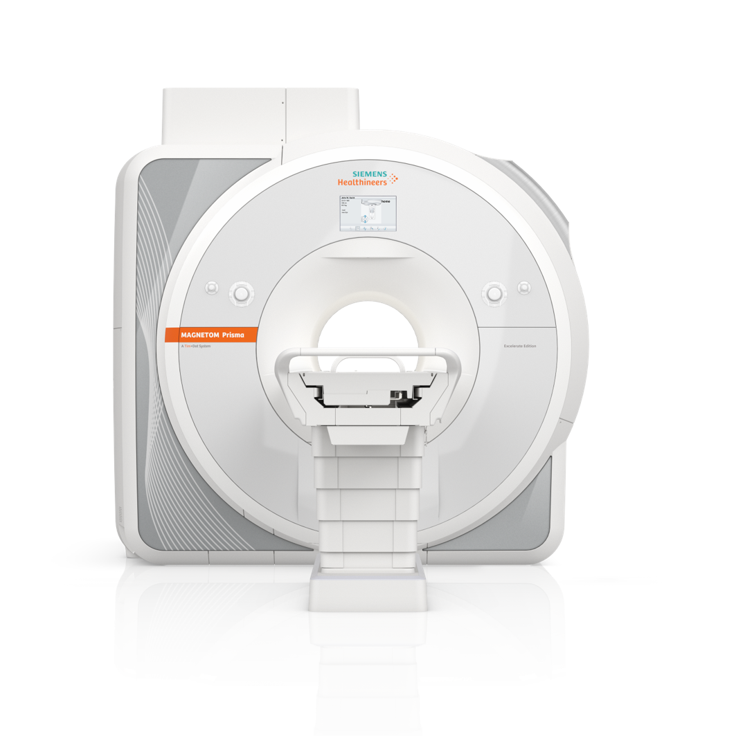
MAGNETOM Prisma1The 3T PowerPack for Exploration.

MAGNETOM Prisma1 offers a unique 3T MRI platform to help you tackle the most demanding MRI research challenges of today and tomorrow. Its breakthrough design delivers maximum performance under prolonged high-strain conditions. Exciting new MRI applications deliver high anatomical detail and provide opportunities to expand imaging capabilities. MAGNETOM Prisma has a proven track record of research and clinical success. It is a 3T PowerPack for exploration.
MAGNETOM Prisma Excelerate Edition
The large MAGNETOM Prisma community has made great contributions to the progress of MRI –driving research and translating research into clinical practice.
MAGNETOM Prisma is a system with the DNA of a champion. Since its beginnings, it allows its users to enter new areas of research with innovative applications and drive the advancement of human health. We are committed that this innovation never stops: we introduce the MAGNETOM Prisma Excelerate Edition. With its new software platform syngo MR XA30, MAGNETOM Prisma allows you to further excelat MR imaging and research. When it comes to tackling the most demanding MRI research challenges, MAGNETOM Prisma Excelerate Edition extends its application scope on its highly appreciated and unique 3T platform. All supported by the right partner, and the right collaboration network.
Features & Benefits
The power to outperform
In the most demanding MRI research projects based on extensive long-term studies, you profit from a powerful, robust MRI machine. MAGNETOM Prisma is your 3T PowerPack: unmatched 3T magnet, XR 80/200 gradient coil, parallel transmit architecture TimTX TrueShape, and receive architecture Tim 4G.
Research Use
The power to explore
Enter new areas of MRI research and set the future trends in healthcare. Exciting applications will open up new possibilities to help you visualize functional processes and better understand diseases.
- Advanced applications such as DWI, SWI, resting state fMRI, dynamic angiography, perfusion, and Diffusion Spectrum Imaging (DSI) – or your own research applications: MAGNETOM Prisma is your companion. It supports you with unparalleled 3T power and offers virtually unlimited imaging possibilities.
- Siemens Healthineers unique Turbo Suite enables greater efficiency with acceleration technologies for all contrasts, orientations, and body regions whilst keeping the quality of your exam.
- With MR Fingerprinting (MRF)5, MAGNETOM Prisma enables diagnostics at the frontier of a paradigm shift. MRF uses quantitative information from scans for decisions based on digital tissue data, not visual impressions, to improve tissue differentiation and enable less invasive diagnostics.

Diffusion Spectrum Imaging
- Up to 514 diffusion directions
- Sensitive to multiple diffusion directions within a voxel
- Potential to characterize crossing fibers

TimTX TrueShape and syngo ZOOMit
- Fine imaging details from the most hidden structures
- High resolution DWI
- DTI Depicting structural CNS connectivity

Most powerful shimming system
- Benchmark magnet homogeneity
- Advanced higher order shim, optional SpectroShim
- Higher specificity in body and multinuclear spectroscopy

Excellent flow sensitivity and time-resolved Angiography with XR 80/200
- Depiction of smaller vessels without contrast agent with ToF angiography
- Enhanced flow effects with XR 80/200 gradient system
- Higher SNR
- Extreme iPAT performance with Tim 4G

syngo DTI with up to 256 directions
- High-resolution isotropic DTI for finer depiction of white matter tracts

Most advanced tools in syngo.via
- Fully integrated fMRI/DTI evaluation
- Interactive track exploration mode with multiple VOI objects

Excellent long-term stability
- Excellent signal stability even for demanding long measurements – thanks to the new XR 80/200 gradient cooling system
And there is so much more...
- Simultaneous Multi-Slice (SMS) for DWI/DTI/BOLD
- SMS TSE, SMS RESOLVE
- Compressed Sensing TOF, CS SPACE, CS SEMAC, CS GRASP-VIBE
- CS Cardiac Cine
- Cardiac Flow Imaging
- High-resolution joint imaging with excellent FatSat
- Zoomed high-resolution imaging of the pelvis
- High-resolution with isotropic 3D imaging
- Up to 2x more SNR in diffusion studies
- Advanced Flow Evaluation
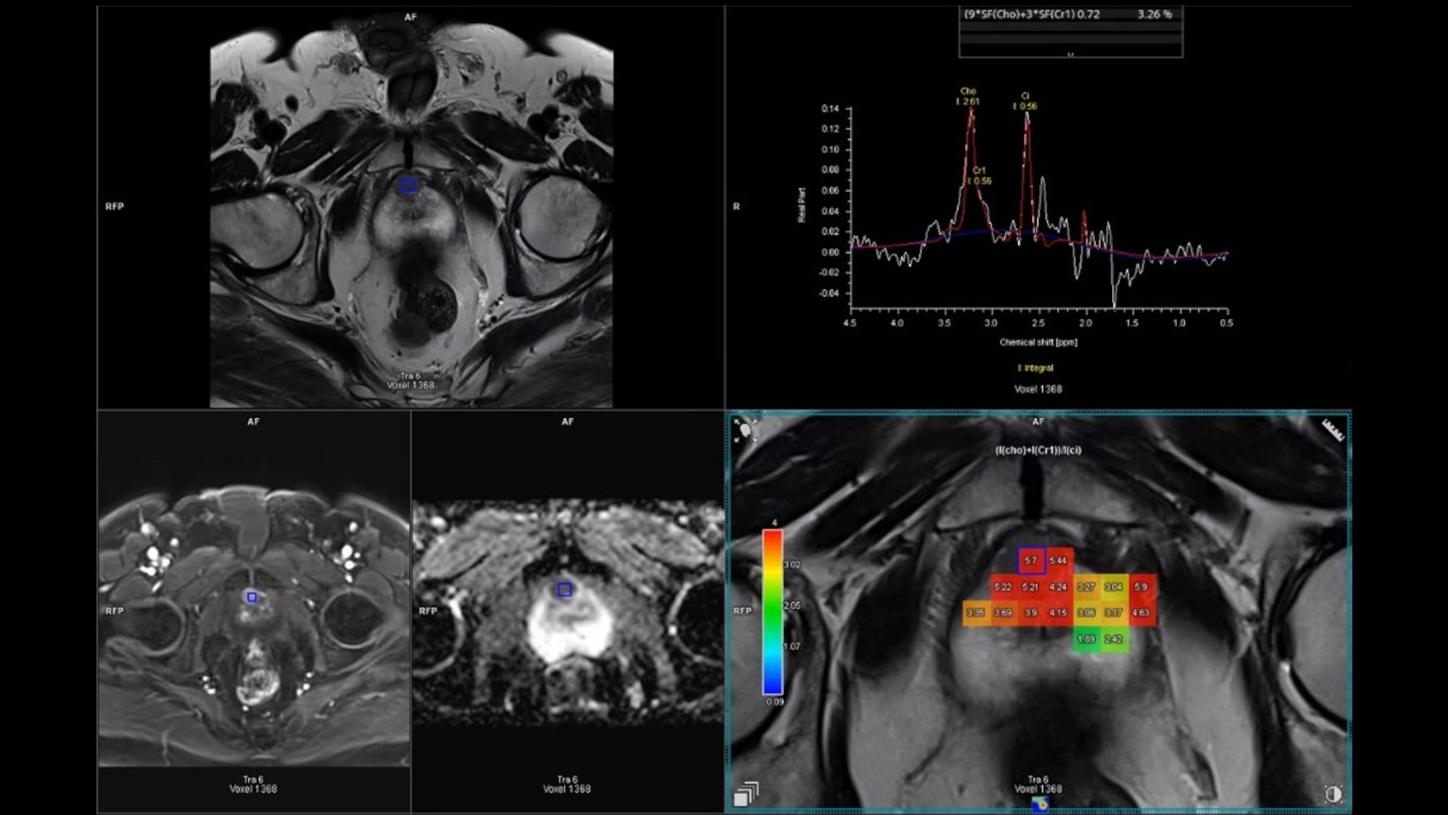
- Advanced multi-nuclear Spectroscopy
- 4D flow of the entire heart
- Single Voxel Spectroscopy with edit method
Clinical Use
Visualize detail in the finest anatomical structures and reveal functional information
Just like a prism, MAGNETOM Prisma1 unfolds the complete spectrum of MRI contrast. When powered by your ideas, scientific knowledge and expertise, the Siemens 3T MRI Scanner MAGNETOM Prisma builds a perfect match to open up a broad range of applications looking to answer the many questions that MR research poses. From brain functionality, to body physiology, organ morphometry, tissue metabolism, and quantitative MRI, MAGNETOM Prisma delivers the answers you are looking for.
The power to succeed
Do you want to make a difference in the lives of others? Fight the most threatening diseases and understand disorders? Advance human health? With the new Siemens 3T MRI scanner MAGNETOM Prisma, you can. A valuable collaboration for your success.
Collaborate and succeed
In MRI research, there are two forces driving the future of healthcare: As a physician, you aspire to transfer ideas from the lab into the clinical routine. Here at Siemens, we strive to translate research results into products like the MAGNETOM Prisma. This shared impulse is basis for a solid partnership founded on collaboration, support, and trust.
Be part of the IDEA network
As part of IDEA (Integrated Development Environment for Applications), the world’s largest and most active research community, you are able to get in touch with like-minded people, discuss ideas, and easily benefit from in-depth exchanges.
www.magnetom.net
MAGNETOM Prisma equips you with the needed competitive edge – the edge that enables you to do what others can’t. It might even facilitate access to research funding and could make your institution more attractive to top talents on the international academic market.
“MAGNETOM Prisma has the power to lead to better expertise and innovative research in the future. Beside the improved gradient system, the 64-ch Head/Neck coil allows us to get better signals from the cortex and also cover the cerebellum and the neck very well. That allows exploring new research fields which are difficult to do on other 3T scanners.”1
Prof. Rainer Goebel6
Professor, Maastricht University & Scientific Director,
Scannexus, Maastricht, The Netherlands
Technical Details
MAGNETOM Prisma’s1 technology sets the trends in the industry. Its ultra-high power impressively shows why Siemens Healthineers is the number one in MRI research.
Overview Technical Details
Magnet technology
MAGNETOM Prisma offers benchmark magnet homogeneity (typ. 1.1 ppm @ 50 cm DSV (diameter spherical volume)) and magnet reliability.
Gradient system
MAGNETOM Prisma comes standard with the XR 80/200 gradient system, the strongest gradients in the industry. The combination of the excellents gradients together with Tim (Total imaging matrix) enables the most demanding applications.
It offers largest anatomical coverage with a FoV up to 50 cm.
RF technology
Tim’s revolutionary all digital-in/digital-out design Direct RF allows for higher SNR and improved stability. All transmit and receive components are located at the magnet.
- Optical links between magnet and equipment room to achieve high RF stability and low noise
- Transmit and receive components are integrated in the magnet housing
- Dual-Density Signal Transfer enables ultra-high density coil designs by integrating key RF components into the local coil
Siting and installation
MAGNETOM Prisma allows for a fast installation. It requires only 33 m2 floor space.
Did this information help you?
http://grants1.nih.gov/grants/guide/pa-files/PAR-14-281.html
https://www.humanconnectome.org/study/human-connectome-project-for-early-psychosis/project-protocol/imaging-protocols-hcp-early-psychosis
R.L. Harms, F.J. Fritz, A. Tobisch, R. Goebel, A. Roebroeck, Robust and fast nonlinear optimization of diffusion MRI micro structure models, Neuro Image, Volume 155, 2017, Pages 82-96.; Slater, DA, Melie‐Garcia, L, Preisig, M, Kherif, F, Lutti, A, Draganski, B. Evolution of white matter tract microstructure across the life span. Hum Brain Mapp. 2019; 40: 2252–2268.
The product / feature is not for sale in the U.S. Its future availability cannot be guaranteed.
Minimum total space requirement for magnet, electronics, and console room.
Channels (coil elements) that can be connected simultaneously.