Prostate MRINew certainty in prostate cancer assessment
Overview

Modern imaging can enhance the entire care pathway of patients with suspected and diagnosed prostate cancer: MRI can help in earlier detection, more precise diagnosis, personalized treatment, convenient follow-up and individual management of advanced disease. We aim to offer a holistic portfolio to support the different stages of this patient journey with high-quality MR imaging.
- Perform a non-invasive prostate MRI exam with surface coils only
- Improve lesion conspicuity in DWI with ZOOMitPRO and RESOLVE
- Improve your reading and reporting with deep-learning based prostate cancer detection and classification with AI-Rad Companion Prostate MR1
- Monitor therapy response earlier with syngo.MR OncoTrend
Highlights

Improve lesion conspicuity in DWI with ZOOMitPRO and RESOLVE
High-quality diffusion-weighted imaging is of utmost importance in multiparametric MRI of the prostate. With ZOOMitPRO and RESOLVE we provide two independent techniques to tailor the exam to every patient's needs:
RESOLVE reduces blurring and susceptibility to artifacts, i.e. in patients with gas in the rectum or artificial implants. The resulting images are largely free of distortions and provide high spatial resolution. With RESOLVE you are able to improve your clinical capabilities by using a technique that achieves diagnostic images even in patients with metal implants.2
ZOOMitPRO is a method for high-resolution, zoomed FOV imaging, allowing diffusion-weighted images with high contrast and resolution in short acquisition times.
Standardize and ease the communication of your findings with PI-RADS
As part of the basic configuration within the syngo.MR General Engine, syngo.MR Prostate Reading allows for intuitive and comprehensive reading and reporting of multiparametric prostate exams. Using the PI-RADS standard, lesions can be rated on a 5-point Likert scale and findings can be visualized intuitively in a prostate pictogram. This eases communication with referring urologists, ultimately improving your institutional workflows.

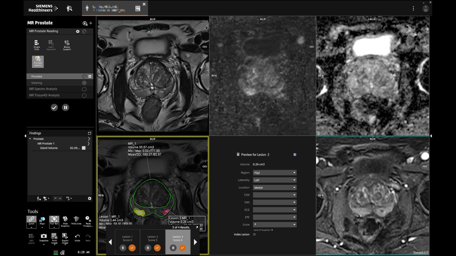
Speed up reading and reporting with Prostate MR1
With Prostate MR1 we introduce a key solution to support consistent, high-quality prostate MRI reading and reporting. By deploying artificial intelligence, Prostate MR1 assists in the interpretation of multiparametric Prostate MRI. The deep neural networks of Prostate MR1 are designed to improve the detection and classification of prostate lesions and to reduce the time needed for reporting by generating a pre-populated report.

Link the MR-reading workflow to the US-monitored biopsy
Prostate MR – For biopsy support provides an automated segmentation of the prostate and estimates the prostate volume. Location and contours of biopsy targets can be added manually. The results can be exported in the DICOM RT Structure Set format to support MR/US-fusion biopsies.

Prostate MRI in the cloud with AI-Rad Companion
Next to on-premise access through syngo.via, MR prostate biopsy support is also available via the cloud-based AI-Rad Companion. The assistant powered by AI helps you to reduce the burden of basic repetitive tasks and may increase your diagnostic precision when interpreting medical images. Find out more about AI-Rad Companion here:

Monitor therapy response early with syngo.MR OncoTrend
syngo.MR OncoTrend as part of the syngo.MR Oncology Workflow enables accurate monitoring of oncological lesions, helping determine whether treatment is effective or needs to potentially be reconsidered. With an intuitive traffic-light color coding, it is possible to visualize functional changes during therapy.