
By Partha Ghosh, MD, Siemens Healthineers, Hoffman Estates, Illinois, USA
Data and images courtesy of Queen Elizabeth Hospital Birmingham, Birmingham, United Kingdom
History
An approximately 75-year-old male with history of posterior spinal stabilization surgery presented with severe lower back pain. Routine X-rays were inconclusive, and the patient was referred for a 99mTc HMDP bone SPECT/CT study to determine the site of bone stress and related pathology.
The study was performed on Symbia Pro.spectaTM[a] SPECT/CT 3 hours following 20.9 mCi (776 MBq) intravenous (IV) injection of 99mTc HMDP. Following anterior and posterior planar whole-body acquisitions, multibed SPECT/CT acquisition of the entire cervical, thoracic, and lumbar spine as well as the pelvis was performed.
CT was performed with 130 kV with 30 reference mAs. 1.5-mm-reconstructed CT slices were obtained for evaluation and fusion with SPECT. Three-bed SPECT was performed with 60 stops per detector and 10 seconds per stop. SPECT was reconstructed using OSEM3D 7i15s with 128 x 128 matrix. SPECT/CT data was reviewed using syngo®.via.
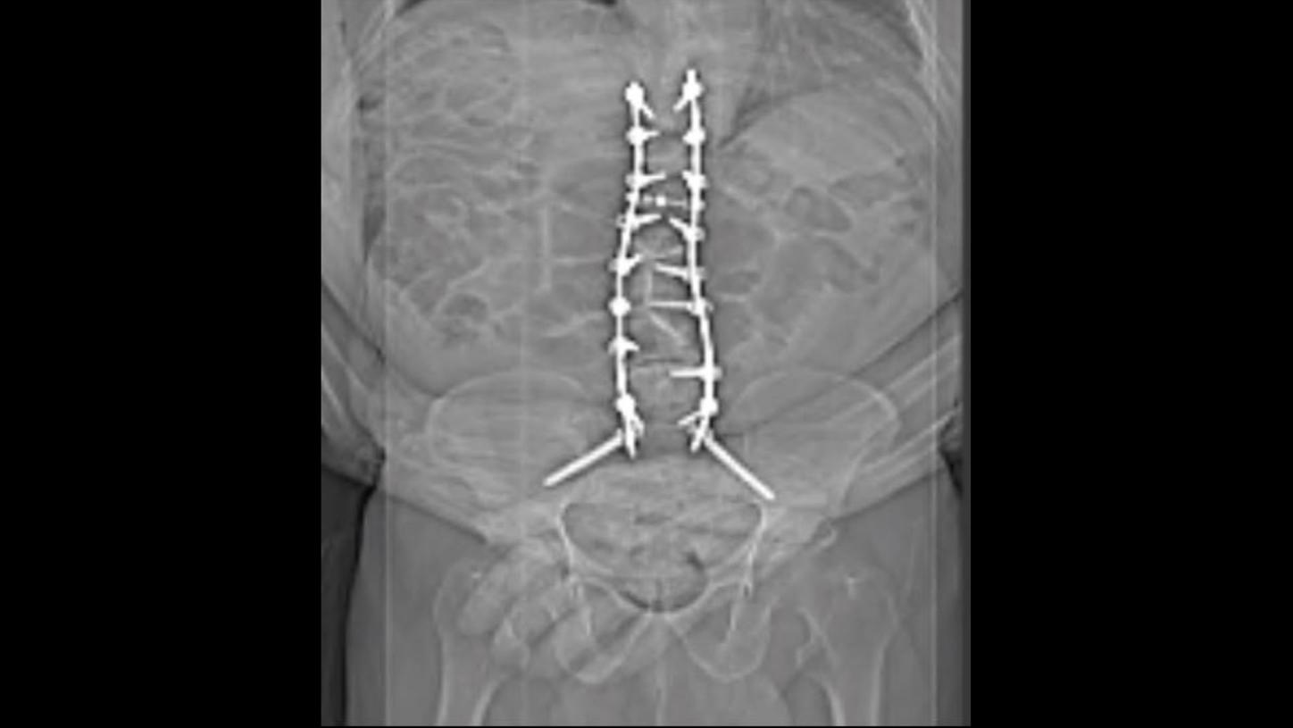
Figure 1: Routine X-ray of the lower thoracic and lumbar vertebrae shows bilateral spinal stabilization rod and screws without any clear evidence of displacement or fracture.


Findings
These findings clearly highlight the importance of high-quality CT with accurate SPECT fusion to enable the accurate interpretation of bone SPECT/CT in situations of spinal stabilization prosthesis fracture, instability, or loosening.
Thin-slice CT images and volume rendering sharply define the left and right posterior spinal stabilization rods and the fixation screws attaching the rod to the vertebral bodies, extending from T10 to S2. Although Symbia Pro.specta is capable of CT metal-artifact reduction (iMAR)[b], it was not applied with this patient. However, the high-quality, thin-slice CT provided optimum diagnostic information without excessive metal-related beam hardening, especially regarding the sites of fracture of the right spinal stabilization rod and the left S1 fixation screw and adjacent facet joints, particularly the sclerosis and focal lysis adjacent to the prosthesis.
CT images show fracture of the right vertical rod at the level of the mid-L4 vertebral body without significant displacement. There is no related hypermetabolism, suggesting absence of any instability on the right side due to this fracture. However, the scoliosis and instability related to the fracture of the left S1 fixation screw is the cause of severe L4-L5 facet arthropathy and associated bone stress around the fixation screw, which is reflected as focal hypermetabolism on SPECT images. The left vertical rod appears intact. The fracture of the left S1 screw distally within the vertebral body is sharply defined on thin-slice CT, although there appears to be no displacement or loosening. Increased uptake within a small lytic area in the left ilium adjacent to the sacroiliac joint associated with subchondral lucency with margin sclerosis likely represents a subchondral bone cyst or geode. Other transpedicular screws show normal alignment and absence of abnormal focal hypermetabolism, which suggests an absence of loosening. The lucency surrounding the left trans-sacroiliac joint screw is suggestive of early loosening. The low-grade uptake at the distal end of the screw reflects abnormal motion of the screw related to loosening.
Discussion
Spinal arthrodesis involving the fixation of vertebrae using metallic rods or plates with pedicular screws inserted within vertebral bodies through vertebral pedicles and lamina, often with intervertebral disc cages, are common surgical procedures for treating spinal instability in various spinal pathologies such as disc degeneration, spondylolisthesis and scoliosis, or kyphosis. A substantial percentage of patients experience recurrent pain following spine stabilization surgery with surgical re-intervention rates around 14% over 4-year follow-up.1 Common conditions causing post-spinal-fusion-surgery pain are loosening and fracture of stabilization rods and screws, vertebral non-union, pseudo-arthrosis, adjacent facet arthropathy, vertebral disk degeneration, and infection.
99mTc HMDP bone SPECT/CT has been shown to have a high accuracy in the detection of the cause of post-spinal-surgery pain and instrumentation failure. Hudyana et al demonstrated a sensitivity of 100% and specificity of 89.7% for bone SPECT/CT for lumbar vertebral prosthesis loosening.2 Overall, loosening was seen in 18.8% of patients undergoing lumbar stabilization surgery. However, in nearly 50% of patients with pain without evidence of loosening, SPECT/CT revealed other causes of symptoms including facet arthropathy, disc degeneration, and sacroiliac joint-degenerative arthropathy.
In this patient, SPECT/CT was instrumental in defining the exact cause of patient symptoms with clear visualization of fracture of the right vertical rod and left S1 pedicular screw. Symbia Pro.specta provided high-quality CT with 32-slice per sub-second rotation (0.33 seconds per rotation) at a collimation of 0.7 mm. The 1.5 mm-reconstructed CT slices with different kernels provided optimum visualization of fracture of the stabilization rods and pedicular screws, as well as the lucency and sclerosis around the screws. The lucency around the trans-sacroiliac joint screw on the left side visualized by the thin-slice CT together with the mild hypermetabolism around the screw was instrumental in detecting the early loosening of the screw. The presence of an osteolytic cyst lateral to the left sacroiliac joint with intense hypermetabolism and sclerotic margins is suggestive of a subchondral bone cyst, which is typically associated with severe sacroiliac joint degenerative changes.
The sclerosis medial to the insertion of the left L4 pedicular screw to the left-sided vertical stabilization rod visualized with thin-slice CT was precisely the region of hypermetabolism seen on SPECT/CT images, reflecting reactive bone stress due to instability, although there was no sign of loosening of the L4 pedicular screw. The intense hypermetabolism in the left L4/L5 facet joint associated with increased facet joint space and periarticular sclerosis reflects the actual cause of pain, which is related to the bone stress on the facet joint due to scoliosis, peri-prosthetic bone loss, and instability related to abnormal motion from the left-side screw fracture and loosening. Other degenerative changes in L2 and L4-5 disc spaces are also related to the scoliosis and instability.
Bone scintigraphy after lumbar fusion surgery can show hypermetabolism at the operative site for up to 1 year following surgery. However, the high intensity of uptake at the facet joint clearly identifies the site of active arthropathy to be the origin of the pain. The exact localization and characterization of multiple coexisting pathologies in this patient, including fracture of stabilization rod and screw, facet arthropathy, degenerative-disk disease, trans-sacroiliac screw loosening, and associated sacroiliac joint degenerative changes, is achieved through a combination of high-quality CT and SPECT with the exact co-registration of both leading to optimum visual and interpretational clarity.
Conclusion
This case demonstrates 99mTc HMDP bone SPECT/CT imaging in the evaluation of pain following thoracolumbar spinal fusion surgery to identify facet arthropathy along with fracture of the spinal stabilization rod. The high-quality CT provided by Symbia Pro.specta was instrumental in defining prosthetic fractures, and when paired with the co-registered SPECT, resulted in SPECT/CT findings that provided clear evidence of the facet arthropathy being the cause of symptoms.
Examination protocol
Scanner: Symbia Pro.specta
The outcomes achieved by the Siemens Healthineers customers described herein were achieved in the customer’s unique setting. Since there is no “typical” hospital and many variables exist (eg, hospital size, case mix, level of IT adoption) there can be no guarantee that others will achieve the same results.