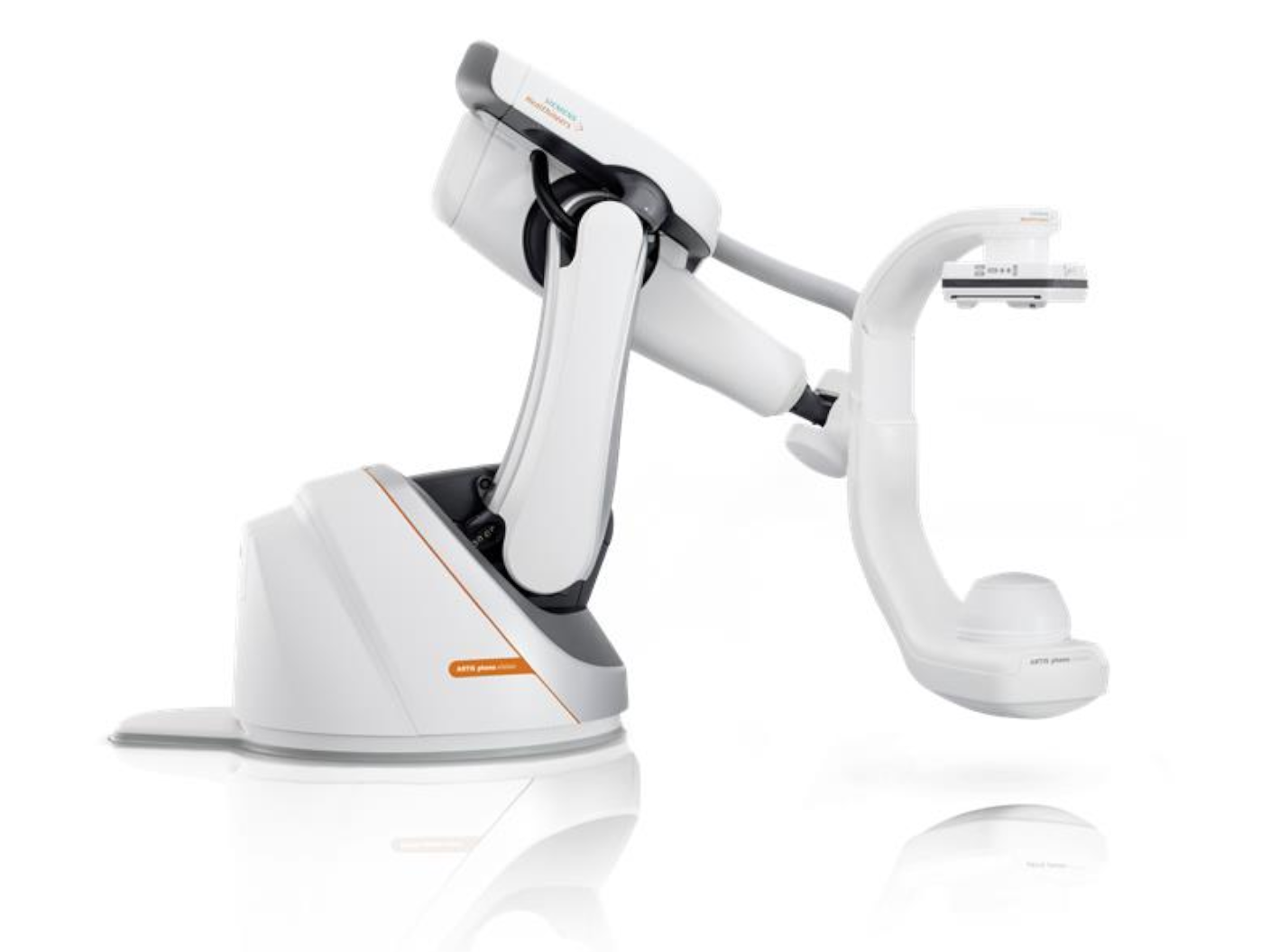
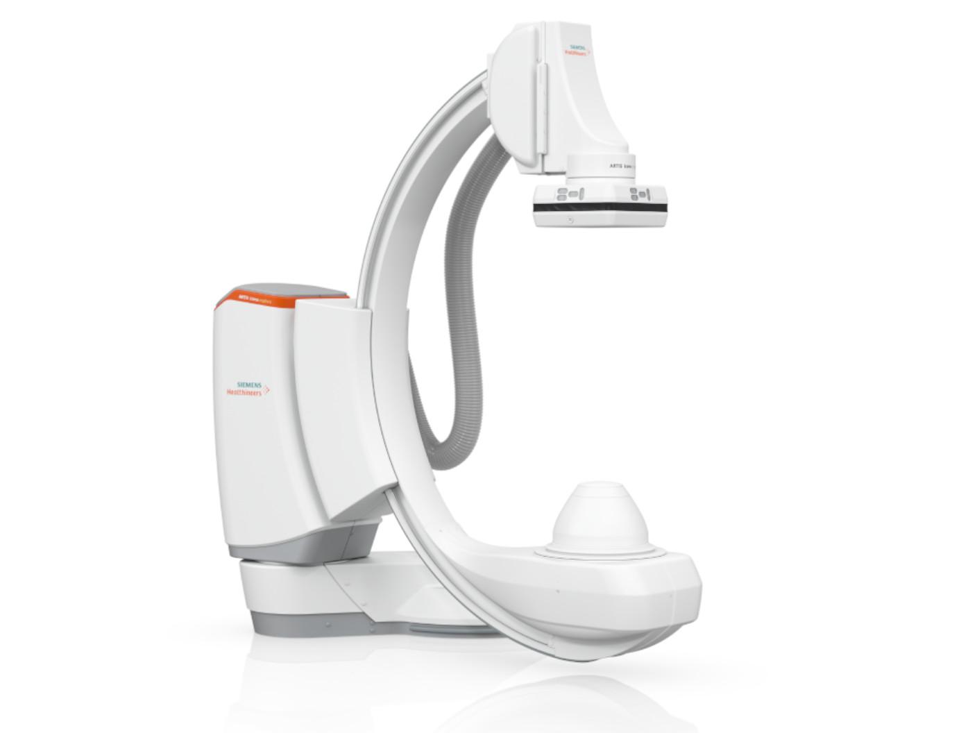
ARTIS pheno.visionInnovation. Powered by AI.
ARTIS pheno.vision1 is the advanced robotic imaging system designed to support a highly diverse procedure mix – empowering multiple interventional and surgical disciplines, both today and into the future. ARTIS pheno.vision is powered by OPTIQ AI2, our next-generation imaging chain combining CNR-driven exposure regulation with AI based image noise reduction. Turn your vision into reality - with ARTIS pheno.vision.


Benefits

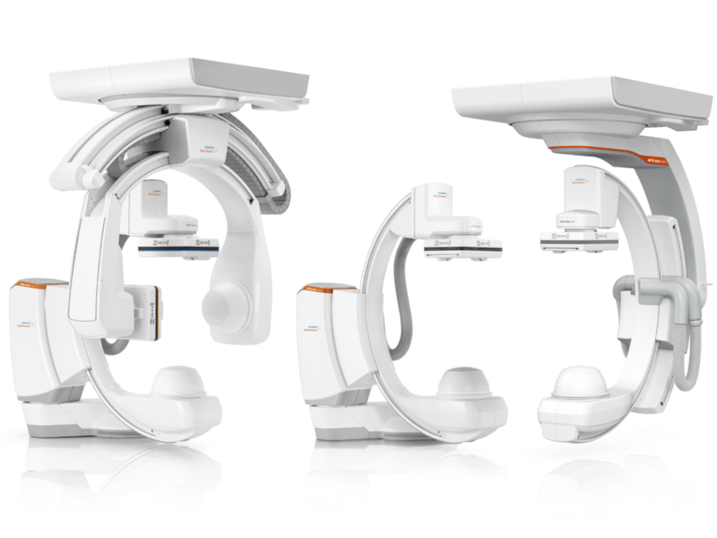
ARTIS pheno.vision is a versatile system for your Hybrid OR, interventional suite or lath lab
- Robotic technology with 9 degrees of freedom
- Wide-space C-arm offering sufficient room
- Flexible isocenter for adjustable working height in 2D imaging
- 3rd party surgical integration enables procedure specific patient positioning

Discover the range of possibilities supported by ARTIS pheno – whether performing standard or complex surgical or interventional procedures, like:
- EVAR, TAVI, iVATS, Deep Brain Stimulation (DBS)
Iliosacral Screw Fixation, Spinal Fusion
Aneurysm Treatments
TACE, PAE, and Laparoscopic Liver Surgery
Needle Procedures, Radiofrequency Ablation
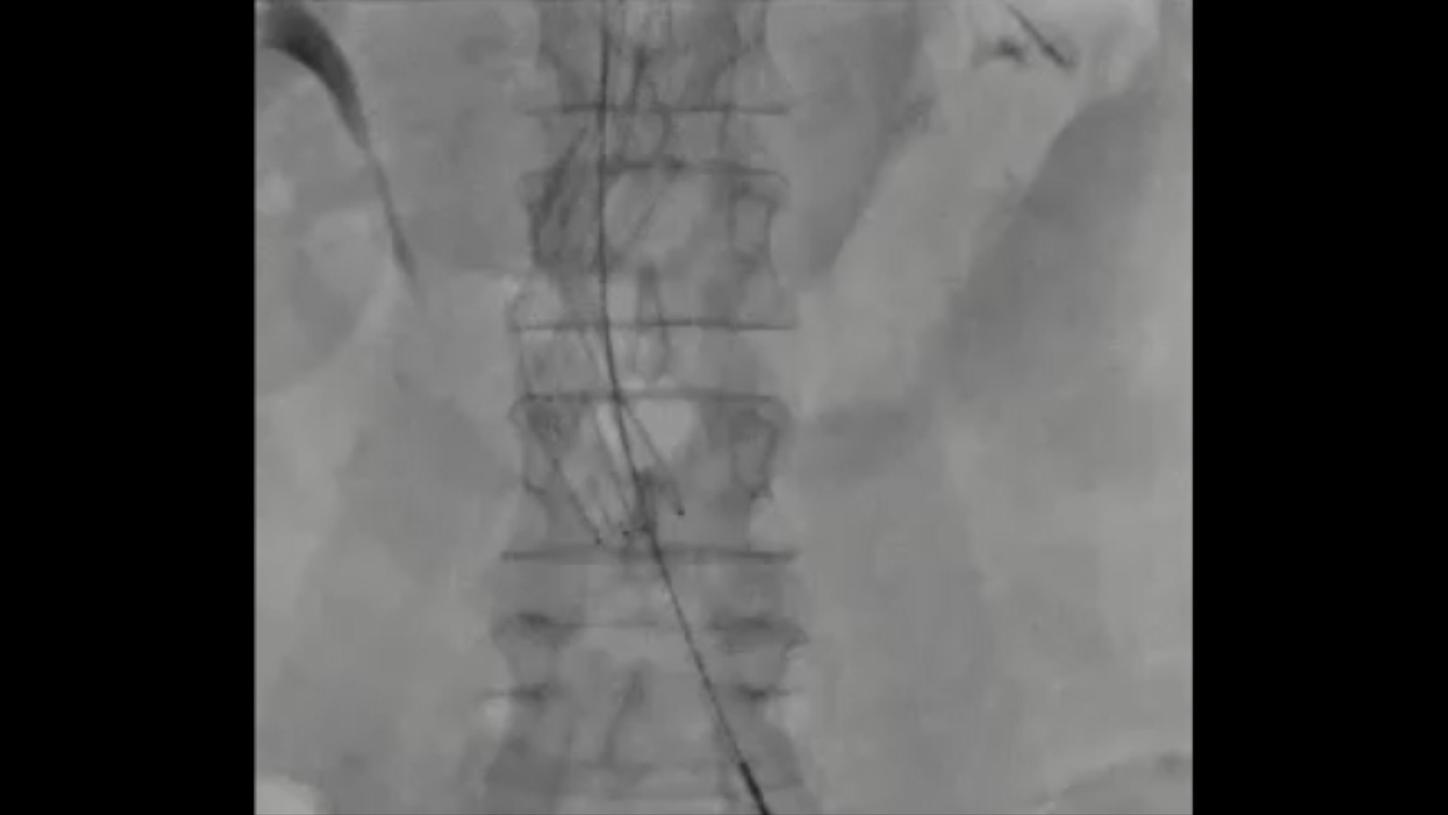 without OPTIQ AI
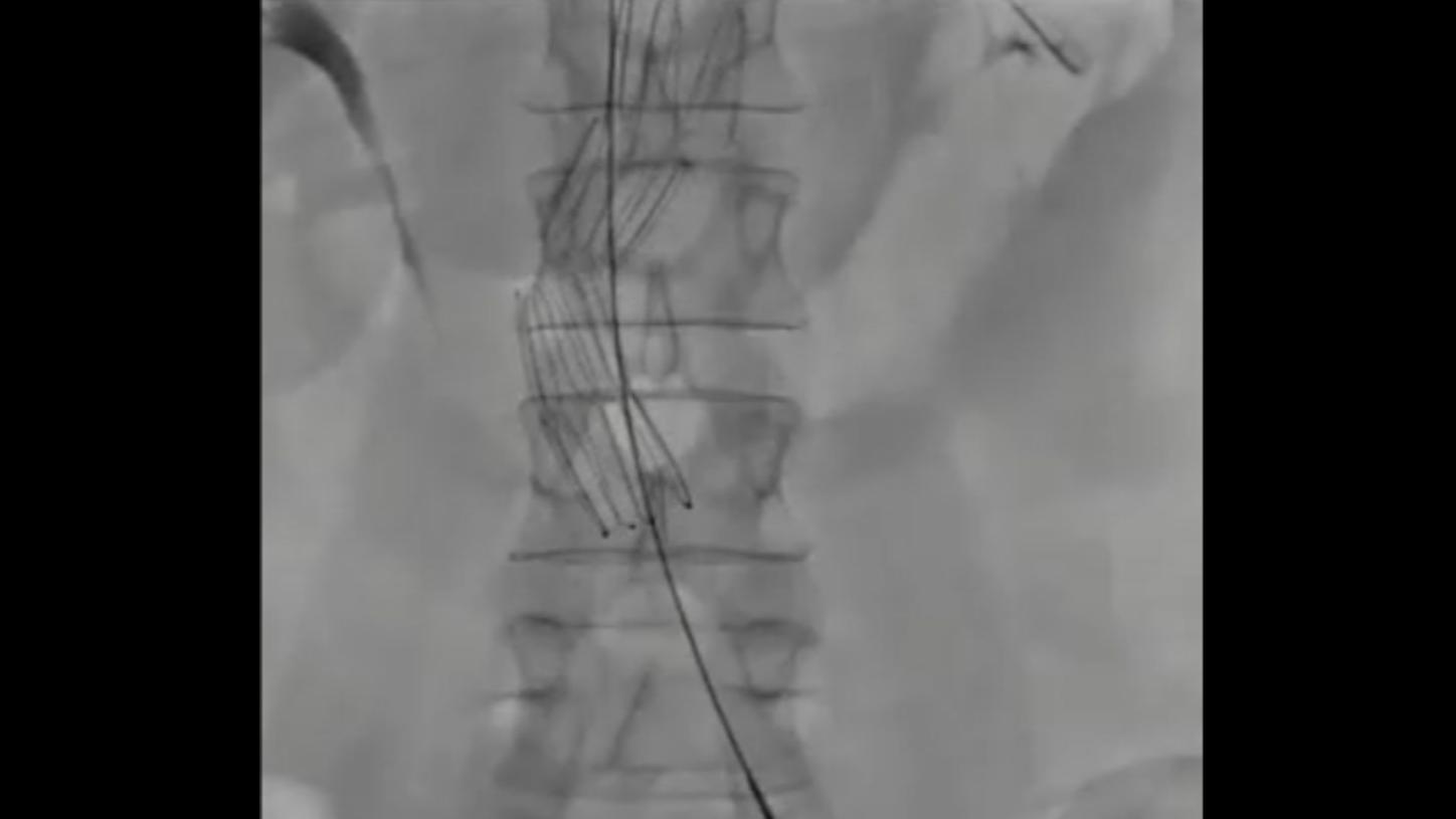 with OPTIQ AI
OPTIQ AI combines CNR-based exposure regulation with AI-based noise reduction.
- Constant image quality (CNR based) supporting ALARA13 principle
- Optimized device visibility based on the X-ray absorption properties of the respective material
- AI-powered algorithm to reduce image noise in real time across different 2D imaging modes and body regions

For each procedure step you can combine a multitude of system settings and turn them into a timesaving, individualized Case Flows, available with one click.
- Set the system to the requirements of each procedure step once: C-arm position, angulation and SID, imaging parameters and zoom, display layout and input selection and others
- Activate these settings with a single touch
- Eliminate up to six manual system interactions

With a system designed with infection control in mind:
- Comprehensive cleaning concept
- Smooth and spill-sealed surfaces
- Quick and easy draping

Sufficient space to navigate complex setups
- Free space of 95.5 cm3
- Facilitates imaging in steep angulations
- Suitable for procedures requiring large instruments and devices
- Good access to for the surgical or interventional team

syngo DynaCT Large Volume and syngo DynaCT 360 can visualize large anatomical areas
- Spine: Height of up to 23.5 cm (9.3”)
- Thorax, abdomen: Diameter of up to 43 cm (16.9”)4
ARTIS pheno.vision is a versatile system for your Hybrid OR, interventional suite or lath lab
- Robotic technology with 9 degrees of freedom
- Wide-space C-arm offering sufficient room
- Flexible isocenter for adjustable working height in 2D imaging
- 3rd party surgical integration enables procedure specific patient positioning
Evidence
Imaging for multiple surgical disciplines
Discover the range of different procedures supported by ARTIS pheno.

Ready for Endovascular aneurysm repair (EVAR)
ARTIS pheno with procedural intelligence helps standardize – and potentially speed up – EVAR procedures. This is possible thanks to assisted preparation of the CT dataset, simplified image acquisition, guided stent deployment with 3D imaging, and immediate assessment of results.


What your peers are saying
“ARTIS pheno offers consistent, excellent DynaCT image quality from head side, left or right patient side for imaging in thorax, abdomen and pelvis. This provides important information during technically challenging procedures, helps us to avoid major complications and ensures complete treatment and documents outcomes."

Director of the Institute for Diagnostic and Interventional Radiology at Hannover Medical School, Hannover, Germany


Product and Customer Videos
Case Studies & Publications
Thoracic Surgery
Image-guided video-assisted thoracoscopic surgery with Artis Pheno for pulmonary nodule resection J. Thoracic Surgery, 2020
Ya-Fu Cheng, Heng-Chung Chen, Pei-Cing Ke, Wei-Heng Hung, Ching-Yuan Cheng, Ching-Hsiung Lin, Bing-Yen Wang
See more
Neurosurgery
Diagnostic performance of intraoperative cone beam computed tomography compared with postoperative magnetic resonance imaging for detecting hemorrhagic transformation after endovascular treatment following large vessel occlusion Journal of Stroke, 2022
Naoki Kato, Katharina Otani, Yukiko Abe, Tomonobu Kodama, Toshihiro Ishibashi, Yuichi Murayama
See more
Intraoperative cone-beam CT with metal artifact reduction for assessment of the electrode position and the intracranial structures during deep brain stimulation procedure Acta Neurochirurgica, 2022
Toshinari Kawasaki, Takayuki Kikuchi, Katharina Otani, Yuto Mitsuno, Yukihiro Yamao, Nobukatsu Sawamoto, Ryosuke Takahashi, Susumu Miyamoto
See more
Spine Surgery
Generating patient‐matched 3D‐printed pedicle screw and laminectomy drill guides from Cone Beam CT images: Studies in ovine and porcine cadavers Medical Physics, 2022
Andrew Kanawati, Alex Constantinidis, Zoe Williams, Ricky O'Brien, Tess Reynolds
See more
TIPS Procedure
syngo DynaCT 360 with intravenous injection to evaluate portal vein patency in TIPS procedure: A study protocol
Ulf Teichgräber, MD, Renè Aschenbach, MD, Jena University Hospital, Germany
See more (pdf)
TIPS Procedure with syngo DynaCT High Speed
syngo DynaCT High Speed: Targeting portal vein with reduced radiation exposure. Use of Cone-Beam Computed Tomography (CBCT) for Targeting the Portal Vein in Transjugular Intrahepatic Portosystemic Shunt (TIPS) Procedures: Comparison of Low-Dose with Standard-Dose CBCT
University Hospital Tuebingen, Germany. Estler A, Nikolaou K, Hoffmann R, Herrmann J, Grosse U, Ketelsen D, Seith F, Artzner C, Grözinger G. Iran J Radiol. 2021 July; 18(3):e111704.
See more
Embolization - TACE
syngo DynaCT for post lipiodol-embolization imaging in TACE. Value of Latest-generation Cone-beam Computed Tomography for Post Lipiodol-embolization Imaging in Hepatic Transarterial Chemoembolization in Comparison with Multi-detector Computed Tomography
University Hospital Frankfurt. Leona S. Alizadeh, MD, Vitali Koch, MD, Thomas J. Vogl, et al. Acad Radiol 2021.
See more
Technical Details
Product Specifications
Installation
Floor-mounted to keep the ceiling free
Minimum technical room size
35m² (377 ft²)
Robotic C-arm
9 axes for positioning of the C-arm, SID lift and Detector + Collimator rotation
Wide space C-arm
130 cm (51") SID and 95.5 cm (37.5") usable clearance for easy patient access and C-arm positioning
Intraprocedural 3D imaging position
Head side and lateral
Working height
Flexible isocenter9 104 cm (40.95") - 150 cm (59.1")
Max. 3D volume size (diameter x height)
43 cm x 17.5 cm (17" x 7.9") or 32 cm x 23.5 cm (13" x 9.3")
Detector
30 x 40 detector
Recommended room size
>= 68 m2 (732 ft2)
Related topics
Meet our new ARTIS solutions

Hybrid OR Imaging Solutions
Perfecting minimally invasive surgery with procedural intelligence


Downloads

|
DownloadServices

Guardian Program with TubeGuard
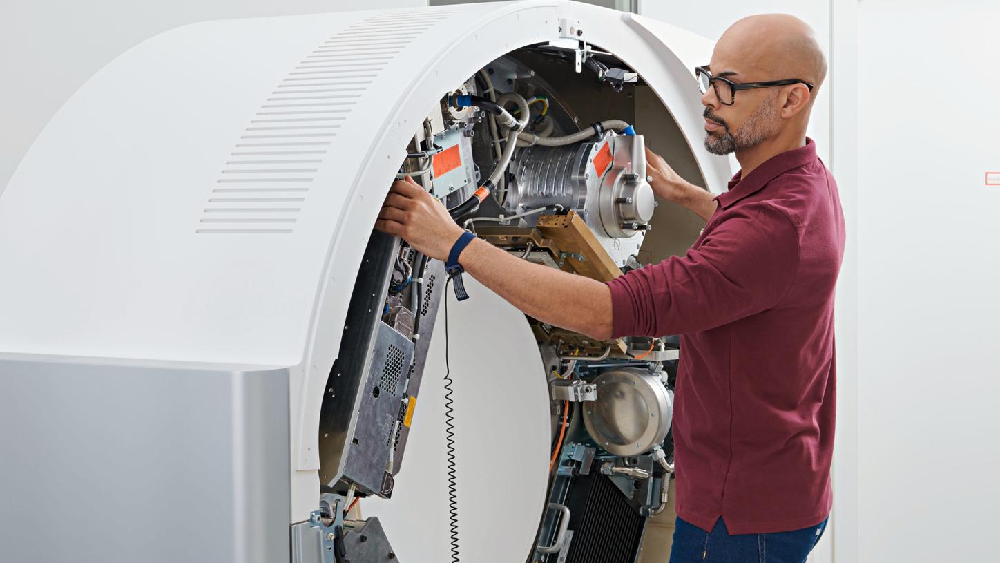
The X-ray tube is your most critical system component, so it is vital that it functions at all times. The Guardian Program with TubeGuard monitors and addresses predictable and detectable tube failures.
Performance Plans
Performance Plans are covering regulatory, quality, and performance needs. They come at fixed costs, offer a wealth of service options, and can be tailored to your needs and budget.

Virtual Education Solutions for ARTIS systems
Active learning experience in a virtual clinical environment for interventional radiology, cardiology, and surgery.
Clinical images courtesy of University Hospital Ulm, Germany (left) and Jikei University, Tokyo, Japan (right).