Follow these patients on their stroke health journeys
The characters and scenes shown are fictional. The character has been generated or altered by artificial intelligence

Follow Mary on her
Stroke Prevention Journey
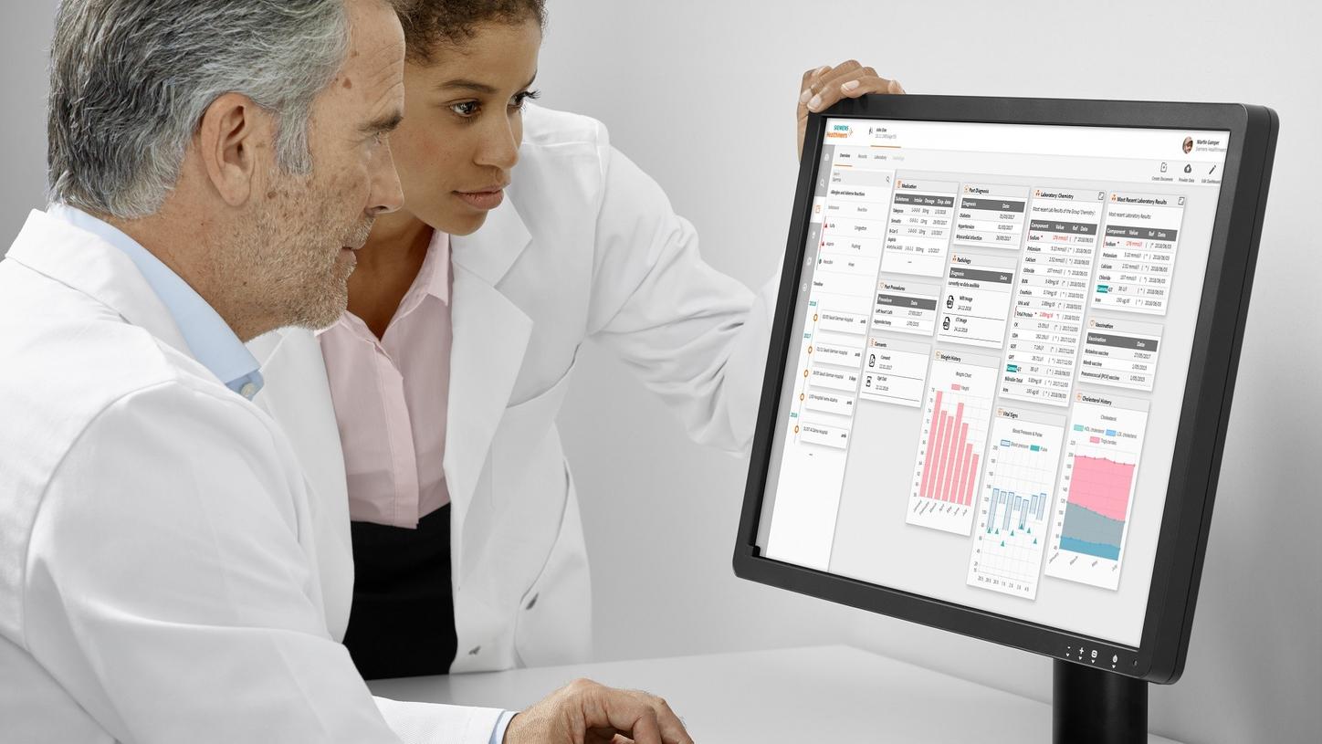
After a TIA, targeted imaging and diagnostics identify the cause early to guide timely prevention.

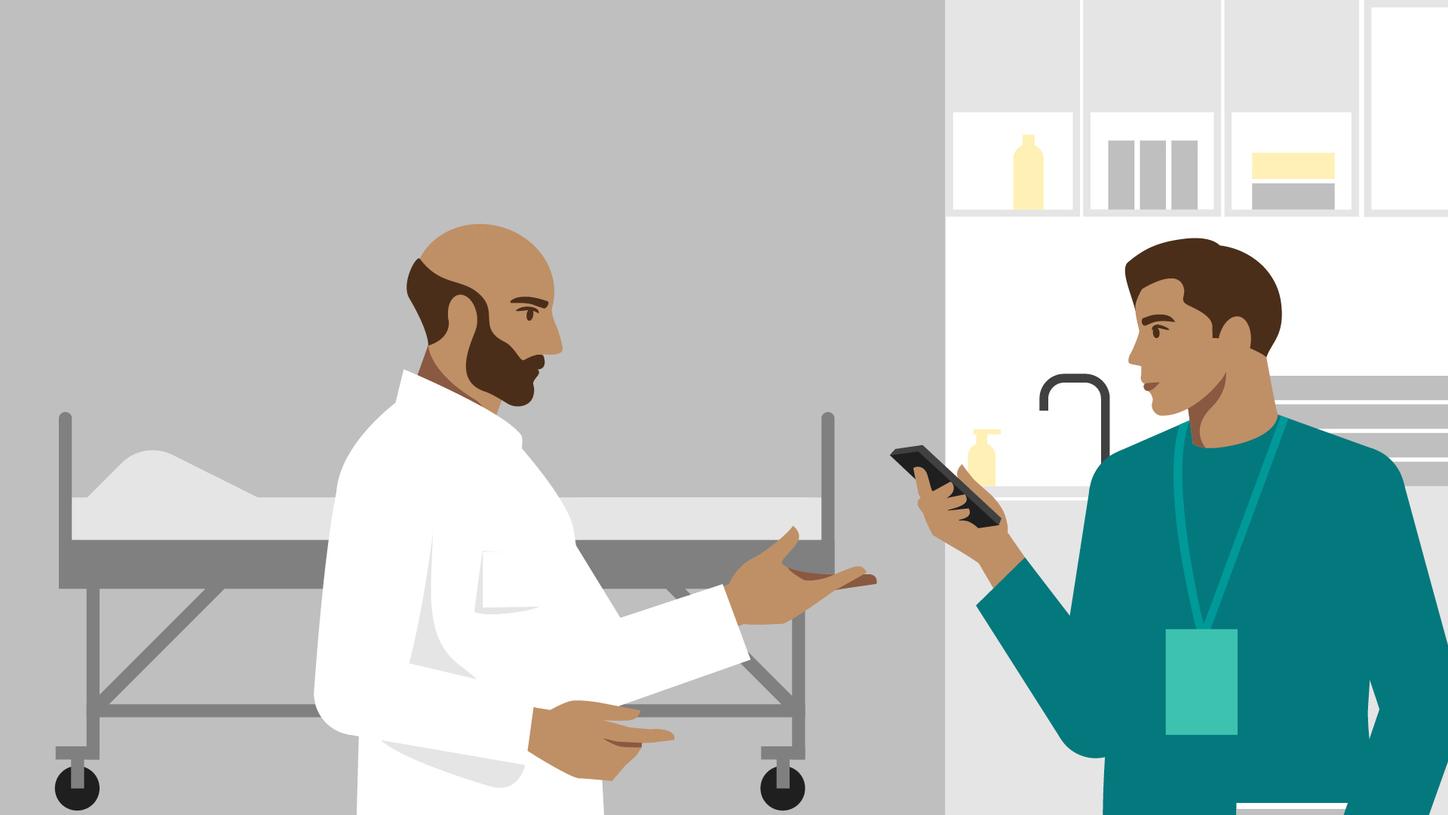
Follow Stéphane on his
Acute Stroke Journey
Sudden neurological symptoms trigger emergency response, rapid imaging, and time‑critical treatment decisions.
Follow Mike on his
Acute Stroke Journey
Ongoing monitoring supports early detection of progression and timely action to reduce stroke risk.

Follow Jenny on her
Stroke Care Pathway
Pre‑hospital stroke assessment begins immediately as the MSU can save critical minutes before hospital arrival.