
Finding meaningful answers without compromise
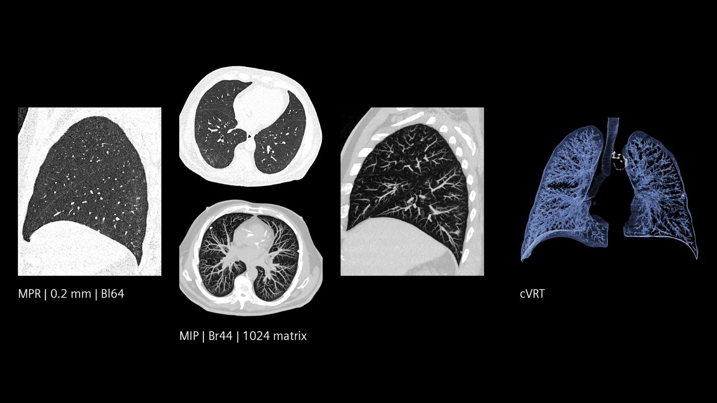
The NAEOTOM® Alpha class with Quantum Technology opens the doors for breakthroughs in diagnosing lung diseases and advancing pulmonology. Thanks to Quantum HD resolution with 0.2 mm slice thickness, improved CNR, and inherent spectral information in every scan, this technology lets clinicians see the lung as they have never seen it before. It offers a comprehensive understanding and access to adequately depicted, fine-tuned detail – from reticulations to honeycombing.
High spectral resolution and image quality underpin confident decision-making by delivering meaningful answers, potentially sparing patients unnecessary exposure to radiation, and enabling more targeted decisions in patient management, including treatment alteration and pre- and post-operative evaluations. Quantum Technology has the potential to transform the field of pulmonology, paving the way for improved treatment and patient outcomes.
Setting new standards in pulmonology
Professor Martine Rémy-Jardin, head of the thoracic imaging department at the University Hospital Center of Lille, France, and Professor David Launay, head of the clinical immunology section, have experienced Quantum Technology as a diagnostic revolution and gained new confidence.
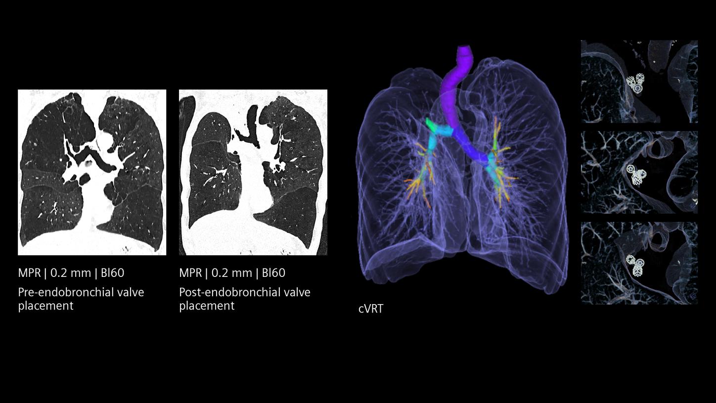
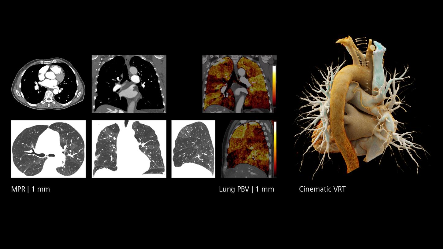
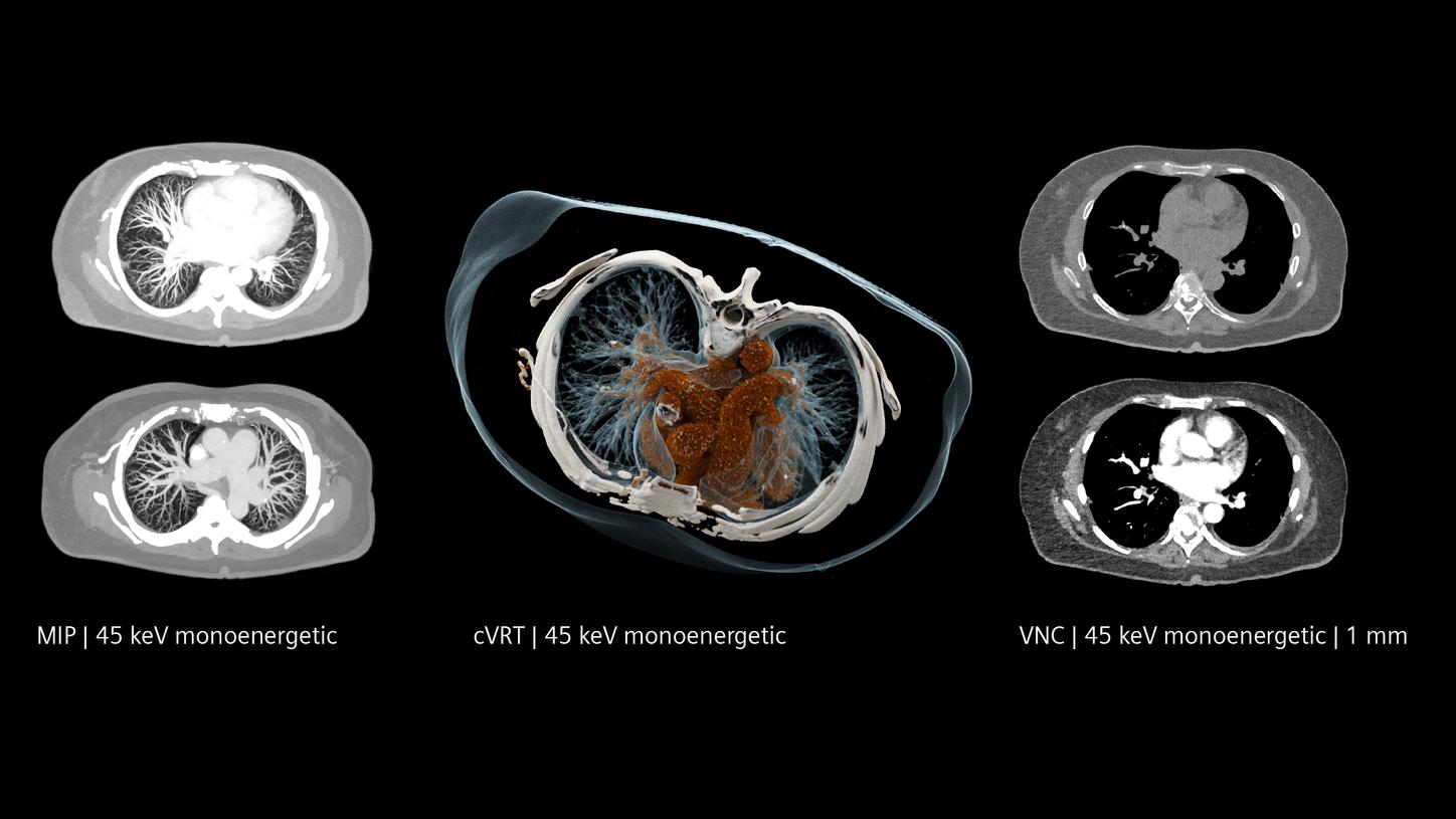
Low dose and Quantum HD lung imaging
NAEOTOM Alpha.Pro | 100 kVp | CTDIvol 1.04 mGy
Courtesy of University Hospital Pilsen, Pilsen, Czech Republic
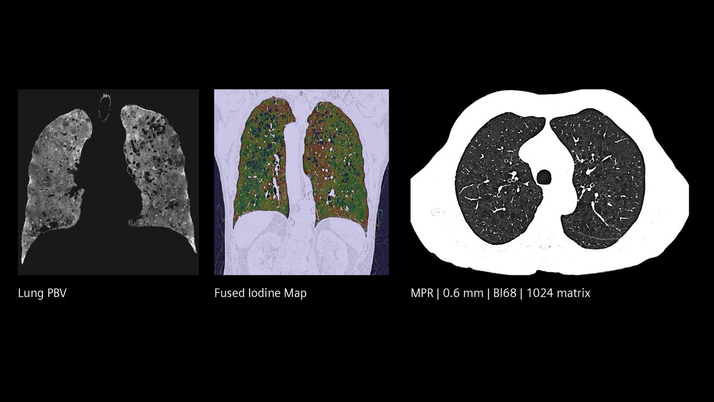
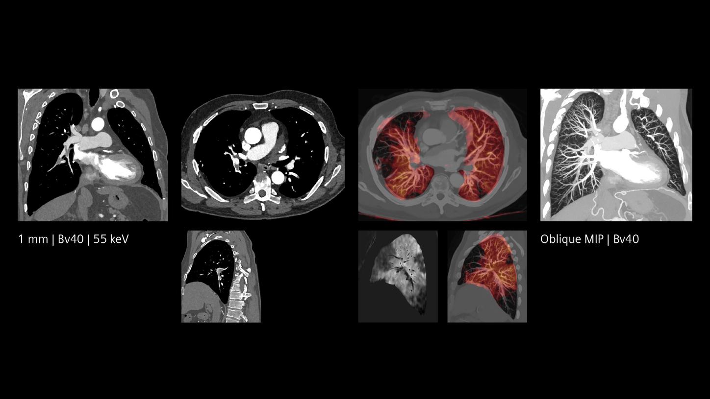
Spectral information with every scan
With Quantum HD images, a single scan has the potential to deliver relevant CT information. Detailed spectral maps enable precise functional evaluation, aiding in the classification of various diseases in daily clinical practice. See for yourself.

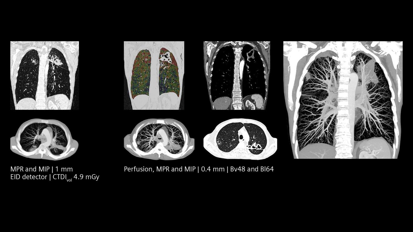
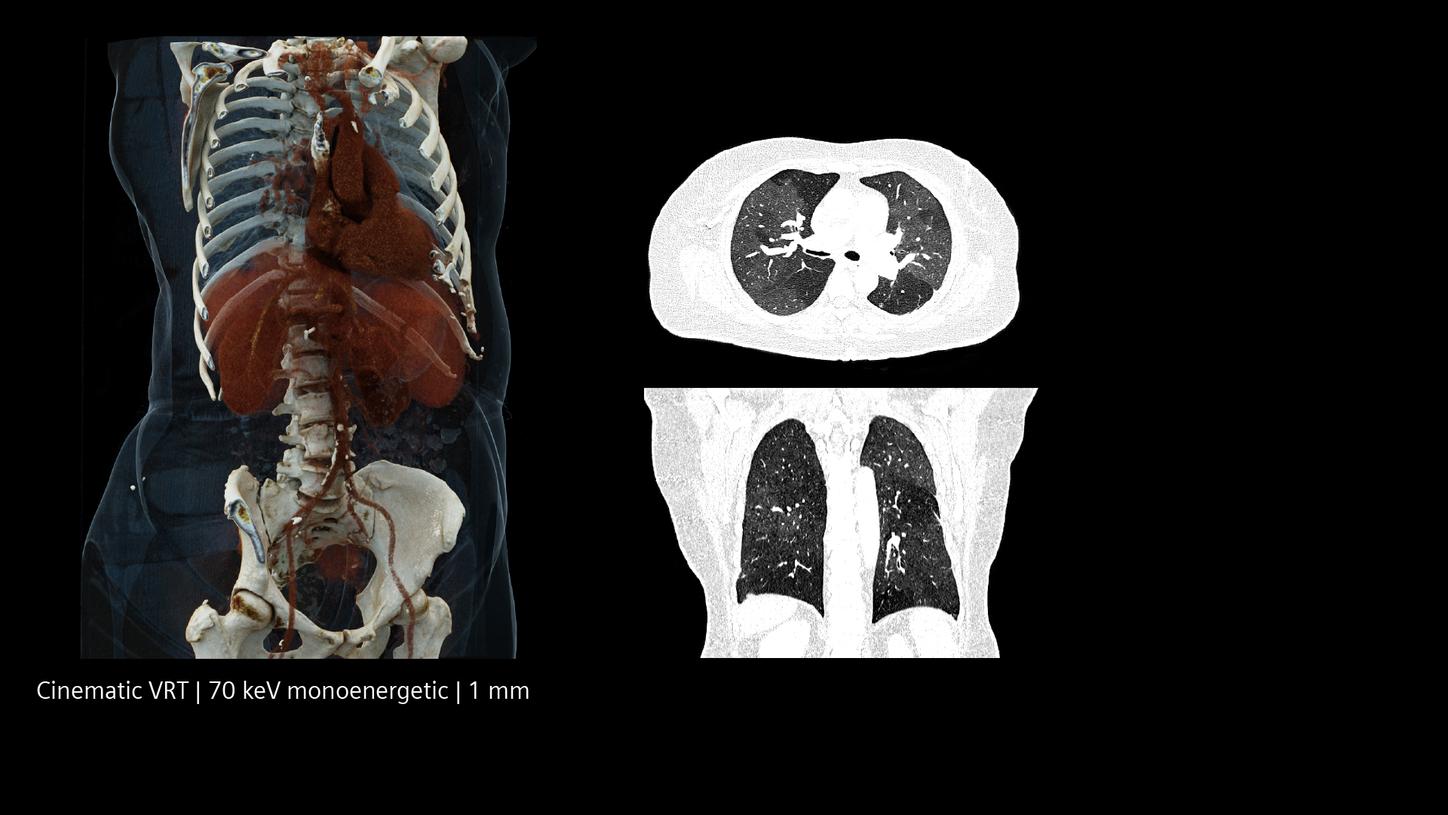
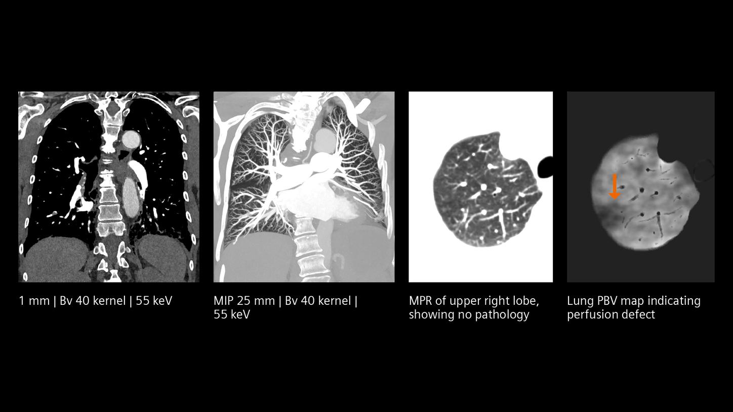
A new era for morphology and function
Quantum Technology offers an easy and streamlined way to obtain quantitative information in addition to morphology. By potentially overcoming the limitations of scintigraphy, matching the appropriate protocols to patients and combining necessary acquisitions into a single scan, Quantum Technology allows for the assessment of lung perfusion that contributes to a comprehensive understanding of lung diseases, aiding in personalized treatment strategies and optimizing patient care.

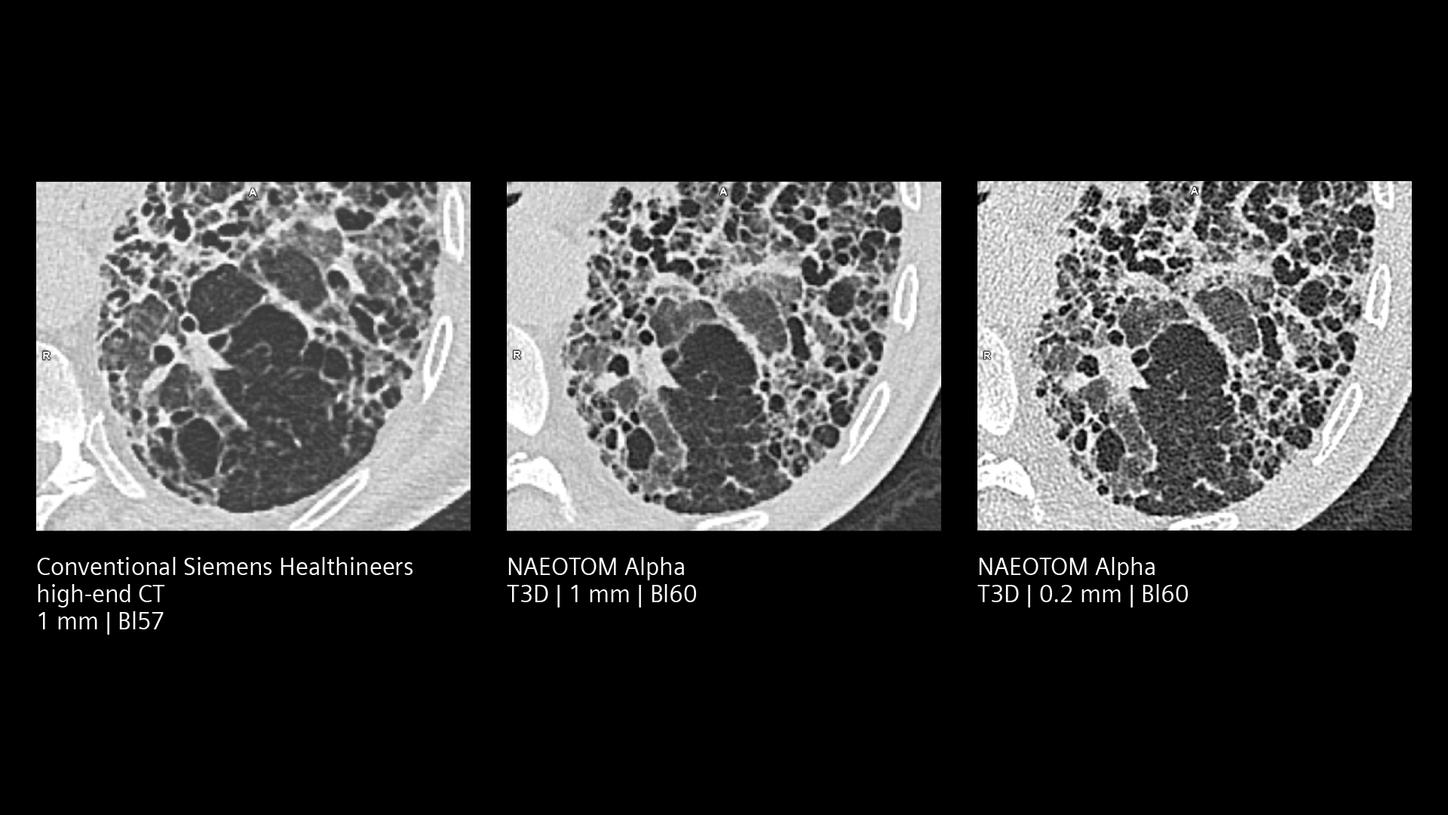
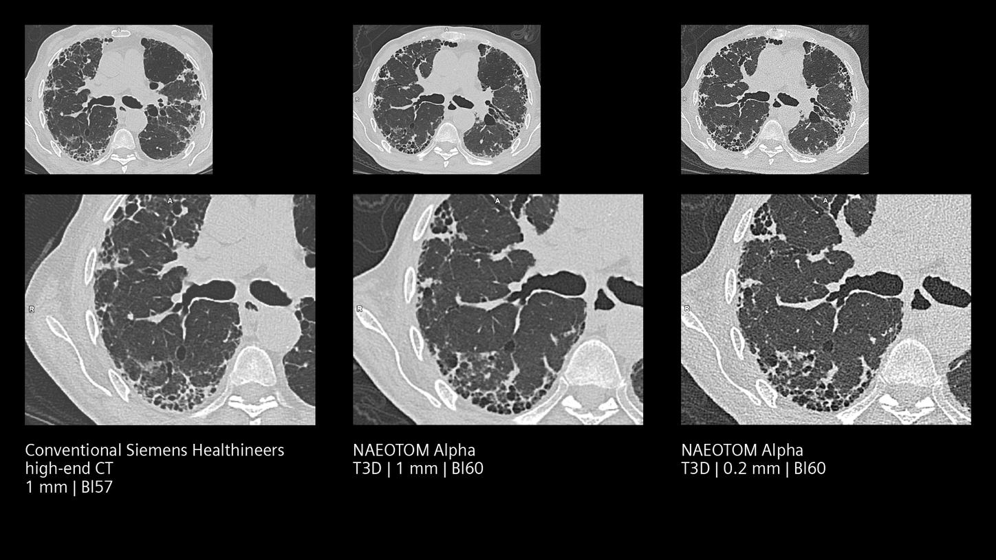
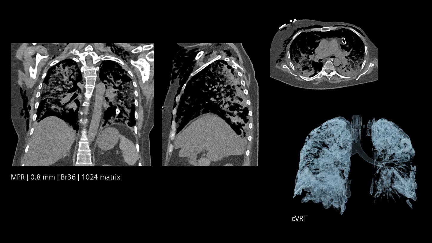
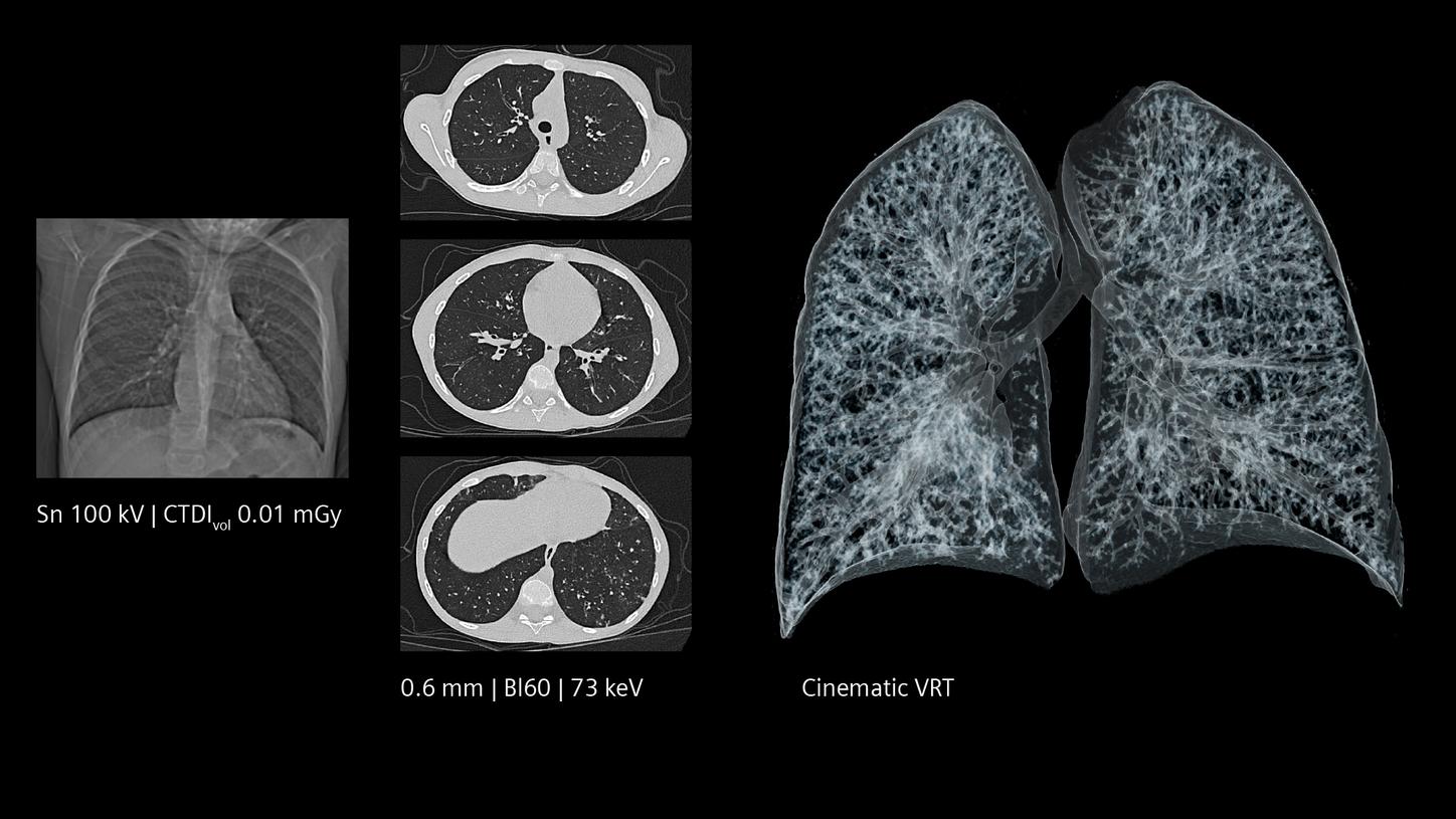
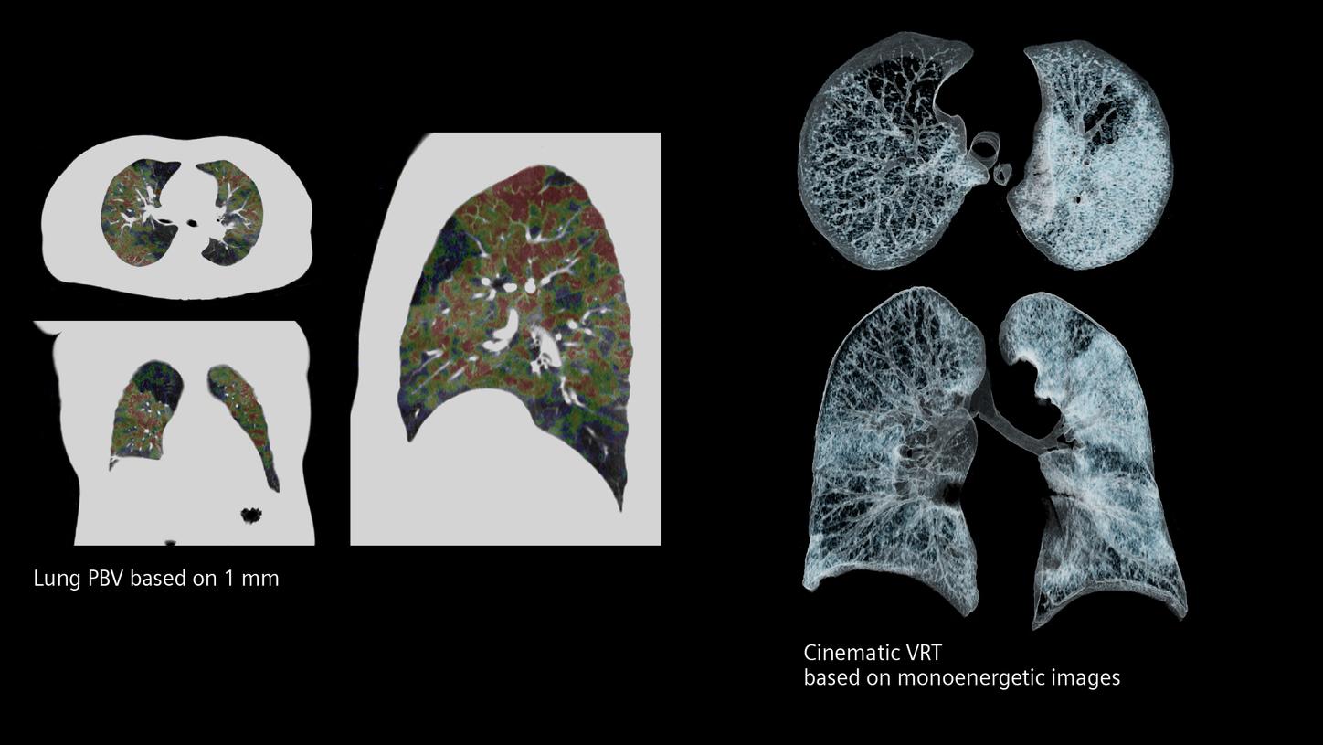
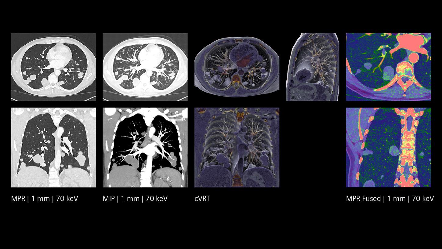
Quantum HD image quality at standard dose levels
By providing spatial resolution with 0.2 mm slice thickness and high contrast, the NAEOTOM Alpha class with Quantum Technology enables precise detection and characterization of small pulmonary nodules, subtle lung abnormalities, and intricate anatomical structures. This can help radiologists increase diagnostic confidence without increasing the radiation dose and enhance early diagnosis and treatment planning – ultimately resulting in improved patient outcomes.

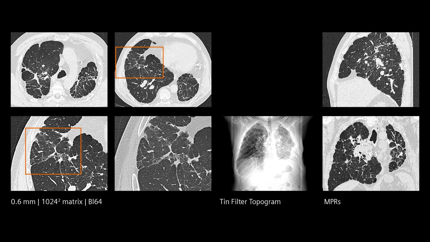
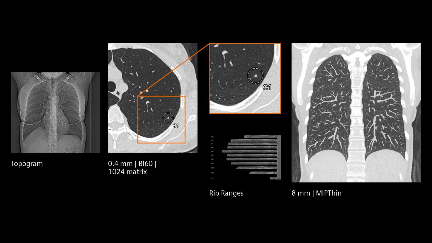
Reduced radiation dose with better image quality
Quantum Technology provides less electronic noise, better CNR, and 0.4 mm slice thickness as a standard. This brings the benefit of minimizing radiation dose exposure while keeping the image resolution at its best for patients who require frequent monitoring or lung cancer screening.
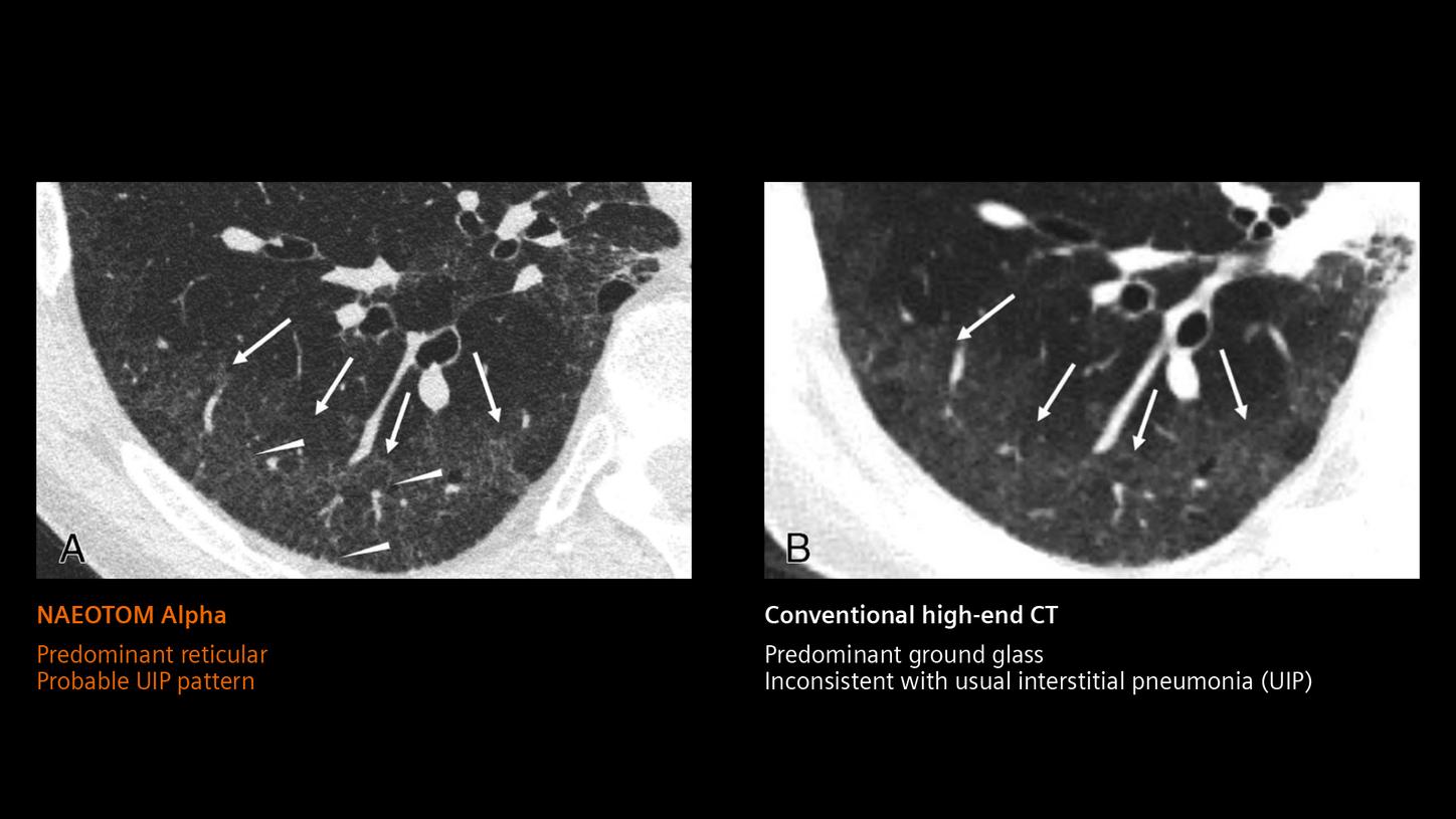
NAEOTOM Alpha will undoubtedly contribute to a redefinition of subtle fibrosis seen in lung images.1

University Hospital Center of Lille,
Lille, France
Scientifically proven technology
Discover the full potential of the NAEOTOM Alpha class.
The NAEOTOM Alpha class in other clinical fields


