syngo.via RT Image SuiteEmpower your decisions – Look closer. See further.

Many treatment decisions are increasingly based on imaging but available software solutions have not been designed with this need in mind.
Based on Siemens’ expertise in imaging, syngo.via RT Image Suite addresses these shortcomings. Specifically built to maximize RT imaging intelligence and expand your clinical capabilities, syngo.via RT Image Suite allows you to look closer at what you need to see today – so that you can see further into the future of individualized therapy.

  • See clearly
  • Contour efficiently
  • Advance your practice

Características e Benefícios


syngo.via RT Image Suite - see clearly
Contouring using the planning CT, T1- and T2-weighted MRI

Drive improved outcomes through a clear and comprehensive view of your patient

Radiation Therapy clinics are striving to provide high-quality individualized care. A wide variety of images – 3D and 4D, CT, MR and PET – acts as a basis for treatment decisions, and therefore have the potential to optimize the outcomes for each patient. Unfortunately, many existing tools such as treatment planning systems and virtual simulation solutions have not been specifically designed to manage the increasing diversity and volume of images in RT.

syngo.via RT Image Suite has been designed to fulfill this need. It displays clinical images in the proven syngo quality and allows the concurrent display of up to 8 image series1 on up to 2 monitors1. Rigid and Deformable1 Registration ensures confidence when using images such as MR and PET, even when these were not acquired in the treatment position.

By providing Radiation Oncologists with a clear and comprehensive view of their patients, clinicians are empowered with a solution that results in easier and more intuitive clinical decision-making.

Main features:

  • Broad image type support: 3D to 4D, CT, MR, PET, and Linac CBCT
  • Drag&Drop image loading and preview functions for straightforward image handling
  • Rigid and Deformable1 Registration
  • Advanced Visualization1: concurrent display of up to a total of 8 image series (4 single or 4 fused series) over 4 image panels
  • syngo image visualization quality, that is known from Siemens imaging devices
syngo.via RT Image Suite - contour efficiently
Contouring moving tumors using the AverageCT, t-MIP and 4D CT individual breathing phases

Increase process efficiency with an elegant and easy-to-use contouring solution designed for Radiation Oncologists

Many Radiation Therapy clinics are forced to meet increasingly high quality demands while providing care to more and more patients despite cuts in budget and staffing. Achieving these objectives requires clinicians to become more efficient than ever.

syngo.via RT Image Suite is an easy-to-use software solution that helps clinicians to efficiently contour both routine and advanced cases. A set of modern tools support fast 3D and 4D CT, MR, and PET delineation. From simple image and structure import and export, to semi-automatic contour propagation over breathing phases2 or contouring in any orientation and any displayed image, syngo.via RT Image Suite offers a broad range of features that support you in achieving high-quality results efficiently.


Main features:

  • Intuitive syngo user interface
  • Easy-to-use contouring tools (freehand, brush/nudge)
  • Contour on any orientation, including oblique
  • Parallel contouring: contouring performed on any image is immediately reflected on all other images
  • Advanced contouring tools including semi-automatic propagation of contours over breathing phases2, ITV generation, and 3D image-based Smart Freehand segmentation
  • “CT-free” contouring: native delineation on MR or PET images
  • Data pre-fetching from PACS, simple import from CDs and DVDs, patient data reconciliation
  • DICOM standard compliant structure set export to Treatment Planning Systems

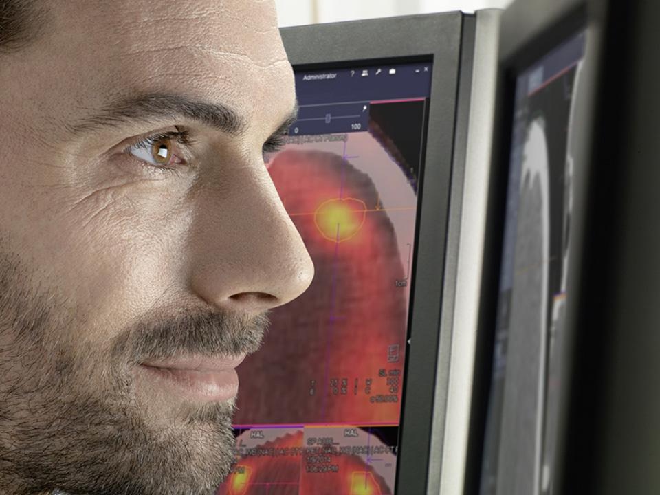
Radiation Therapy - syngo.via RT Image Suite - Evaluation of PET-based tumor physiology changes over time
Evaluation of PET-based tumor physiology change over time

Enable image-based care pathways to expand your clinical capabilities
Clinical teams consistently strive to advance their capabilities and require tools that enable them to achieve this objective. Imaging is a determining factor in achieving optimal results, whether it is when planning the treatment of a recurrence, adapting strategies during treatment, monitoring therapies, or for investigating the added value of functional imaging. However, many clinics looking to provide image-based care pathways find it impractical due to the lack of adequate tools.

syngo.via RT Image Suite enables you to integrate imaging into your daily clinical and research practices. For a wide range of applications from advanced segmentation features such as contour warping, to Advanced Visualization1 as well as a client-server architecture that facilitates collaboration, syngo.via RT Image Suite offers the tools to support you and your team in advancing your clinical practice.

Main features:

  • Broad anatomical and functional image type support including PET and multi-parametric MRI
  • Visualization of previously drawn structures on current images to facilitate assessment of changes
  • Semi-automatic contour warping2 to support efficient re-contouring
  • syngo.via client-server architecture for efficient collaboration across different locations
  • Expandable with powerful syngo.via oncology applications1


Uso Clínico


SOMATOM Definition AS Open - RT Pro edition with syngo.via RT Image Suite

Start the RT challenge with an advantage
syngo
.via RT Image Suite supports you in getting more out of your CT images.

  • Streamlined workflow with data pre-fetching and easy export of images and contours
  • Easy review and selection of the right images with the ability to display up to 4 series side-by-side3
  • Flexible and confident contouring of moving tumors, using 4D CT data, AverageCT, t-MIP, and individual breathing phases
  • Efficient contour propagation over breathing phases2 and fast generation of ITV
  • Expandable with further syngo.via CT applications1

MAGNETOM RT Pro edition for MAGNETOM Aera and MAGNETOM Skyra

syngo.via RT Image Suite helps leverage MRI information in Radiation Therapy by enabling you to:

  • Visualize MR images with the proven syngo image quality
  • Easily select the appropriate MR series thanks to the image preview function
  • Load and register MR sequences reconstructed in different orientations
  • Contour in any orientation, including oblique, and on a variety of anatomical and multi-parametric images, such as T1, T2, Diffusion-weighted, and more - allowing you to utilize all information at hand
  • Display up to 4 series or fused-image series side-by side3, to get the complete picture of the patient
  • Support the use of MR information with Rigid and Deformable1 Registration, even when the images are not acquired in the treatment position
  • Start contouring on MRI without the need of a planning CT, supporting a flexible “CT-free” workflow in external beam RT planning and brachytherapy
  • Efficiently involve expert research collaborators, thanks to the syngo.via client-server architecture

Visualize the molecular change of cancer with Biograph mCT

Visualize the molecular change of cancer with Biograph mCT:
syngo.via RT image Suite enables you to smoothly integrate PET functional information in your Radiation Therapy workflow.

  • High quality syngo-based visualization of PET and PET/CT
  • Threshold-based contouring of Molecular Imaging data such as PET for example
  • Ability to display and contour on up to 4 images or image series side-by side1xxxx, to get the complete picture of the patient’s condition
  • Rigid and Deformable1 Registration to support the use of PET images, even when not acquired in the treatment position
  • Easier management of complex cases thanks to efficient collaboration with other experts such as Nuclear Medicine Physicians, with the syngo.via-based client-server architecture
  • Expandable with syngo.PET&CT Oncology1, for example to monitor treatment progress
  • Import of RT structure sets including GTVs created in syngo.PET&CT Oncology1


Especificações Técnicas

A variety of syngo.via RT Image Suite deployments allows you to customize the solution to meet your needs and your budget.

  • syngo.via workstation
    A great solution for smaller sites with one concurrent floating user license, giving you the flexibility of installation on multiple PCs.
  • syngo.via server
    An ideal solution for sites that need maximum flexibility and the comfort of multiple concurrent floating user licenses installed on multiple PCs, inside or outside of the department.
  • Adding syngo.via RT Image Suite to existing syngo.via installations
    A solution that adds syngo.via RT Image Suite floating user license(s) to existing syngo.via server deployments in the institution and that is deployed on the PCs of the Radiation Therapy department or anywhere it is needed.

Ease-of-use and general features

  • Data pre-fetching from PACS and imaging devices, simple import from CDs and DVDs, patient data reconciliation
  • Image preview function
  • Drag&Drop image loading
  • Automatic send to TPS configuration
  • DICOM and IHE-RO standard compliance

Contouring features

  • Freehand 2D, 3D image-based Smart Freehand segmentation, 2D/3D brush/nudge
  • Contour on any arbitrary plane including oblique planes
  • Organ algebra (union, intersection, exclusion)
  • Symmetric and asymmetric structure growth or contraction
  • Geometrical and smart image-based contour interpolation
  • Multiple structure set support (1 per image series)
  • Molecular imaging data such as PET, threshold-based and skin, gray-value-based, segmentation
  • “CT-free” contouring: native PET or MR contouring
  • Parallel contouring: contouring performed on any image is immediately transferred on all other images
  • Semi-automatic contour propagation over 4D CT breathing phases2 and ITV generation
  • Visualization of previously drawn structures on the current image series
  • Contour copy and warping between image series1

Imaging Features

  • 3D/4D CT, PET, PET•CT, MRI, and Linac Cone Beam CT (CBCT) image support
  • Advanced Visualization: concurrent display of up to a total of 8 image series (4 single or 4 fused series) over 4 image panels1
  • Dual monitor support1
  • Rigid and Deformable1 Registration1
  • Cine-loop visualization of 4D CT dataset


1
2
3