The combination of modern hardware and powerful AI-powered software is changing how nuclear diagnostics are utilized in daily routine. The results: better images, more usability, and addresses quality of care across various disciplines.
Photography by Simon Lau and Annika Daria Neubauer.
Article content reflects MI World Summit 2026 Session 3, Copenhagen, Denmark.
Modern nuclear imaging is not only about scanning, but also about extracting more information out of imaging datasets with the help of information technology, including artificial intelligence (AI). The result, according to Prof. John O. Prior, head of Nuclear Medicine and Molecular Imaging at Lausanne University Hospital (CHUV), Switzerland, can be more comfortable and more efficient workflows. But digital algorithms can also be “quality-relevant,” for example, when they are used to reduce artifacts or to minimize radiation exposure.
Prof. Prior delved into this topic during the Molecular Imaging World Summit 2026 in Copenhagen, Denmark. The “Faster Way to Reporting” session featured multiple global molecular imaging leaders who focused on AI and data-driven patient adaptation, AI-assisted reading and reporting, ventilation and perfusion from SPECT/CT to improve respiratory health, and dosimetry.

Prior discusses OncoFreeze AI and AI denoising.
Fewer artifacts, less noise for respiratory care
Prior presented two examples of lesions in lung and liver. This is a field where PET/CT imaging for such lesions can be challenging because of respiratory motion artifacts. The Siemens Healthineers solution OncoFreeze AI can address this issue by providing data-driven respiratory gating for continuous bed motion.
While OncoFreeze AI technology can reduce scan time and enhance patient comfort, Prior explained that it also can improve image quality. He shared a case of a cancer patient who, in an external PET, showed an unclear hot spot in the liver. It disappeared once the imaging was repeated on a Biograph Vision PET/CT. Thanks to motion correction, it became clear that the issue was overlapping structures which mimicked a hotspot that was not actually there. “This is something we really appreciate. Oncofreeze AI improves delineation and quantification of small lesions considerably.”a
What is AI denoising?
Another example of AI assisting with PET/CT imaging is AI denoising.b It does what it sounds like: Convolutional Neural Networks are used to improve the signal-tonoise ratio of Fludeoxyglucose F-18 (18F-FDG) image datasets, the overall goal being high-quality reconstructions with lower radiation exposure. Does it work? Prior and colleagues explored this in the context of PET/CT-based pulmonary lesion detection in lung cancer screening. Low radiation exposure is mandatory here, given that PET/CT competes with low-dose CT imaging. A cohort of 49 patients with 141 lesions was meticulously analyzed. For Prior, the available evidence supports the use of low-dose PET/CT in lung cancer screening―something, he emphasized, that would also benefit patients. “By using low-dose PET/CT instead of low-dose CT, there is potential for earlier diagnostic clarification. Patients would not have to wait three months with uncertainty in case of abnormal findings.”
AI-based V/Q SPECT/CT
Trishul Siddharthan, MD, from the Medical Department, Division Pulmonary Care, at the University of Miami’s Miller School of Medicine, Florida, USA, discussed how AI can be used with nuclear imaging to deliver meaningful results for the patients. Together with Siemens Healthineers, Siddharthan introduced an AI-based lung parcellation software for ventilation and perfusion (V/Q) SPECT/CT. The software helps to calculate ventilation heterogeneity (VH).c
During the first step, the experts in Miami showed that VH is a good predictor for perceived lung function in chronic respiratory disease patients― better in fact than spirometry. Further down the line, it could be shown that improvement in VH correlates nicely with treatment effects not only of lung surgery and lung valve procedures, but also of antibody therapies. “Ventilation can explain a significant portion of why and how those therapies work,” said Siddharthan.
From a patient perspective, Siddharthan sees AI-assisted VH measurements as a promising tool to better personalize respiratory care. “VH is associated with patient-centered measures of respiratory health. We could use these kinds of tools to assess therapy response and optimize therapies.” Notably, the Miami V/Q AI project illustrates the value of close partnerships between medical institutions and industry.
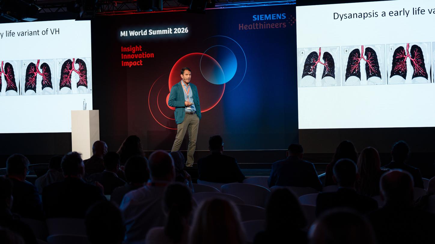
Siddharthan explains the AI-based lung parcellation software for ventilation and perfusion SPECT/CT.
AI-assisted PET reading
AI can also be used in various ways to assist reporting in nuclear medicine and radiology. Prof. Mohsen Beheshti, head of the Division of Molecular Imaging and Theranostics at the Department of Nuclear Medicine and Endocrinology, Paracelsus University Salzburg, Austria, stated that when it comes to PET/CT reporting, specifically, AI can assist in total lesion assessment, in lesion detection, and in lesion classification. “Much of this is already implemented in clinical routine,” said Beheshti. He also sees AI assisting in differentiating new lesions from lesions that already existed. AI-assisted lesion detection is especially helpful for smaller lesions in atypical places. To illustrate this, Beheshti told of a woman with ovarian cancer. PET/CT revealed three metastatic lesions in the peritoneum, but only the automatic detection algorithm found another on the spinal cord. “This is a very atypical location. It usually would have been interpreted as an artifact.”
Lesion detection is not restricted to FDG-PET. There are also tools in use for PSMA-avid lesions. They lead to a faster and more standardized interpretation of prostate cancer scans. But, said Beheshti, there were limitations to lesion detection software. Depending on the scenario, there was still a considerable mismatch between automatic detection and physician readings, for example, although these mismatches were not clinically relevant most of the time. “We clearly need guidance for successful implementation of these tools.”
From a workflow perspective, a reduction in reporting times is the big asset of AI-assisted lesion detection. “AI is already here”, said Beheshti. “It requires human oversight, good governance, and proper training. And most importantly: AI doesn’t replace physician’s judgement― it sharpens it.”
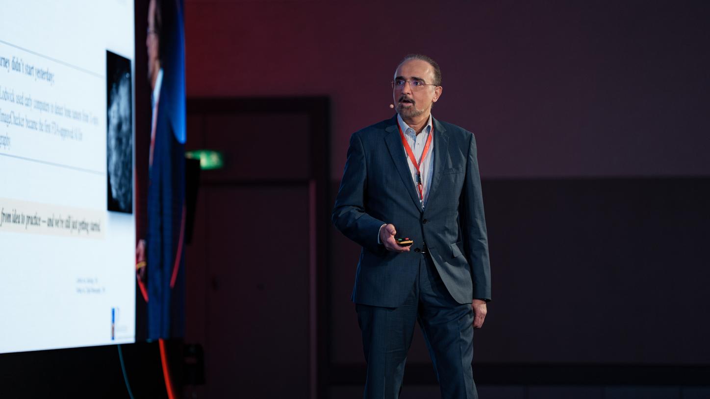
Beheshti states that when it comes to PET/CT reporting, specifically, AI can assist in total lesion assessment, in lesion detection, and in lesion classification.
About the author
Philipp Grätzel who has been working as a healthcare and technology Primafila Correspondent in Berlin, Germany, for many years.