King’s College London & Guy’s and St Thomas’ PET Centre are using their identical total-body PET/CT scanners to explore promising research avenues and succeed with their TOTEM study.
Photographs and data courtesy of King’s College London & Guy’s and St Thomas’ PET Centre, UK.
When Prof. Alexander Hammers looks out of one of St Thomas Hospital’s countless riverside windows, he can see Big Ben and the Houses of Parliament right across the river Thames. Beautiful as it might be, the view is not the main attraction for Hammers, who is head of King’s College London & Guy’s and St Thomas’ PET Centre, run jointly by King’s College London (KCL) & Guy’s and St Thomas’ NHS Foundation Trust (GSTT). What he really is excited about are his twins.
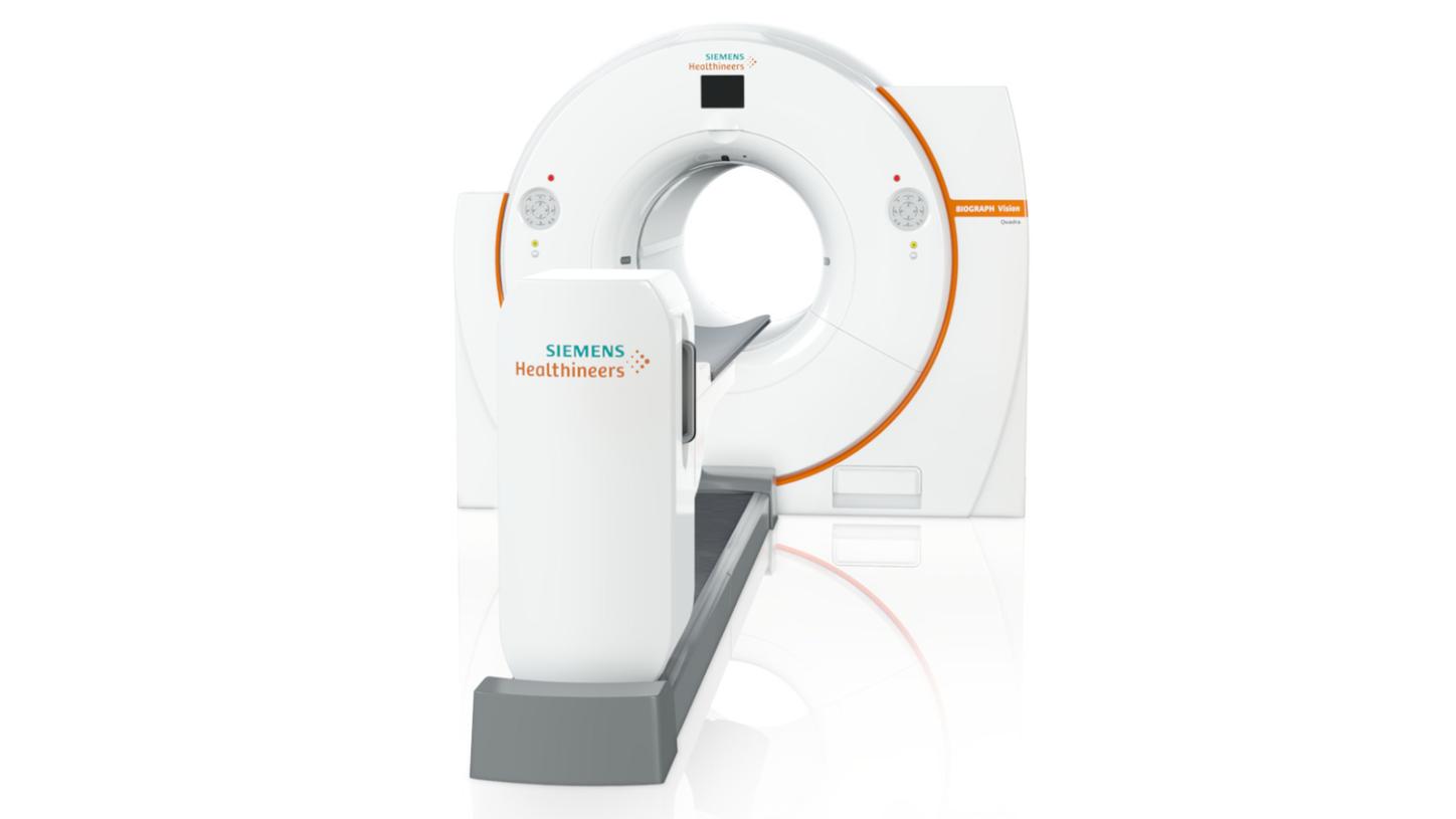
“Twins” is a nickname used at KCL & GSTT PET Centre for two identical Biograph Vision Quadra PET/CT scanners that were acquired in late 2024 and early 2025. The investment, according to Hammers and to Gary Cook, professor of molecular imaging at KCL School of Biomedical Engineering & Imaging Sciences, was a major milestone for the PET Centre.1,2,3 It made clinical routine diagnostics way more efficient. And it opened a lot of promising research avenues that are now being explored.
Why two?
KCL & GSTT PET Centre is one of the major PET/CT hubs in the UK, if not in Europe. One of the two new scanners was competitively awarded, in partnership with Imperial College London, through a grant scheme provided by the Medical Research Council National PET Imaging Platform. The second was financed by KCL.4,5 But why have two identical total body scanners?
It was a strategic decision, according to Hammers, based on experiences that other PET/CT centers had made. The Biograph Vision Quadra’s high sensitivity means that less tracer and less radiation is needed, and scans can be done quickly.a,b “Now we can split the benefit of Biograph Vision Quadra between patients and the Centre. We can scan patients much faster, and they are getting roughly half of the dose they were getting before―which also means our staff are getting less dose.” a,b
How did the economics factor in? Cook co-authored a scientific paper on the economics of total-body PET/ CT scanners in various settings all over the world.6 The study analyzed the overall costs of using one or two total-body PET/CT over a period of 10 years, compared to extending the working day with standard short axial field of view PET/CT scanners. “In most situations, the total-body PET/CT turned out to cost less, despite the scanner’s initial costs. This is because of increased throughput, and because of savings on staff, radioactivity, and so on.”
Expanding the research spectrum
There is also a research case that can be made for having two total-body PET/CTs instead of one, according to Hammers. It is about using GMPmanufactured radiopharmaceuticals more efficiently. “Regulations are getting tighter and tighter for making GMP radiopharmaceuticals. There is an absolute dearth of radiotracers in the UK. If we do make a batch of research tracer, we need to capitalize on this as much as we can.” Hammers’ example is [11C]methionine PET/CT. “We have managed to do one Carbon-11 synthesis and scan four dynamic [11C]methionine patients, thanks to the two scanners. That’s absolutely astounding. Other centers need [18F]fluoroethyltyrosine ([18F]FET) to achieve the same.”
“We are talking about a second or even less, which allows us to look at membrane transport and intermediate compartments. This is all very exciting stuff.”

Putting the two total-body PET/CT scanners into operation in early 2025 led to a substantial expansion of research projects and research areas. On the technical side, advanced reconstruction has become a focus topic, says Hammers. “We get much, much better data now, and this means a step change for reconstruction.” In addition, ultra-low-dose PET/CT is a totally new research area that hadn’t been addressed at the center before. There are various projects on novel tracers. But researchers are also digging deeper into what can be done with existing tracers. “Prof. Simon Cherry from the University of California-Davis joined us and is doing work on early short and ultra-short frames, together with our own reconstruction experts (Prof. Andrew Reader). We are talking about a second or even less, which allows us to look at membrane transport and intermediate compartments. This is all very exciting stuff.”
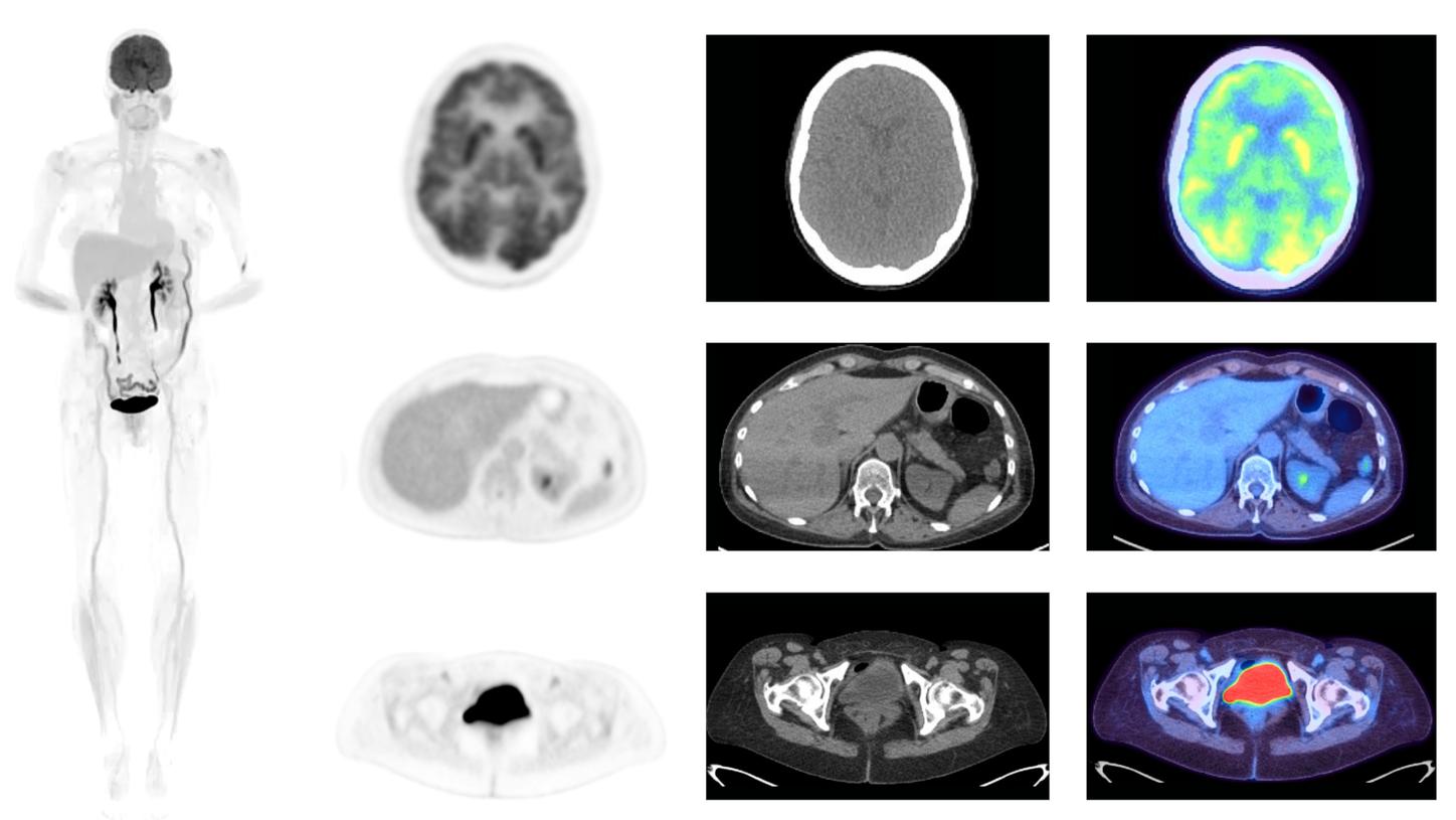
Full-body [18F]FDG PET image (left) acquired with the Biograph Vision Quadra PET/CT. Selected slices (PET, CT, fused; right) show the brain, liver, and pelvic region.
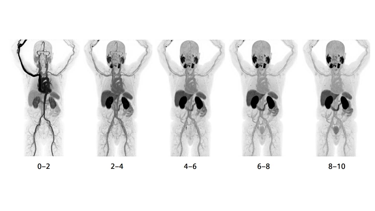
Example of whole-body dynamic PET imaging using [18F]PSMA with the Biograph Vision Quadra PET/CT. The images show the first ten minutes of a scan reconstructed in two-minute frames
TOTEM study: turning clinical routine into a knowledge factory
A project which Hammers and Cook are particularly proud of is the TOTEM (Total Body PET―Exploring new iMaging protocols for clinical research) study.7 Recruitment has recently reached 1400 patients. Cook explains that TOTEM was designed to try and make as much research use as possible of clinical examinations. The goal is to develop and validate standardized protocols for the total-body PET/CT scanner―a process still in its infancy, since long axial field of view (LAFOV) scanning is a recent technology. To get proper data as quickly as possible, TOTEM research is deeply embedded into routine workflows. “For example, we can ask a patient to have a dynamic scan after their injection rather than waiting in the uptake room, so we can develop protocols for kinetic analyses. Or we can do delayed scans with a low-dose CT.”
A trial like TOTEM may sound straightforward, but in the context of highly regulated PET/CT research, designing it was a major effort. Initially, the study’s ethics committee required that patients have their patient information sheet for the study 24 hours in advance. This turned out not to work, Cook recalls. “The total-body PET/CTs had such a profound effect on our waiting lists that we were booking patients in faster than Royal Mail could deliver the sheets.” The study ethics needed an amendment. It is now allowed to approach patients on the day they arrive to get permission to use their data for research. If extra scans are planned, the patients will have to be contacted in advance by the research nurse.
Low-dose imaging: more to come
All in all, there are currently eight substudies running within the TOTEM study framework, and at least eight others are in preparation. One specific example is testing various ultra-low-dose CT protocols that involve using tin filters to minimize radiation exposure. These projects were made possible by the sensitivity of the total-body PET/CT scanners and, according to Hammers, were extremely successful. “We could verify, in real patients, the effects we had already predicted together with our colleagues from Siemens Healthineers, who are our embedded scientists.”
Testing such protocols would normally have taken ages, says Hammers. “But within TOTEM, we have been able to acquire the necessary data within six weeks or so. To my mind, it is the first time that talking about PET pilot studies really makes sense. And I don’t expect things to stop there. There will be other innovations and other ideas of getting really low-dose protocols. We look forward to trialling those as well.”
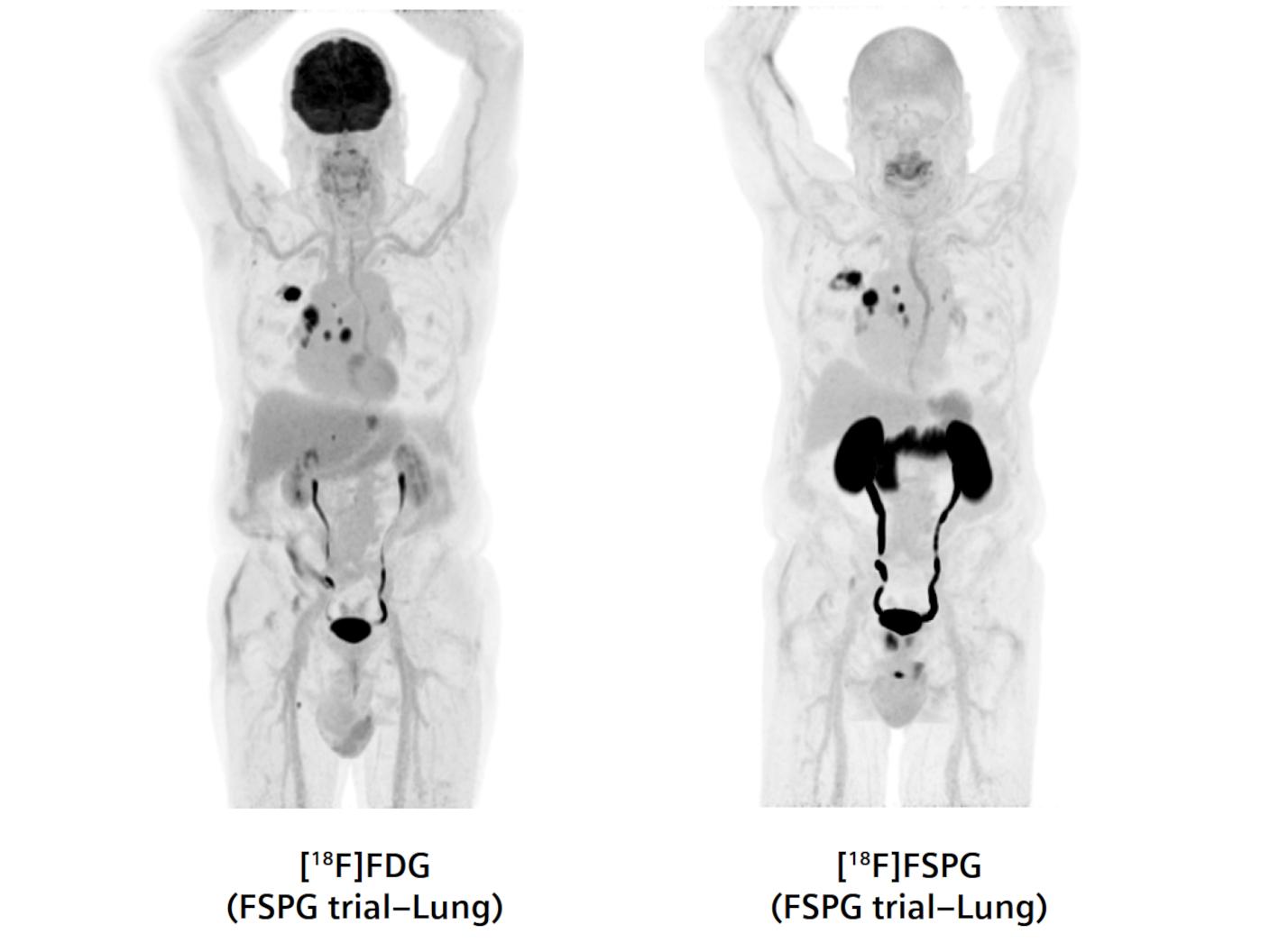
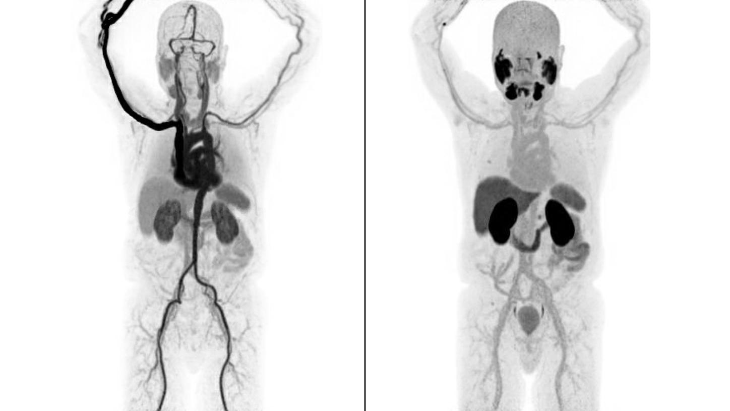
Comparison between the tracer uptake for [18F]FDG (left) and [18F]FSPG (right). Whole-body PET images were acquired with a Biograph Vision Quadra PET/CT.
Rethinking therapeutic monitoring in oncology
Equally exciting, and already approaching the finish line, is an oncology trial that uses a cutting-edge imaging tracer, [18F]fluoropropyl-L-glutamic acid ([18F]FSPG), in patients with head and-neck cancers. With [18F]FSPG, according to Cook, the ability of the cancer cells to evade oxidative stress during treatment can be measured. The better cancer cells can evade oxidative stress, the worse for the patient, because it is more likely the cancer cells will be treatment resistant. The idea is to use FSPG imaging to predict early on which cancers will be resistant. And it works. “We now know that within four weeks of starting the treatment, we can predict what the clinical treatment outcome will look like on a standard FDG-scan after 12 weeks.”
In the longer run, FSPG-imaging might revolutionize therapeutic monitoring in oncology, although more research must be done. Without highly sensitive scanners like Biograph Vision Quadra PET/CT, this type of research would not have been possible, says Cook. “The trial involved examining the kinetics of FSPG uptake into the tumor―something that had never been done before.8 The ability to acquire high data, fast kinetic scans over an hour has allowed us to do some quite innovative kinetic analyses. We have come up with a model of FSPG uptake into tumors which perhaps challenges assumptions we had before from animal work.”
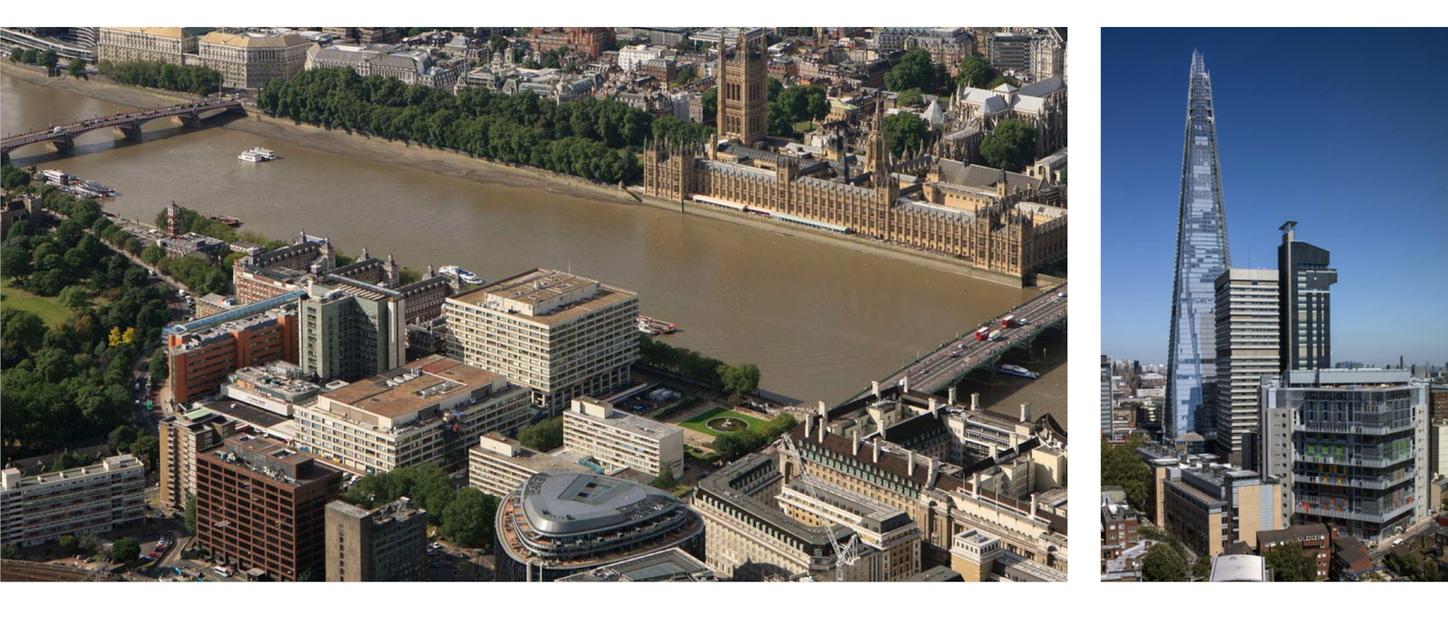
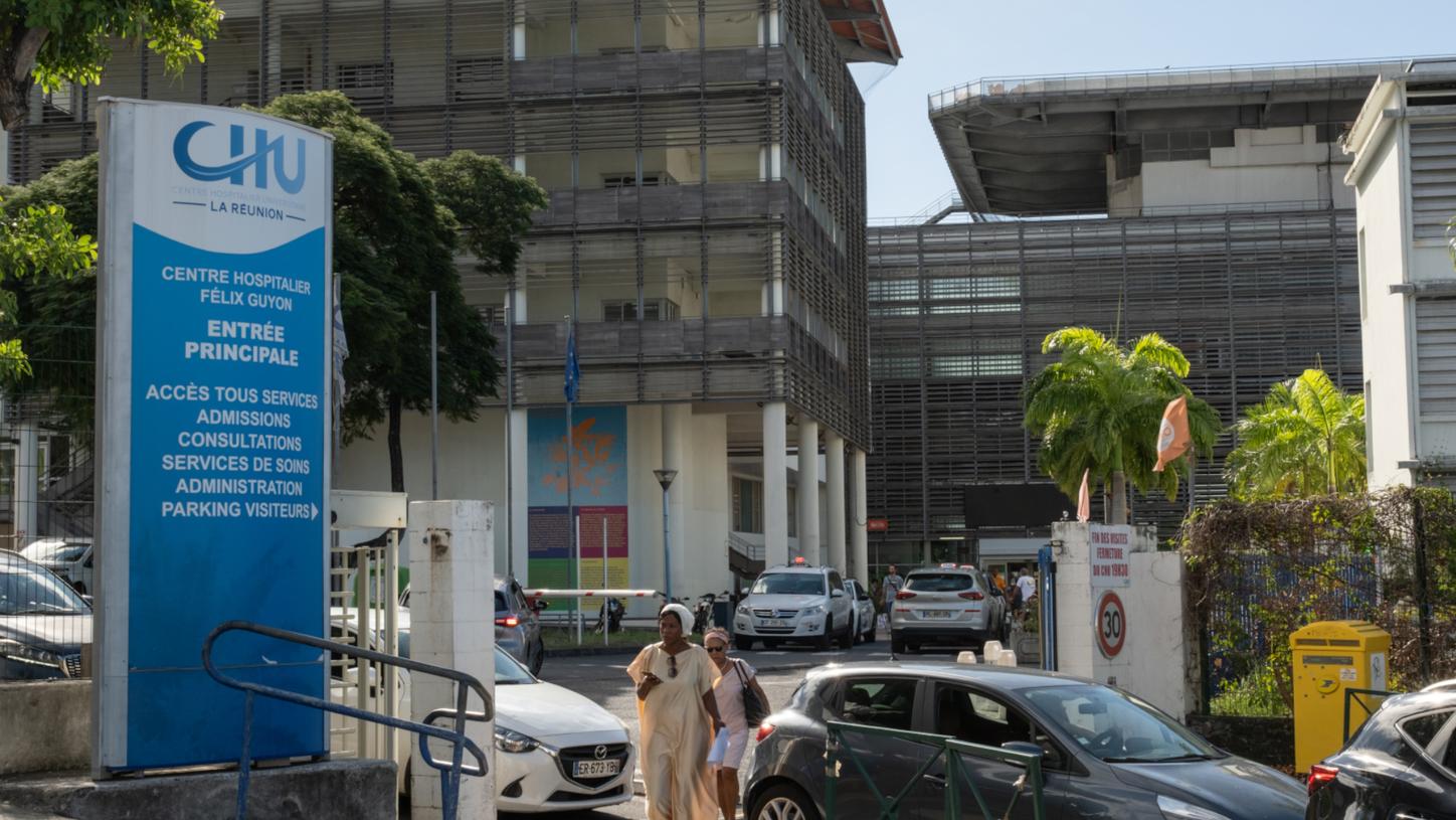
King’s College London and Guy’s & St Thomas’ PET Centre, UK.
(Left) St Thomas’ Hospital (Right) Guy’s Hospital.
“The ability to acquire high-data, fast kinetic scans over an hour has allowed us to do some quite innovative kinetic analyses. We have come up with a model of FSPG uptake into tumors which perhaps challenges assumptions we had before from animal work.”

Looking ahead
Both Hammers and Cook could continue talking about the opportunities their “twins” create because there is much more to come at KCL & GSTT PET/CT Centre. A major advancement will be the rollout of cardiac PET/CT using rubidium-82 (82Rb). The centre is also planning to launch imaging with Fluorine-18- labeled sodium fluoride ([18F]NaF) which can be used for bone lesion detection in oncology, but also for infectious disease and atherosclerosis imaging. Their first study (MRC NPIP funded) is to improve quantitative kinetics of skeletal metabolism to enable measurements of regional skeletal blood flow. Starting in 2027, GSTT will provide a dedicated pediatric oncology service for South London, adding another 300 to 400 scans per year to the 10,000 already being done. “It is really a very exciting time to be in PET,” says Hammers, with Cook nodding affirmatively.
About the author
Philipp Grätzel is a medical doctor by training, who has been working as a healthcare and technology journalist in Berlin for many years.