
AI COVID-19Joint efforts towards precision medicine
The coronavirus pandemic continues to spread, causing unprecedented system disruption and confronting healthcare professionals around the world with clinical and operational challenges. We believe the COVID-19 pandemic will significantly change healthcare systems. Specifically, it will serve as a catalyst for the development of more effective, efficient and, above all, more humane medicine.
Coronaviruses (CoV) are a large family of viruses that cause diseases ranging from the common cold to more severe diseases such as Middle East Respiratory Syndrome (MERS-CoV) and Severe Acute Respiratory Syndrome (SARS-CoV). COVID-19 is caused by the severe-acute respiratory syndrome Coronavirus 2 (SARS-Cov2) and has a high case fatality rate of up to 4%.1 Common symptoms include fever, dry cough and breathing problems. In severe cases, COVID-19 can cause pneumonia and multiple organ failure. Due to a highly efficient replication and the infectious nature of the disease, tools for rapid testing and evaluation are vital to track and mitigate the spread.
At Siemens Healthineers we understand the urgency and complexity for healthcare systems and providers to handle this situation. In a collaborative approach along with our research network and partners, we have developed a CT Pneumonia Analysis algorithm2.
AI Covid-19 Symposium
At the digital European Congress of Radiology (ECR) 2020, Professor Martine Remy-Jardin, Professor Philippe Grenier and Dr. Dorin Comaniciu (VP Artificial Intelligence at Siemens Healthineers) participated at the symposium “The future of lung disease assessment: How a global pandemic, international collaboration and rapid AI development are creating a ‘new normal’”. They talked on the topics of
- Interstitial lung diseases (ILD): from visual analysis of CT scans to quantitative CT approaches (Professor Martine Remy-Jardin)
- Chest CT in COVID-19 Pneumonia: Automatic Quantification versus Visual Assessment by Radiologists (Professor Philippe Grenier)
- AI COVID-19 – Joint efforts towards precision medicine (Dr. Dorin Comaniciu)
Watch the recording of the symposium and active discussion to receive insights on, overcoming the challenges radiologists face in daily practice, AI-based Automatic Quantification and the research algorithm CT Pneumonia Analysis².
COVID-19: A challenge in diagnosis and therapy planning


Although the role of CT and X-ray for diagnosis is currently being debated, preliminary studies showed that Chest CT imaging of the lung provides improved sensitivity when associated with RT-PCR for individuals suspected of having COVID-19.5 The primary features seen on a lung affected by COVID-19 are peripheral focal or multi-focal ground glass opacities, consolidations and crazy-paving patterns.
- Opacity lesions in the peripheral and posterior lungs on CT images are indicative of COVID-19 pneumonia
- CT can play an important role in the evaluation of COVID-19 providing advanced imaging evidence
Non-contrast chest CT has been useful not only to detect, quantify severity, and assess the progression of the disease, but also to evaluate the potential response to therapy alternatives. Similar results can be achieved with chest X-rays, e.g., for tracking the disease progression and follow-ups.
A collaboration as an answer to the complex situation
Our aspiration to drive innovations forward so people live healthier and longer lives is more valid today than ever before. Our collaboration partners and our inhouse expert teams are aware of the urgency and complexity of the current situation. That is why we decided to jointly focus on the development of a new algorithm to support the fight against the COVID-19 pandemic. For their collaboration we gratefully acknowledge the contributions of
- Hôpital Foch, Paris, France
- Northwell Health, New York, NY, USA
- University Hospital Basel, Clinic of Radiology & Nuclear Medicine, Basel, Switzerland
- Vancouver General Hospital, Vancouver, Canada
- Clínica Universidad de Navarra, Navarra, Spain
- Health Time, Jaén, Spain
- Houston Methodist, Texas, USA
- and multiple other frontline hospitals
And we are truly grateful for and look forward to all future contributors who will join us in this journey.
With our unique AI research and development team in Princeton, NJ, USA our software development center in Bengaluru, India, our CT product experts in Forchheim, Germany, our customer collaboration partners in Paris, France and the power of our Sherlock supercomputer, we were able to enhance our AI portfolio one step further with an algorithm dedicated to CT imaging. We are stepping up as a partner to support healthcare systems deliver high-value care to patients and families by developing an AI algorithm for CT pneumonia analysis.
How could AI be beneficial for radiology in the context of COVID-19?
To improve COVID-19 therapy, there is an urgent need to increase global knowledge about the new coronavirus. The inclusion of radiological findings in confirming the COVID-19 diagnosis of a patient has significantly increased the workload of radiologists. More and more cases must be read, annotated and prioritized. An AI-powered analysis of radiological images has the potential to reduce this growing burden on radiologists, speed up their reading time and accuracy
- Human efficiency and accuracy are compromised as a result of the overwhelming workload. The automation aspect of AI can offer support.
- AI can be a powerful aid in recognizing lesions in CT images, quantitatively characterizing findings and comparing changes between exams, which is crucial for precision medicine.
- AI may support clinicians to answer several questions such as patient triaging, diagnosis (in combination with RT-PCR tests and epidemiological risk), assessment of severity and progression, and response to therapy alternatives in patients exhibiting COVID-19 symptoms.
How does the CT Pneumonia Analysis2 work?
The algorithm is designed to automatically identify and quantify abnormal tomographic patterns in the lungs from chest CT for research purposes. The system takes as input a non-contrasted chest CT, identifies and 3D segments the lungs and lobes before segmenting the abnormalities. It outputs two combined measures of the severity of lung/lobe involvement, quantifying both the extent of COVID-19 abnormalities and presence of high opacities. High opacity abnormalities were shown to correlate with severe symptoms. The first disease severity measure is global, while the second is lobe-wise.
First global measure:
- Percentage of Opacity (PO): Percentage of predicted volume of abnormalities compared to the total lung volume
- Percentage of High Opacity (PHO): Percentage of predicted high opacity volume compared to the predicted volume of abnormalities
Second lobe-wise measure:
- Lung Severity Score (LSS): The extent of abnormalities across each lobe
- Lung High Opacity Score (LHOS): The extent of high opacity abnormalities for each lobe

The computed results could be used to analyze the severity and monitor the progression of abnormalities in patients exhibiting COVID-19 symptoms.
The performance of the method in estimating PO, LSS, PHO, and LHOS is evaluated on a database of 100 COVID-19 cases and 100 controls from multiple institutions from Canada, Europe, and the U.S. Ground truth is established by computing the same measures from manual annotations of the lesions, lungs, and lobes.
Science publication
Get deeper insights on the research approach and results: "Automated Quantification of CT Patterns Associated with COVID-19 from Chest CT" published on rsna.org.
Summary: Automated quantification of abnormalities associated with COVID-19 from chest CT could help clinicians evaluate the disease and assess its severity and progression. This study proposes measures of disease severity and a deep learning and deep reinforcement-based method to compute them.

Customer Voices/ Collaboration partners:
Dr François MELLOT – Hôpital FOCH – Head of Radiology Department - Suresnes – France
The software for Lung Infection at CT very promptly developed by Siemens was very useful to evaluate the extent and the mapping of the COVID-19 pneumonia. The tool is very easy to use and allows to have a separate quantification of ground glass opacities and consolidations.6
Pr Philippe GRENIER, Head of Implementation and Development of AI – Hôpital FOCH – Suresnes – France
The automated quantification of abnormalities associated with COVID-19 from chest CT provided through a new AI-based software developed by Siemens Healthineers has been a very appreciated boon for radiologists. After the experience of using the software on more than one hundred CT scans from patients with COVID-19 pneumonia we may testify the tool is efficient, accurate and quite user-friendly. Such a tool was particularly expected by radiologists as there is an urgent and insisting demand to compute the extent and severity of COVID-19 pneumonia in a reproducible fashion for clinical trials that are assessing treatment response.6
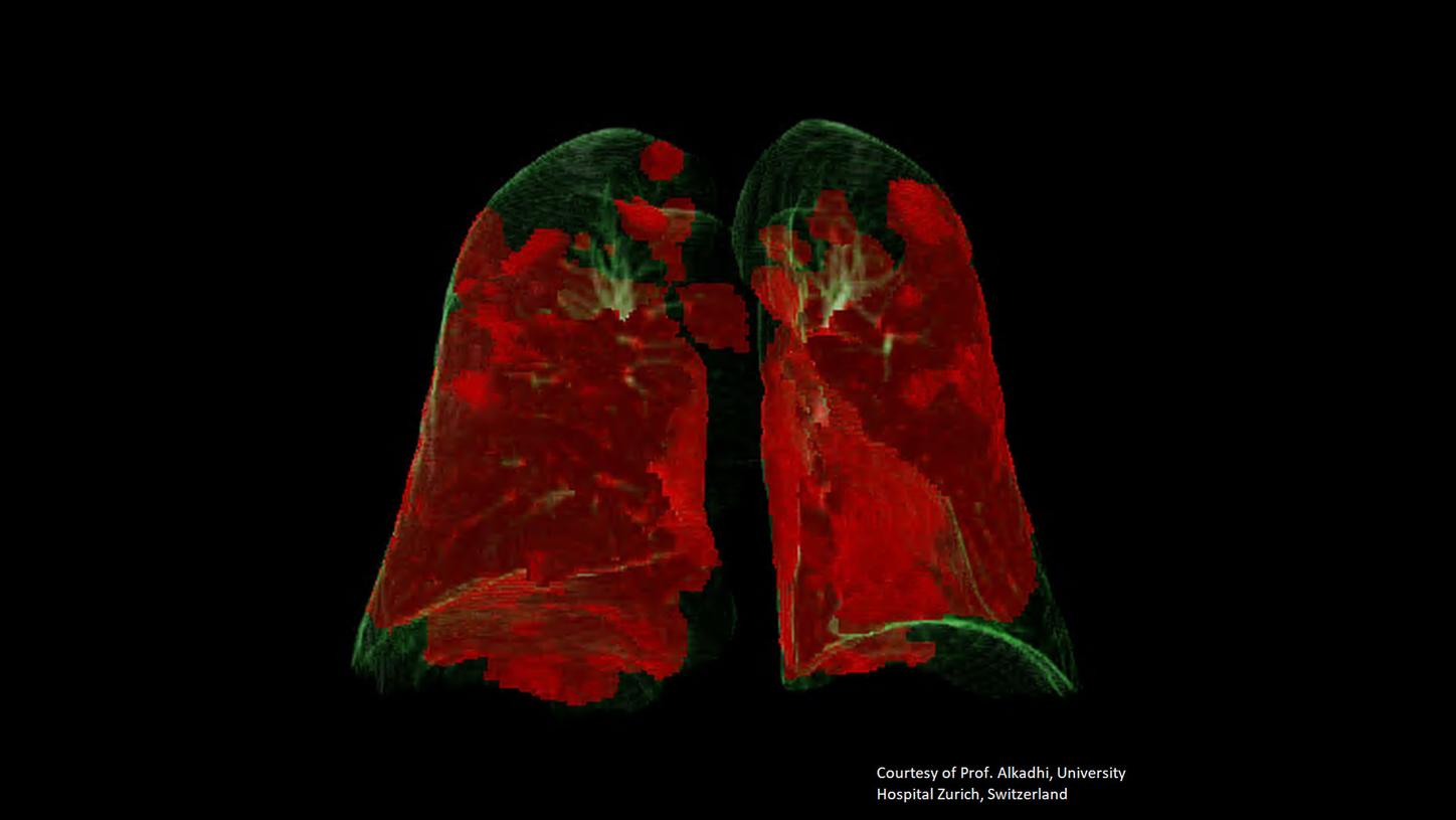
Impressions of the Prototype
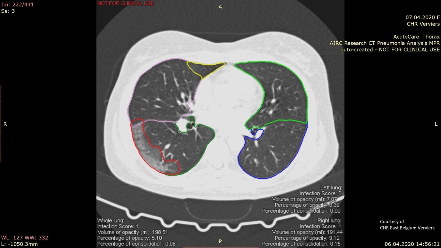
Courtesy of CHR East Belgium Verviers


CT Pneumonia Analysis is currently available for a free download to all syngo.via Frontier users.
syngo.via Frontier CT Pneumonia Analysis2: syngo.via Frontier allows you to strengthen your clinical opinion leadership with easy access to numerous post-processing prototypes that are seamlessly integrated with your routine syngo.via environment. The new CT Pneumonia Analysis2 prototype is included in the dedicated syngo.via Frontier Prototype Store and the Digital Marketplace, which is the continuously enriched with new contributions.

The Pulmonary-Density Plug-In is now available!
Based on certain parts of the “CT Pneumonia Analysis”-prototype, the “Pulmonary-Density Plug-In” offers:
- Quick & Easy overview of the lung with a complementary color-coded pictogram and key
- Segmentation and Quantification of ground-glass opacities and high densities in the lung
- VRT (Volume Rendering) for additional overview of opacity spatial distribution
- 2D axial views - overlaid with delineations of the opacities and the lungs
The “Pulmonary-Density Plug-In” is CE-labeled has also been approved under the FDA enforcement policy during the 2020 COVID-19 pandemic. It is currently available for syngo.via, syngo.via View&GO, and the AI-Rad Companion Chest CT. Please find more information on the plug-in in our two-pager or contact you local Siemens Healthineers representative.
Beyond research, AI algorithms may indicate signs of COVID-19 on chest X-rays embedded in the clinical workflow.
Artificial Intelligence plays an important role today, and its support can contribute to the radiologist’s workflow. The AI-Rad Companion Chest X-ray7, a new part of the AI-Rad Companion product family is another solution where deep learning is embedded in a dedicated software. The AI-Rad Companion Chest X-ray7 automatically characterizes radiographic findings for a lung X-ray (Upright patient position and PA direction).
It works like a second or third reader to support radiologists in differential diagnosis and clinical decision-making. It is capable of characterizing pneumothorax, pulmonary lesions (nodules, masses, and granuloma), atelectasis, consolidation, and pleural effusion. Atelectasis and consolidations are findings that show a correlation with signs of pneumonia caused by the SARS-CoV-2 virus.


Did this information help you?
The AI-Rad Companion, syngo.via, syngo.via View&GO, syngo.CT Extended Functionality (of which the Pulmonary Density Plug-In is a part) are not yet available in all countries. Due to regulatory reasons, future availability cannot be guaranteed. Please contact your local Siemens Healthineers organization for further information. The CT Pneumonia Analysis prototype and syngo.via Frontier are Research Only. Not for Clinical Use.