
Intraoperative quality control in orthopedic trauma surgery
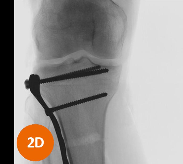
Conventional 2D intraoperative imaging does not always provide enough information to achieve the best possible results in orthopedic trauma surgery. Depending on the type and location of fractures, it can be tricky to assess whether a screw or wire is positioned correctly. If detected postoperatively, revision surgery will be necessary for some patients. Intraoperative 3D imaging enables the surgeon to correct malpositioned implants intraoperatively and not in a revision surgery.

New standard in intraoperative imaging
Intraoperative 3D imaging with Cios Spin shows more clearly if screws have been placed correctly. See the difference!


