
English translation by Omnilingua Worldwide, LLC
Photography by Michel Campfens
Data courtesy of Northwest Clinics, Alkmaar, The Netherlands
Northwest Clinics has a remarkably large nuclear medicine department with a long track record that began in 2004 when it became one of the first hospitals in the Netherlands to install a PET/CT scanner. Fast forward to 2019 and the PET/CT center, located in the city of Alkmaar, is one of the departments for which Northwest Clinics received a top clinical ranking in the STZ (Cooperating Top Clinical Training Hospitals) register. Northwest Clinics additionally received another top clinical ranking for cardiac imaging, a special focus for the nuclear medicine physicians in Alkmaar. In recent years, more than 6,000 patients have been examined for myocardial perfusion using PET/CT, which makes Northwest Clinics one of the cardiac-imaging leaders in the Netherlands. Yet, on a national basis, SPECT/CT is often still the standard technique for this type of examination.
Advantages of PET/CT
To many, the advantages of cardiac PET/CT are obvious. “In simple terms, you need less radiation, and you get high image quality with the potential for accurate diagnosis,” says medical physicist Sergiy Lazarenko, PhD, who made the transition from University Medical Center Groningen (UMCG) to Northwest Clinics in Alkmaar almost ten years ago to help establish a high-quality nuclear medicine department. Together with nuclear medicine physician Remco Knol, MD, PhD, the pair are two of the organizers of the Alkmaar Cardiac PET/CT Workshop. Knol says, “a key advantage of PET/CT is that you can quantify, which makes an important difference in cardiological evaluation. With SPECT, we lack that quantitative information and can only produce diagnoses using images. Sometimes that gives an incomplete view of the situation.” In other words, when using SPECT/CT, image interpreters can often see that the heart muscle is perfused, but the absolute myocardial blood flow cannot be assessed.
“A key advantage of PET/CT is that you can quantify, which makes a huge difference in cardiological evaluation.”

Predictive value
The precise information PET/CT offers can be used to determine the effect of narrowing on cardiac blood flow—even in small blood vessels—which is not clearly visible on a SPECT image. In addition, using a PET/CT scan, physicians can identify tissue that is no longer viable following a heart attack, as well as tissue that is damaged but can still recover with restored blood flow. PET/CT scans are thus an excellent method for assessing whether interventions, such as stent placement, can be advantageous.

The Northwest Clinics in Alkmaar, The Netherlands
Logistical challenges
PET/CT is far from being in use everywhere, partly because it is a more complex technique. The radioactive materials decay much more quickly than those used in SPECT examinations, which have a half-life of hours or even days. The radiotracer used by Northwest Clinics is ammonia labeled with the radioactive isotope Nitrogen-13 (13N). “This is a great substance for visualizing heart muscle blood flow,” says Knol. “But the logistics are a major challenge, as it has a half-life of 10 minutes.” Northwest Clinic’s onsite cyclotron helps them address these logistical challenges.
Despite the logistical challenges of using tracers with a short half-life, Lazarenko says the approach with PET/CT has its advantages. “It means that, in one study, the patient can have both rest and stress examinations, whereas SPECT requires two examinations on separate days due to the long time the substance remains in the system.” On the other hand, there is the downside that you must make the tracer on site because, due to the short half-life, road transport is simply not an option. Lazarenko says, “ever-smaller cyclotrons make it feasible for more hospitals to do this. You need a good business case for this, with sufficient patient numbers. Logistically, given the time pressure, all steps in the process need to be coordinated in detail.” Knol adds, “in addition, producing and assessing cardiac scans also requires specific experience.”

Sergiy Lazarenko, PhD, and Remco Knol, MD, PhD, stand in front of their department’s Biograph Vision PET/CT scanner.
Benefits of technology
Alkmaar’s top-ranked cardiac imaging approach is achieved, in part, because of Northwest Clinic’s commitment to providing advanced scanner technology, software, and protocol optimization.
Significant time savings have been achieved in cardiac PET/CT examinations thanks to a protocol developed in Alkmaar. Lazarenko explains, “normally, an examination would take about an hour, mainly because you needed a certain waiting time between the rest and stress images. Without that waiting time, the residual radiation from the first examination would disturb the subsequent second scan. However, together with Siemens Healthineers, we have developed an algorithm that filters out that ‘residual activity.’ As a result, we can start the second examination much sooner, which means the total examination time is just 25 minutes.” In addition to the shortened examination times, image quality has also improved. Lazarenko reveals, “thanks to the improved software the risk of artifacts, for example, is much lower. In older systems, crucial ‘blind spots’ in the image can be caused by a mismatch of the CT and PET images.” Knol emphasizes, “that is the major issue you can experience with this type of examination. Our Biograph Vision™ has additional software [CardioFreeze and OncoFreeze] that reduces motion artifacts. This constitutes a huge leap forward in quality.”
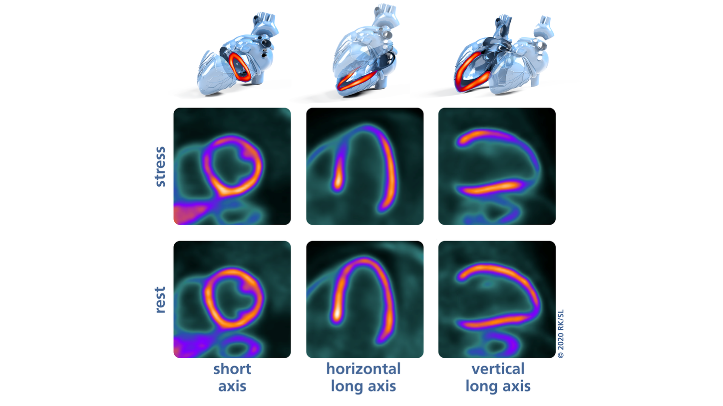
Adenosine stress (top) versus rest (bottom) 13N-ammonia PET/CT images in short-axis, horizontal long-axis, and vertical long-axis slices. A moderate-to-severe reversible perfusion defect is present in the anteroseptal and apical myocardium due to a significant LAD stenosis.
Sharing knowledge
Knol, Lazarenko, and colleagues now want to share the knowledge and possibilities of cardiac PET/CT that were developed in Alkmaar. According to Lazarenko, “last year, we got into a conversation with international colleagues at a conference and noticed there was a lot of interest.” Northwest Clinics thus organized a two-day workshop on cardiac PET/CT in September 2020; the workshop covered a range of sub-topics, from specific clinical applications to the possibilities of AI and the development of a business case. The intent is for the workshop to be an annual event, with the next workshop occuring in September 2021.
The objective of the workshop was to offer a practical approach to cardiac PET. Knol says, “you need to get a handle on it for yourself. With the help of Siemens Healthineers, we have been able to obtain workstations and licenses, and have selected anonymized case histories from our own database. In addition, the participants—cardiologists, nuclear medicine physicians, and radiologists—can get to work on real examinations during the afternoon sessions. That is really the essence of what we want to achieve. We have experienced for ourselves how the imaging and diagnostics of cardiac perfusion can be improved with PET/CT. We want to give people with no experience the tools to get them started.”