Computed Tomography plays a vital role in pediatric diagnosis, treatment planning, and follow-up, but it also comes with unique challenges. Greater sensitivity to radiation, higher heart rates, patient motion, and the emotional impact of the examination – all require special attention.
Our CT portfolio combines patient-centered solutions with advanced imaging technologies, including photon-counting CT. Designed for children, their families, and the clinical team, our systems help create a more reassuring imaging experience while supporting confident clinical decision-making.

PediatricsShaping the future of pediatric care with CT
Highlights & Innovations
Innovating in pediatric imaging with the NAEOTOM Alpha class
With NAEOTOM Alpha, the world's first photon-counting CT, we have introduced a technology that can contribute to a new role for CT in pediatrics. With low radiation dose, high spatial resolution and improved image contrast, it helps you address the challenges of pediatric imaging.
The dose reduction and speed enabled by the photon-counting detectors in the NAEOTOM Alpha class are of particular importance in the diagnosis and treatment planning of complex pediatric cases, especially in infants and young children, at Erasmus University Rotterdam in the Netherlands.
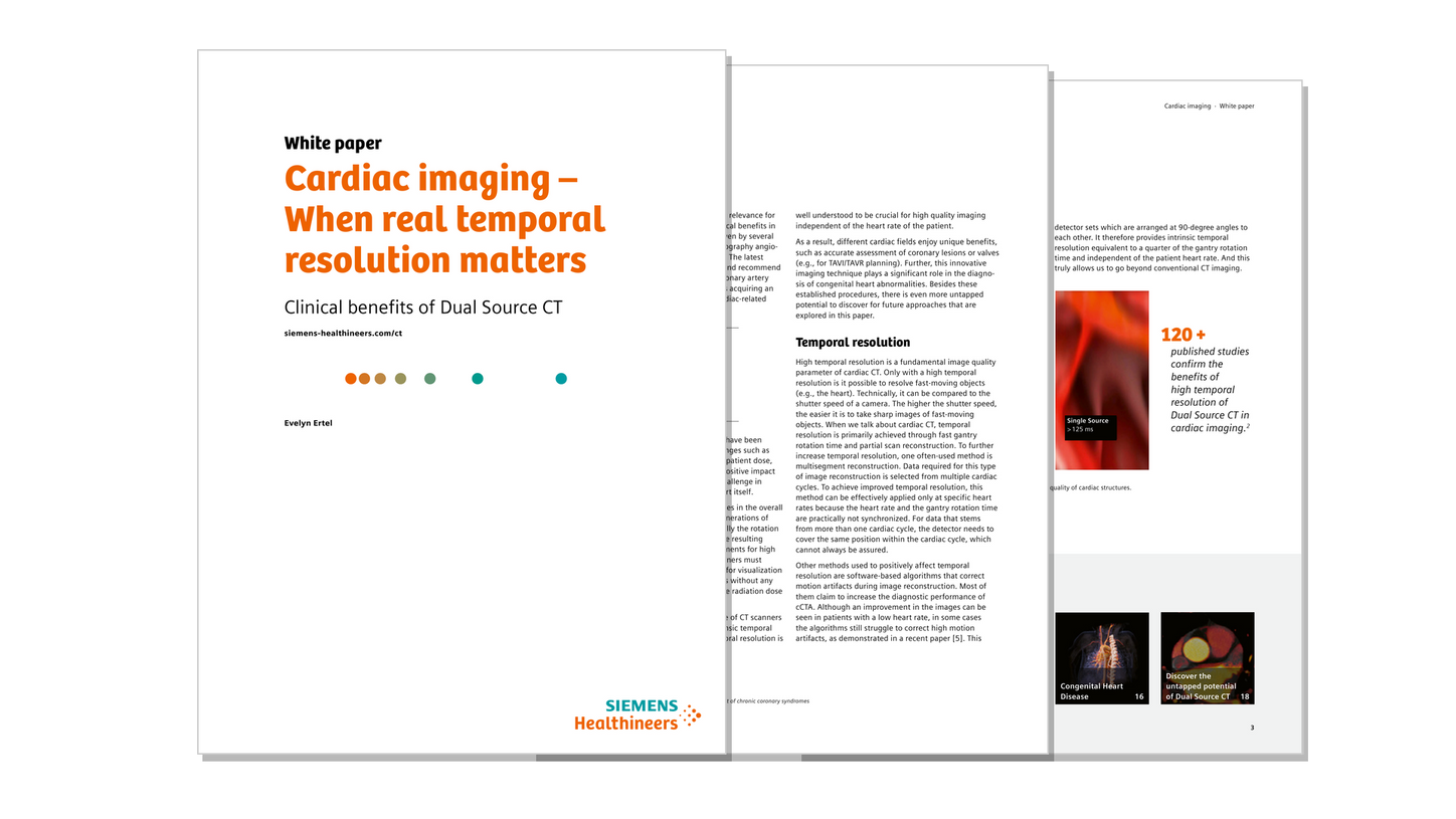
Dual Source CT in pediatric imaging
SOMATOM Pro.Pulse is a smartly designed Dual Source CT scanner that combines the strengths of Dual Source technology with simpler scanning workflows and a range of dose reduction features. Ideal for pediatric applications, it delivers fast, low-dose scans and reduced motion artifacts, while streamlining installation and maintenance efforts for healthcare facilities.
Holistic patient experience
Children differ fundamentally from other patient groups in terms of their emotional and cognitive needs. To ensure smooth pediatric imaging exams and best possible treatment, Siemens Healthineers and its partners have developed a holistic approach. This includes child-friendly preparation and supporting materials – designed to help reduce the perceived stress level of medical staff, children, and parents.
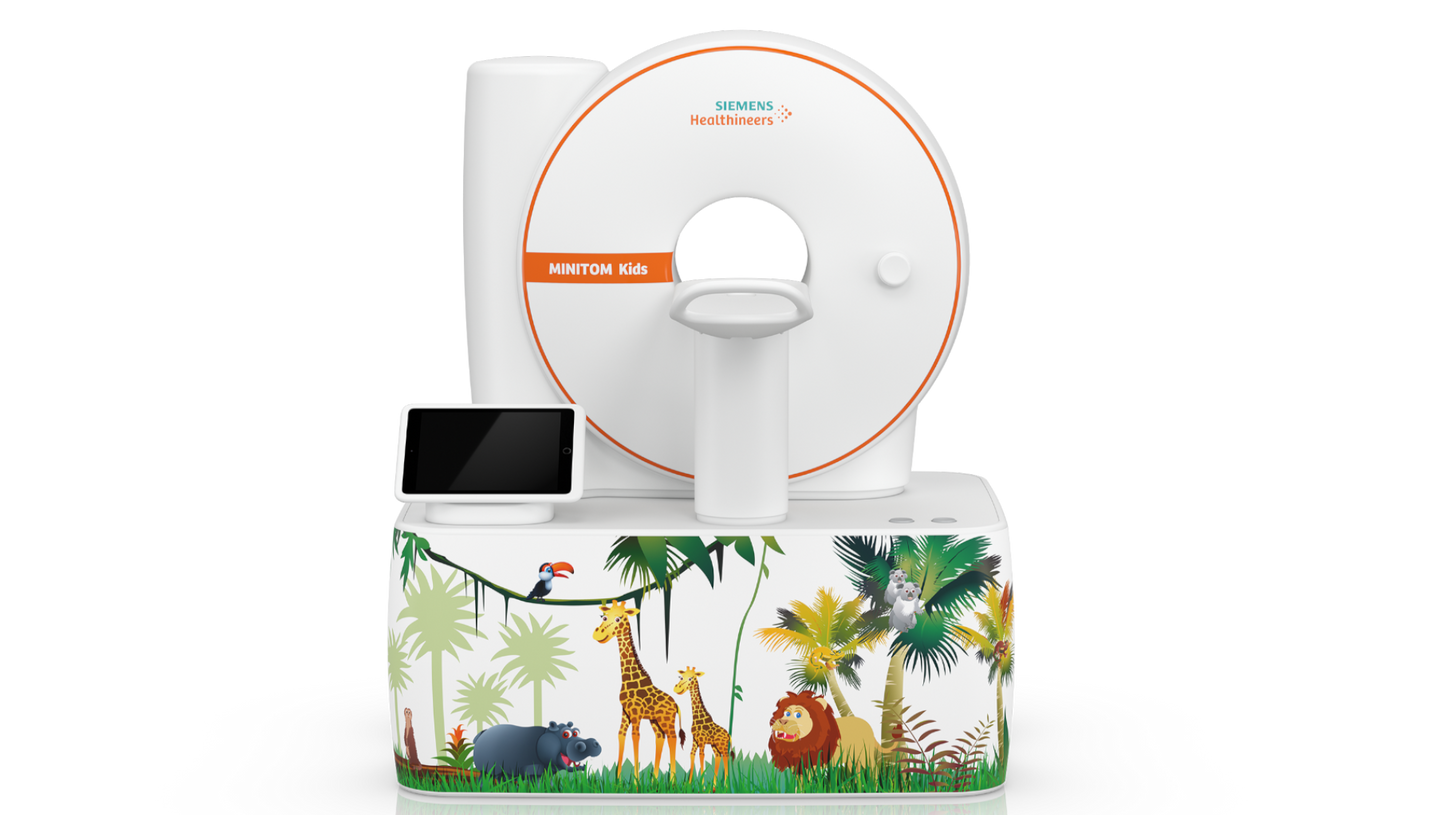
Redefine the pediatric patient experience with MINITOM Kids
MINITOM Kids as a child-sized model scanner allows children to become familiar with the scan process through interactive exploration. It includes a moveable table, a button that creates scan noises, and a tunnel under the scanner which children can use to practice lying still in the bore. The attached tablet demonstrates how Gerda, the Brave Giraffe is scanned. Gerda is also the protagonist of our children’s book for pediatric patient preparation. The interactive parts of the book and the integrated audio book help children to reassure themselves in the unfamiliar situation of an examination.
Clinical cases
/

Sn100 kVp | CTDIvol 0.93 mGy | DLP 25mGy*cm | Pitch: 3.2
Low dose and sub-second lung imaging for pediatrics
Courtesy of University Hospital Augsburg, Augsburg, Germany
Upper left image: Sn100 kV | CTDIvol 0.01 mGy
Down left image: 0.6 mm | BI60 | 73 keV
Middle image: Bronchial wall thickness visualization
Right image: cVRT


/
Clinical practice
Explore more pediatric imaging possibilities with two in-depth cases.
The products/features (mentioned herein) are not commercially available in all countries. Their future availability cannot be guaranteed.
The statements by Siemens Healthineers’ customers described herein are based on results that were achieved in the customer's unique setting. Because there is no “typical” hospital and many variables exist (e.g., hospital size, samples mix, case mix, level of IT and/orautomation adoption) there can be no guarantee that other customers will achieve the same results.