
MR NeurologyUnderstanding complexity
MRI is the ultimate tool for diagnostic imaging and neuroscience research, providing morphological images with the highest spatial resolution and unmatched soft tissue contrast as well as unique functional information of the central nervous system (CNS).
The advent of new powerful gradient systems has opened opportunities to explore the microstructure of the brain at an unprecedented level. Deep learning reconstruction is fundamentally transforming MR imaging and will allow higher patient throughput without compromising image quality.
At Siemens Healthineers, we firmly believe that combining the latest hardware innovations with software-based features such as deep learning reconstruction will create great momentum in the neuroradiological and neuroscience community. Let's continue to explore the human brain, together.
Highlights
Be at the forefront of the neuroimaging community with our comprehensive solution portfolio for diagnostic imaging and neuroscience research.

Deep Resolve
At the forefront of the revolution in MRI acceleration, Deep Resolve enables game-changing acceleration by combining parallel imaging, SMS and AI-powered image reconstruction1.
Your benefits:
- Drastically reduce scan times and double your in-plane resolution
- Unlock b-values that were previously difficult to obtain in diffusion-weighted imaging
- Open a new dimension in speed with Deep Resolve 3D


Clinical Applications
Siemens Healthineers exclusive applications
At Siemens Healthineers MR, we are driven to deliver outstanding applications that shift the limits of MRI. The following exclusive features can only be found on our MRI machines and set you apart from competition.

2aaaa2230
Deep Resolve Swift Brain
Based on a multi-shot EPI approach, sophisticated magnetization preparation and deep learning reconstruction, Deep Resolve Swift Brain enables the fastest brain examination ever on our MRI machines. Together with Simultaneous Multi-Slice, net scan times below 2:00 minutes are possible.
Your benefits:
- Fastest standard brain exam on our MRI scanners by multi-shot EPI and deep learning reconstruction
- A new Static Field Correction will further reduce geometric distortions
- Full flexibility: Only acquire contrasts required for your clinical question, save even more scan time


More neurology applications
Address all relevant clinical questions with our rich portfolio of MR neurology applications.

ZOOMit
ZOOMit – the first TimTX TrueShape application for clinical use – uses dynamic excitation pulses to achieve selective field-of-view (zoomed) imaging. Zoom into your image to appreciate the smallest details, improve diagnostics and expand research possibilities with ZOOMit, powered by TimTX TrueShape.
Your benefits:
- Zoomed imaging for EPI (DWI and BOLD) and SPACE
- Reduced susceptibility artifacts and geometric distortions in challenging anatomies
- Higher temporal resolution through faster imaging

ASL (Arterial Spin Labeling)
ASL is an MR technique using the water in arterial blood as an endogenous contrast agent to evaluate perfusion non-invasively. Pseudo-continuous ASL (PCASL) allows larger coverage with more and thinner slices in a shorter acquisition time, resulting in higher perfusion image quality.
Your benefits:
- 2D and 3D PCASL enable higher in-plane resolution and thinner slices
- Automatic generation of relative CBF and Bolus Arrival Time maps
- M0 maps are available for research purposes

SWI (Susceptibility-Weighted Imaging)
SWI is an MRI contrast which exploits the susceptibility differences between tissues. As a result SWI can detect deoxygenated blood, products of blood decomposition and microscopic iron deposits much better than conventional MR techniques.
Your benefits:
- Improved depiction compared to T2* contrast of susceptibility variations such as hemorrhages and microbleeding
- 3D imaging for complete brain coverage
- The perfect CAIPIRINHA: Wave-CAIPI SWI for maximum acceleration in SWI
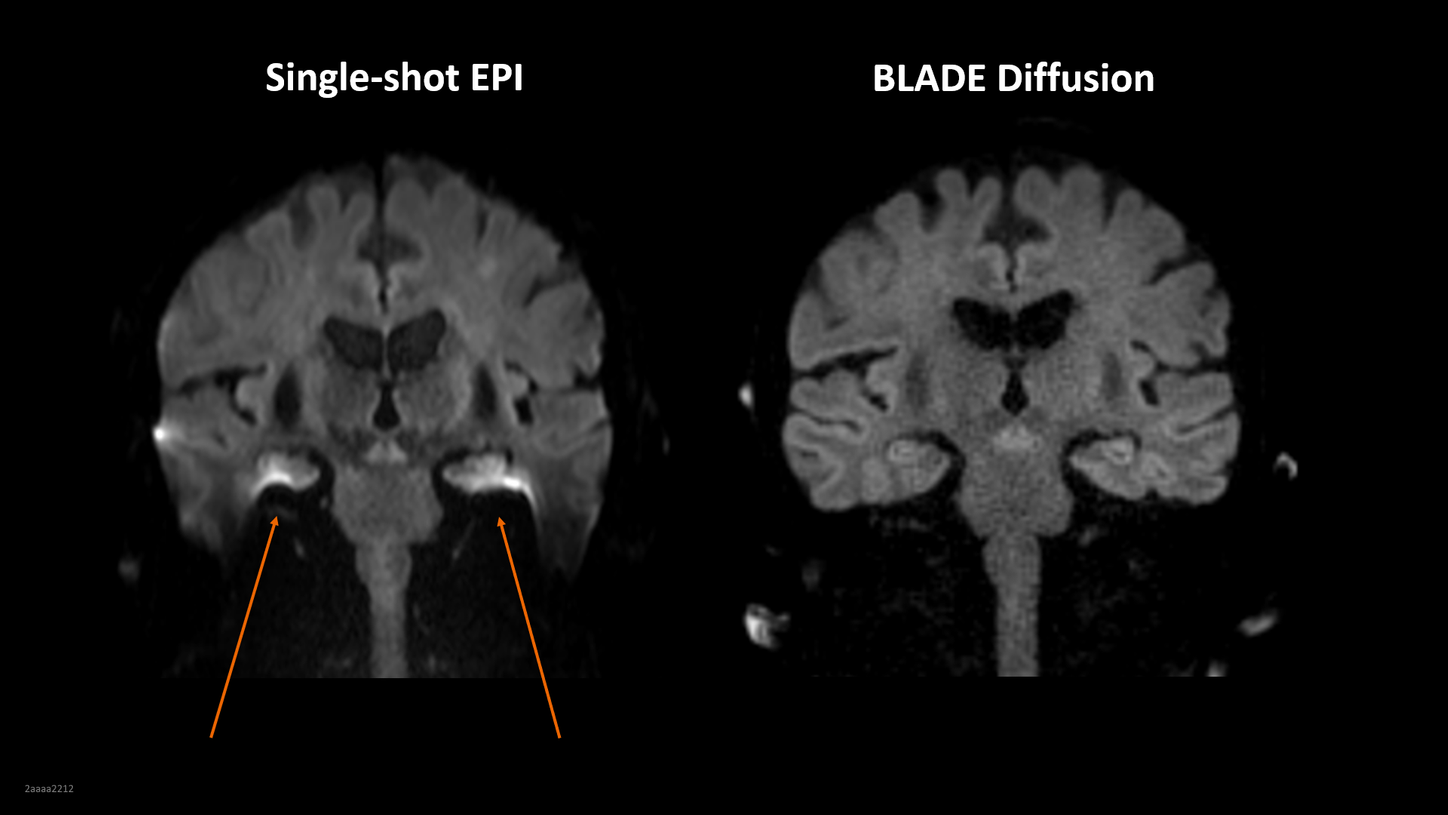
HASTE and BLADE diffusion
HASTE and BLADE diffusion enable virtually distortion-free diffusion-weighted imaging (DWI) by employing non-EPI acquisition methods. These methods have been found helpful for the detection of cholesteatoma6-8 and bring clarity to DWI in challenging anatomies where susceptibility artifacts cannot be avoided9.
Your benefits:
- Non-EPI-based DWI that is virtually free of geometric distortion
- Siemens Healthineers exclusive: SMS capability for BLADE diffusion to keep scan times at bay
- Superb detection of difficult pathologies such as cholesteatoma6-8 and cerebellopontine angle tumors9
Clinical Focus Topics

Headache
In clinical routine, many MRI scans deal with unspecified headache disorders that account for 50% of all neurology related disorders10. MRI is a tool to clarify if abnormalities in the brain are responsible for the symptoms, but 90% of these scans are usually without pathological findings11. So there is a large amount of routine brain MRI scans that require fast and robust technology to deliver reliable results. We have what it takes to address these requirements:
- An industry leading portfolio of acceleration methods, including Deep Resolve
- Software-based features such as AutoAlign, myExam Companion and BioMatrix Motion Sensor
- Hardware innovations such as Frequalizers, BioMatrix CoilShim and our industry-leading BioMatrix Head/Neck 64 channel coil

Emergencies
Stroke is the second leading cause for death among all diseases, only surpassed by cardiovascular disease10. Time is brain in acute stroke, and while CT is typically the method of choice, MRI is considered equivalent by current guidelines12 and has even been shown to be more effective13. Besides stroke and in particular wake-up strokes14, MRI can play a decisive role in vulnerable groups such as pregnant women and cranial trauma15. We support these use cases with innovations such as
- Deep Resolve, our AI-based1 image reconstruction technique
- Deep Resolve Swift Brain for the fastest brain exam ever
- Wave-CAIPI SWI, the perfect CAIPIRINHA
When stroke strikes, be ahead of your time

Neurodegenerative Diseases
Neurodegenerative Diseases (ND) such as Alzheimer’s Disease (AD), dementia, or Multiple Sclerosis (MS) have seen a significant increase in prevalence over the last decade and are the second leading cause of death among neurology related disease, only surpassed by stroke10. With ageing populations in many developed countries, increasing numbers of ND are expected. MRI plays a major role in diagnosing and monitoring diseases such as MS. Additionally, it is also mandated for therapy monitoring of the newest generation of AD drugs that slow the progress of early stage AD.
- AI Rad Companion Brain MR for Morphometry might be potentially helpful in identifying patients with early stage AD
- AI Rad Companion Brain MR for White Matter Hyperintensities automatically determines count, volume, and location of white matter hyperintensities
Clinical Protocols

Access clinical protocols, phoenix images, and application tips including videos by industry experts in our Clinical Corner.