- Home
- Medical Imaging
- Computed Tomography
- Clinical Imaging Solutions
- CT-guided interventions

CT-guided interventionsInnovative CT solutions for efficient procedure planning and needle guidance
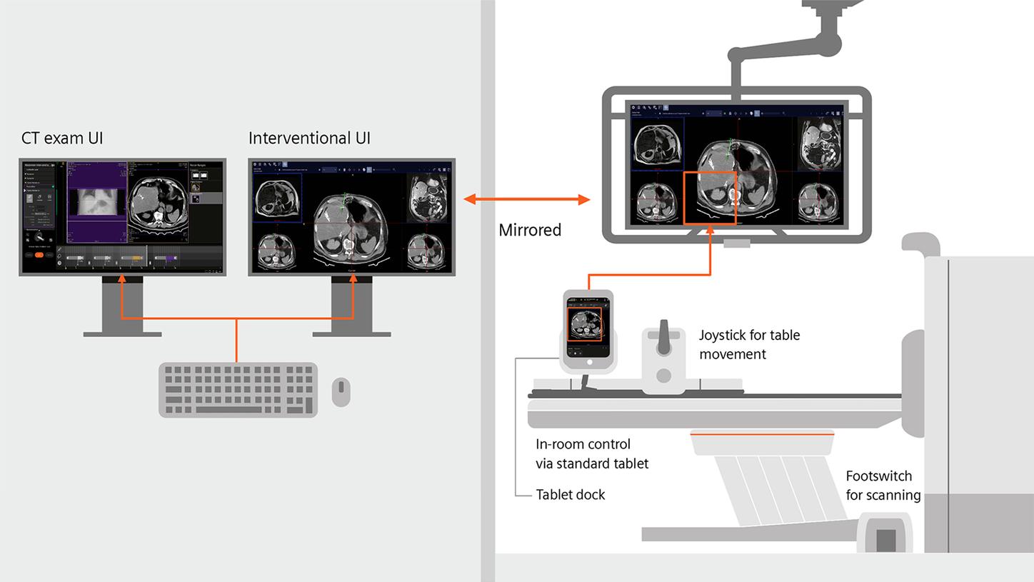
Simplify routine and complex CT-guided interventions with a modular, scalable solution for procedure planning and needle guidance. Advanced path planning, AI-based needle detection, image fusion, laser guidance, and flexible in-room control support efficient workflows with or without an assistant.
Clinical applications
CT-guided interventions support a growing range of diagnostic and therapeutic procedures. Explore key applications for CT-based procedure planning and needle guidance.
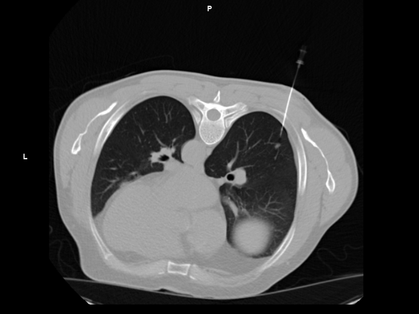
Biopsy
CT guidance improves needle placement accuracy and increases the success rate of obtaining diagnostic tissue during the initial biopsy procedure.1
Image courtesy of Hospital Maennedorf, Switzerland
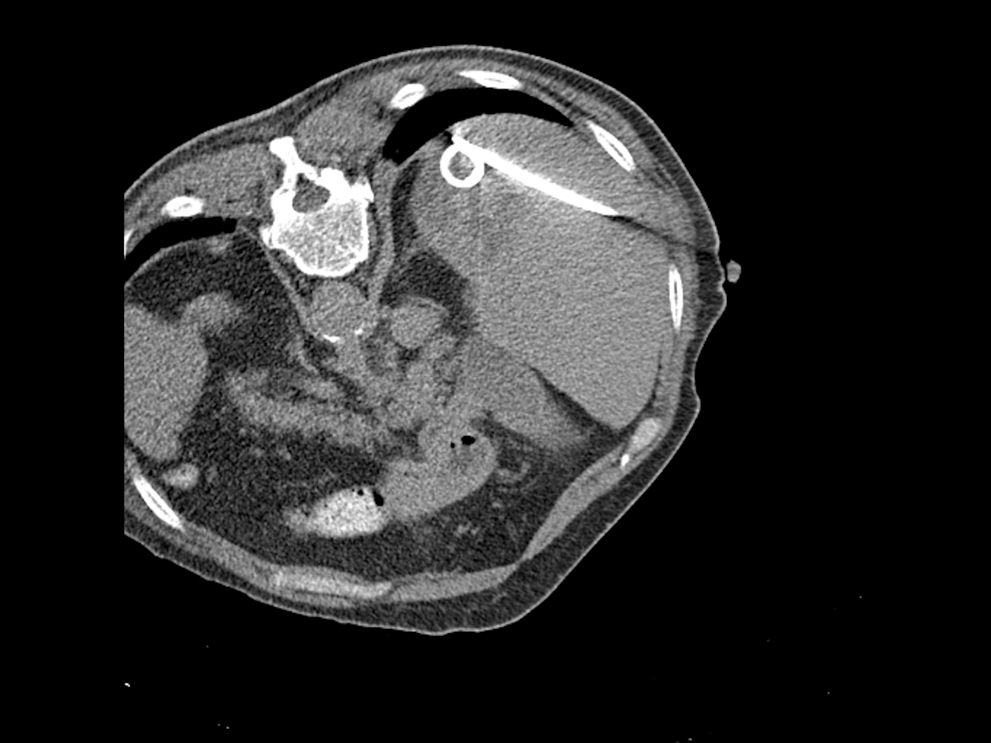
Drainage
CT guided placement during drainage procedures ensures safe, efficient workflows that help patients recover faster2
Image courtesy of The Ohio State University Wexner Medical Center, USA
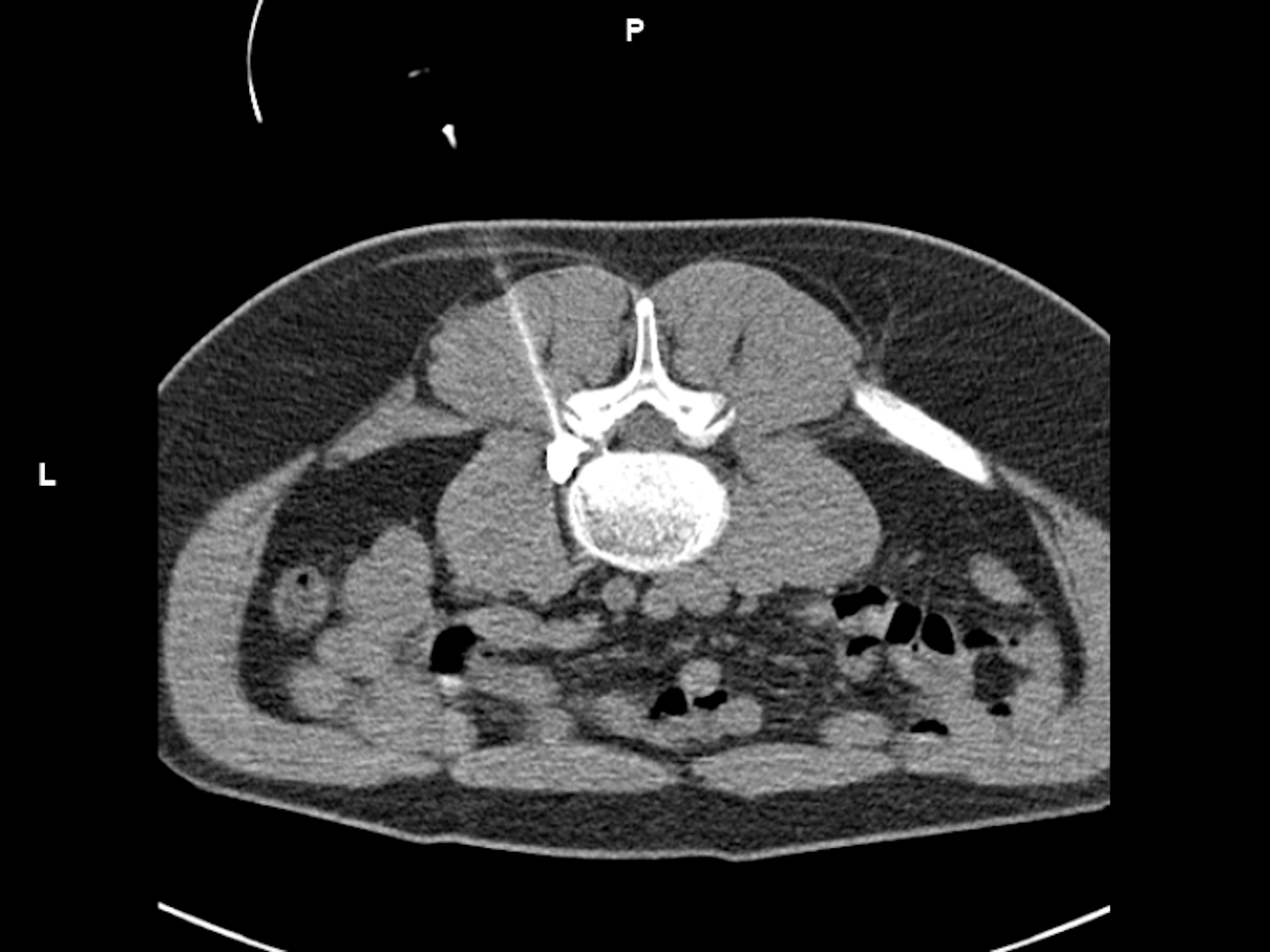
Pain management
CT guided pain medication injections deliver targeted relief and can help avoid more invasive procedures.3
Image courtesy of Benson Mount Gambier Hospital SA, Australia
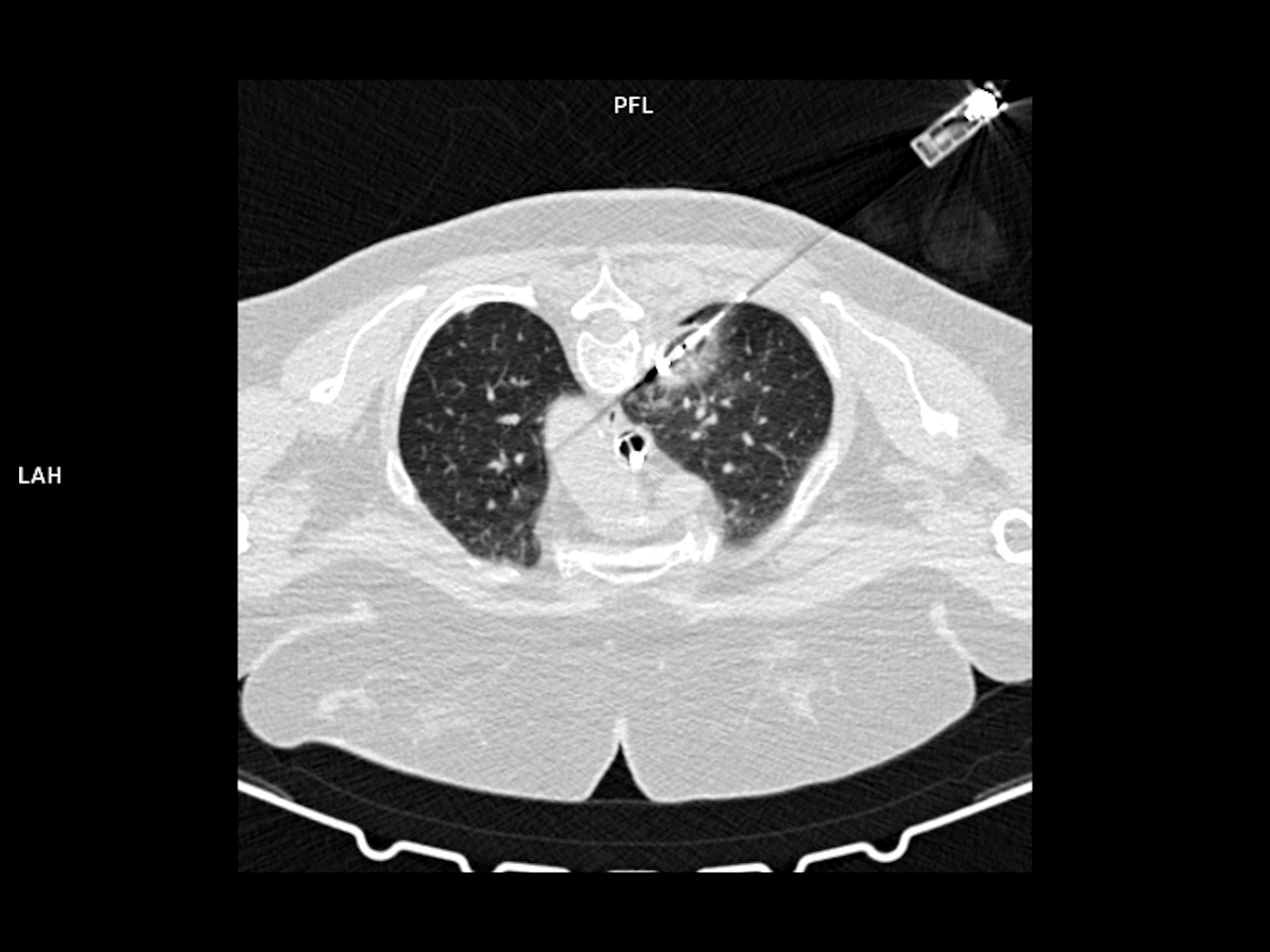
Tumor ablation
CT-guided tumor ablation provides a minimally invasive treatment approach that supports localized tumor control.4
Image courtesy of RNS Group Practice for Radiology and Radiotherapy Wiesbaden, Germany
Stay competitive in a growing and dynamic field
The number of CT-guided interventions performed is expected to grow rapidly. While biopsies will remain the core application, therapeutic procedures are expected to become much more important due to the obvious advantages of minimally invasive therapy: fewer complications, faster recovery times, and shorter hospital stays.
New guidelines
The growing importance of thermal ablations in cancer treatment is highlighted by a number of new guidelines.5,6
Increase in ablations
Ablation procedures in cancer therapy are projected to increase by 8 percent per year between 2020 and 2030.7
Alternative to surgery
CT-guided drainages provide an alternative to surgery with lower morbidity and mortality rates.2
577 million patients
More than half a billion patients worldwide suffered from low-back pain in 2017.8 Image-guided injections can help ease this burden and minimize surgical interventions.
Dedicated solutions for CT-guided interventions
Our CT-guided intervention solutions combine intelligent planning tools, AI-based needle guidance, and flexible in-room control to support efficient workflows for routine and complex procedures.

myNeedle Laser
myNeedle Laser9 is a powerful, fully integrated option designed to make the intervention workflow more efficient by automatically projecting the needle entry point and insertion angle onto the body of the patient – even in advanced double-angulated procedures with multiple needle paths.
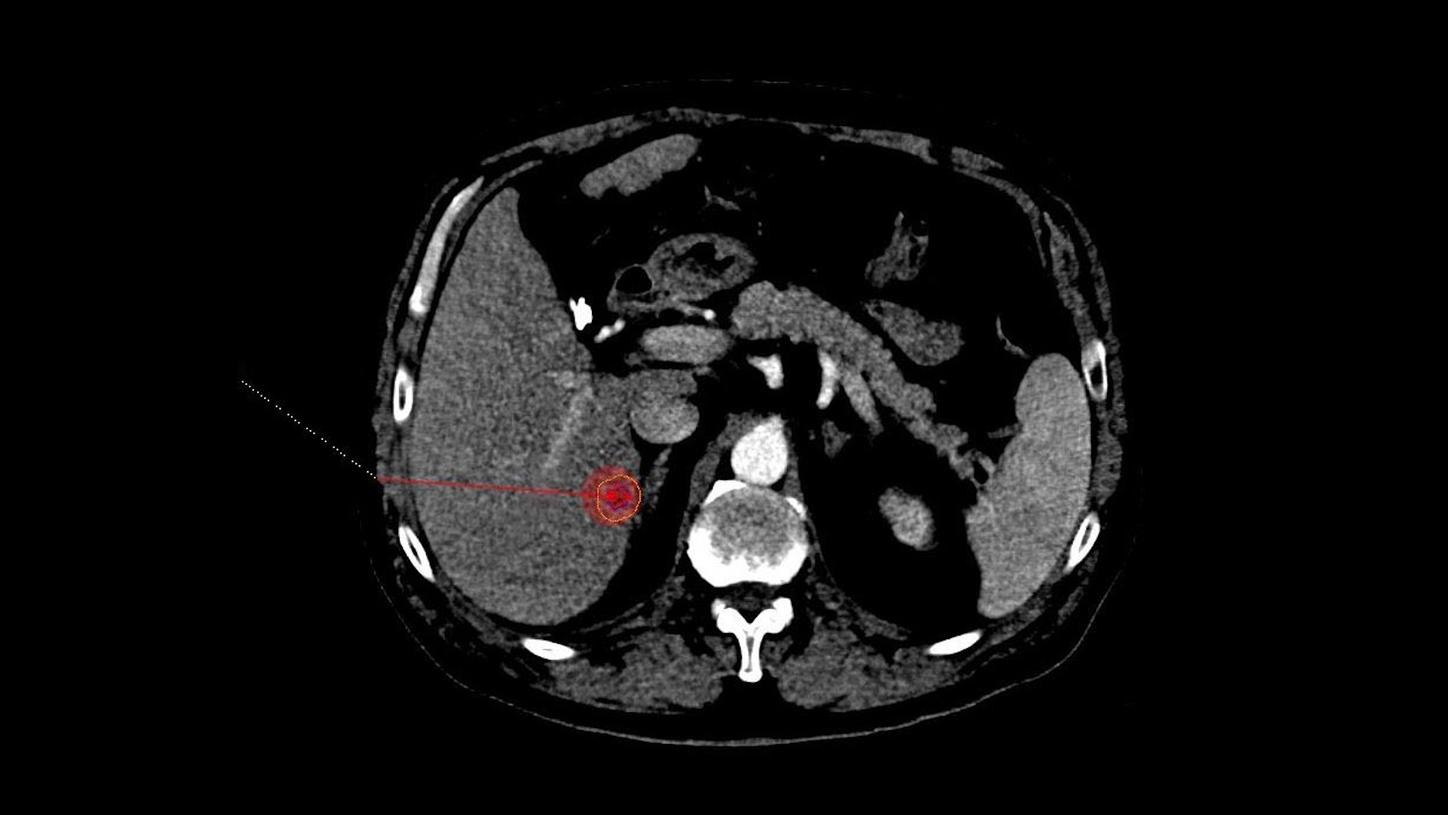
myAblation Guide
myAblation Guide10 brings unprecedented consistency to your microwave liver ablation workflow. With sophisticated planning tools, easy postprocedural assessment, and a coherent user experience across imaging and ablation systems, we’ve got you covered every step of the way.
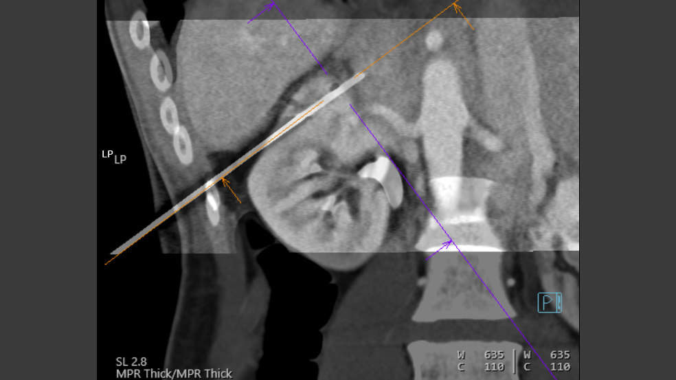
myNeedle Guide 3D
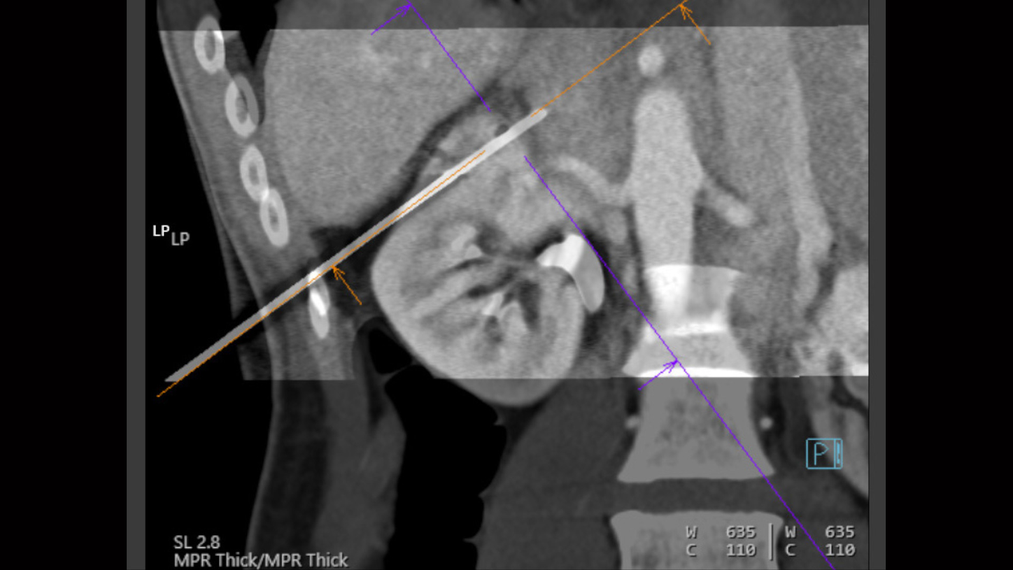
myNeedle Guide 3D11 simplifies the workflow for both routine and complex CT-guided interventions. You can easily plan multiple needle pathways in different cross sections. Both path planning and visually guided insertion of multiple needles are supported within the workflow.
The AI based needle detection algorithm myNeedle Detection12 supports an efficient workflow by minimizing user interaction when progressing the needle.
Integrated Image Fusion allows fusing of 3D images from different modalities or contrast-enhanced prior CT studies to accurately plan and guide needle paths around critical anatomy.
Clinical cases
Courtesy of RNS Gemeinschaftspraxis, Wiesbaden, Germany
Microwave ablation of a liver mass
CT-guided intervention with myNeedle Guide 3D on SOMATOM go.Up
Contrast enhanced planning spiral:
2 events
Exposure time: 12.2 s / 13.6 s
Scan length: 299 mm / 329 mm
130 kV
CTDIvol: 13.9 mGy
DLP: 379 mGy*cm / 420 mGy*cm
FAST i-Sequence:
21 events
Exposure time: 16.8 s
Scan length: 15 mm (each)
80 kV
CTDIvol: 5.57 mGy
Accum. DLP: 379 mGy*cm

Courtesy of Kantonsspital Baden, Baden, Switzerland
Biopsy of a small suspicious lesion at the shoulder blade
CT-guided intervention with myNeedle Guide 2D on SOMATOM X.ceed
Planning and control spiral:
Exposure time: 3.6 s / 1.7 s
Scan length: 131 mm / 41 mm
100 kV
CTDIvol: 3.9 mGy / 3.15 mGy
DLP: 57.6 mGy*cm / 18.7 mGy*cm
i-Fluoro:
3 events
130 kV
Scan length: 9 mm
Exposure time: 0.7 s / 4.3 s / 0.7 s
CTDIvol: 6.48 mGy / 39.3 mGy / 6.48 mGy
Accum. DLP: 47.1 mGy*cm
Complete procedure time: 36 min

Courtesy of University Hospital Erlangen, Erlangen, Germany
cVRT performed with syngo.via
Biopsy of suspicious lesion in the liver
CT-guided intervention with myNeedle Companion on SOMATOM X.cite
Planning spiral:
Exposure time: 4.6 s
Scan length: 215 mm
80 kV
CTDIvol: 10 mGy
DLP: 256
i-Spiral:
8 events
80 kV
Scan length: 70 mm - 98 mm
Exposure time: 1.93 s (each)
CTDIvol: 4.2 mGy / 6.8 mGy
Accum. DLP: 386 mGy*cm

Courtesy of Centro Hospitalar de São João, Porto, Portugal
Biopsy of a small suspicious lung lesion
CT-guided intervention with myNeedle Guide & i-Fluoro on SOMATOM go.Up
Planning and control i-Spiral:
Exposure time: 14 s (each)
Scan length: 341 mm (each)
130 kV (each)
CTDIvol: 5.1 mGy
DLP: 160 mGy*cm
i-Fluoro to monitor needle progression:
2 events
Exposure time: 2.2 s / 12.6 s
Scan length: 9 mm (each)
110 kV
CTDIvol: 14.5 mGy / 83.7 mGy
Accum. DLP: 88.4 mGy*cm

Courtesy of University Hospital Erlangen, Erlangen, Germany
Biopsy of a suspicious lesion between kidney and spleen
CT-guided intervention with myNeedle Guide 3D on SOMATOM X.cite
Planning and control spiral:
3 events
Exposure time: 3.6 s (each)
Scan length: 175 mm (each)
70 kV
CTDIvol: 3.3 mGy
DLP: 65.5 mGy*cm
i-Spiral to monitor needle progression:
12 events
70 kV - 80 kV
Exposure time: 2.3 s - 2.4 s
Scan length: 86 mm - 175 mm
CTDIvol: 2.2 mGy / 2.4 mGy
DLP: 6.75 mGy*cm / 23 mGy*cm
Accum. DLP: 123 mGy*cm
Complete procedure time: 1h 15 min

Courtesy of Luzerner Kantonsspital, Luzern, Switzerland
cVRT performed with syngo.via
Cervical periradicular infiltration for pain management
CT-guided intervention with myNeedle Companion on SOMATOM X.ceed
Planning and control spiral:
5 events
Exposure time: 3-4 s (each)
Scan length: 146 mm (each)
Sn100 kV
CTDIvol: 4.61 / 5.05 mGy
Accum. DLP: 395 mGy*cm
i-Fluoro
5 events
Sn130 kV
Exposure time: 0.35 s - 5.64 s
Scan length: 9 mm
CTDIvol: 3.25 / 52.1 mGy
Accum. DLP: 92 mGy*cm

Courtesy of Centro Hospitalar de São João, Porto, Portugal
Drainage placement in Seldinger technique
CT-guided intervention with myNeedle Guide 2D & i-Fluoro on SOMATOM go.Up
Planning and control spiral:
Exposure time: 9.3 s / 4.53 s
Scan length: 232 mm / 127 mm
130 kV
CTDIvol: 7.42 mGy
DLP: 153 mGy*cm / 75 mGy*cm
i-Fluoro:
2 events
Exposure time: 10.9 s / 21.3 s
Scan length: 15 mm (each)
80 kV
CTDIvol: 58 mGy / 122 mGy
Accum. DLP: 169 mGy*cm
Courtesy of Kantonsspital Baden, Baden, Switzerland
Microwave ablation of a liver metastasis
CT-guided intervention with myNeedle Companion on SOMATOM X.ceed
Contrast enhanced planning spiral:
Exposure time: 4 s
Scan length: 234 mm
80 kV
CTDIvol: 1.64 mGy
DLP: 43.6 mGy*cm
i-Spiral for monitoring the needle progression:
7 events
Exposure time: 2 s
Scan length: 71 mm
Sn100 kV
CTDIvol: 2.2 / 2.4 mGy
Accum. DLP: 224.5 mGy*cm
i-Spiral of arterial phase after treatment:
Sn 100 kV
Scan length: 217.6 mm
Exposure time: 5 s
CTDIvol: 2.18
DLP: 52 mGy*cm
Courtesy of RNS Gemeinschaftspraxis, Wiesbaden, Germany
Microwave ablation of a liver mass
CT-guided intervention with myNeedle Guide 3D on SOMATOM go.Up
Contrast enhanced planning spiral:
2 events
Exposure time: 12.2 s / 13.6 s
Scan length: 299 mm / 329 mm
130 kV
CTDIvol: 13.9 mGy
DLP: 379 mGy*cm / 420 mGy*cm
FAST i-Sequence:
21 events
Exposure time: 16.8 s
Scan length: 15 mm (each)
80 kV
CTDIvol: 5.57 mGy
Accum. DLP: 379 mGy*cm

Courtesy of Kantonsspital Baden, Baden, Switzerland
Biopsy of a small suspicious lesion at the shoulder blade
CT-guided intervention with myNeedle Guide 2D on SOMATOM X.ceed
Planning and control spiral:
Exposure time: 3.6 s / 1.7 s
Scan length: 131 mm / 41 mm
100 kV
CTDIvol: 3.9 mGy / 3.15 mGy
DLP: 57.6 mGy*cm / 18.7 mGy*cm
i-Fluoro:
3 events
130 kV
Scan length: 9 mm
Exposure time: 0.7 s / 4.3 s / 0.7 s
CTDIvol: 6.48 mGy / 39.3 mGy / 6.48 mGy
Accum. DLP: 47.1 mGy*cm
Complete procedure time: 36 min

Courtesy of University Hospital Erlangen, Erlangen, Germany
cVRT performed with syngo.via
Biopsy of suspicious lesion in the liver
CT-guided intervention with myNeedle Companion on SOMATOM X.cite
Planning spiral:
Exposure time: 4.6 s
Scan length: 215 mm
80 kV
CTDIvol: 10 mGy
DLP: 256
i-Spiral:
8 events
80 kV
Scan length: 70 mm - 98 mm
Exposure time: 1.93 s (each)
CTDIvol: 4.2 mGy / 6.8 mGy
Accum. DLP: 386 mGy*cm

Courtesy of Centro Hospitalar de São João, Porto, Portugal
Biopsy of a small suspicious lung lesion
CT-guided intervention with myNeedle Guide & i-Fluoro on SOMATOM go.Up
Planning and control i-Spiral:
Exposure time: 14 s (each)
Scan length: 341 mm (each)
130 kV (each)
CTDIvol: 5.1 mGy
DLP: 160 mGy*cm
i-Fluoro to monitor needle progression:
2 events
Exposure time: 2.2 s / 12.6 s
Scan length: 9 mm (each)
110 kV
CTDIvol: 14.5 mGy / 83.7 mGy
Accum. DLP: 88.4 mGy*cm

Courtesy of University Hospital Erlangen, Erlangen, Germany
Biopsy of a suspicious lesion between kidney and spleen
CT-guided intervention with myNeedle Guide 3D on SOMATOM X.cite
Planning and control spiral:
3 events
Exposure time: 3.6 s (each)
Scan length: 175 mm (each)
70 kV
CTDIvol: 3.3 mGy
DLP: 65.5 mGy*cm
i-Spiral to monitor needle progression:
12 events
70 kV - 80 kV
Exposure time: 2.3 s - 2.4 s
Scan length: 86 mm - 175 mm
CTDIvol: 2.2 mGy / 2.4 mGy
DLP: 6.75 mGy*cm / 23 mGy*cm
Accum. DLP: 123 mGy*cm
Complete procedure time: 1h 15 min

Courtesy of Luzerner Kantonsspital, Luzern, Switzerland
cVRT performed with syngo.via
Cervical periradicular infiltration for pain management
CT-guided intervention with myNeedle Companion on SOMATOM X.ceed
Planning and control spiral:
5 events
Exposure time: 3-4 s (each)
Scan length: 146 mm (each)
Sn100 kV
CTDIvol: 4.61 / 5.05 mGy
Accum. DLP: 395 mGy*cm
i-Fluoro
5 events
Sn130 kV
Exposure time: 0.35 s - 5.64 s
Scan length: 9 mm
CTDIvol: 3.25 / 52.1 mGy
Accum. DLP: 92 mGy*cm

Courtesy of Centro Hospitalar de São João, Porto, Portugal
Drainage placement in Seldinger technique
CT-guided intervention with myNeedle Guide 2D & i-Fluoro on SOMATOM go.Up
Planning and control spiral:
Exposure time: 9.3 s / 4.53 s
Scan length: 232 mm / 127 mm
130 kV
CTDIvol: 7.42 mGy
DLP: 153 mGy*cm / 75 mGy*cm
i-Fluoro:
2 events
Exposure time: 10.9 s / 21.3 s
Scan length: 15 mm (each)
80 kV
CTDIvol: 58 mGy / 122 mGy
Accum. DLP: 169 mGy*cm
Courtesy of Kantonsspital Baden, Baden, Switzerland
Microwave ablation of a liver metastasis
CT-guided intervention with myNeedle Companion on SOMATOM X.ceed
Contrast enhanced planning spiral:
Exposure time: 4 s
Scan length: 234 mm
80 kV
CTDIvol: 1.64 mGy
DLP: 43.6 mGy*cm
i-Spiral for monitoring the needle progression:
7 events
Exposure time: 2 s
Scan length: 71 mm
Sn100 kV
CTDIvol: 2.2 / 2.4 mGy
Accum. DLP: 224.5 mGy*cm
i-Spiral of arterial phase after treatment:
Sn 100 kV
Scan length: 217.6 mm
Exposure time: 5 s
CTDIvol: 2.18
DLP: 52 mGy*cm
Courtesy of RNS Gemeinschaftspraxis, Wiesbaden, Germany
Microwave ablation of a liver mass
CT-guided intervention with myNeedle Guide 3D on SOMATOM go.Up
Contrast enhanced planning spiral:
2 events
Exposure time: 12.2 s / 13.6 s
Scan length: 299 mm / 329 mm
130 kV
CTDIvol: 13.9 mGy
DLP: 379 mGy*cm / 420 mGy*cm
FAST i-Sequence:
21 events
Exposure time: 16.8 s
Scan length: 15 mm (each)
80 kV
CTDIvol: 5.57 mGy
Accum. DLP: 379 mGy*cm


myNeedle Guide 2D
myNeedle Guide 2D simplifies CT-guided interventions along the whole clinical workflow, from imaging through planning and monitoring the current needle position.
i-Fluoro
i-Fluoro13 mode is designed for precise and fast placement in even complex, moving anatomies. At the same time, it offers HandCARE a real-time dose reduction providing significant protection from radiation to both radiologist and patient.14
In-room control
myNeedle | myAblation | myNeedle | myNeedle | i-Fluoro | In-room | |
SOMATOM | x | x | x | |||
SOMATOM | x | x | x | x | x | |
SOMATOM | x | x | x | x | x | x |
SOMATOM | x | x | x | x | x | |
NAEOTOM | x | x | x | x | x | |
NAEOTOM | x | x | x | x | ||
NAEOTOM | x | x | x | x |
Customer voices
Kristina Ringe, Prof., MD
University Hospital Hannover, Germany
Clinical field: Tumor ablation & Pain Palliation
Products: myNeedle Laser & Cryoablation Workflow
Kristina Ringe, MD demonstrates how combining myNeedle Laser with cryoablation elevates precision and efficiency in treating a painful desmoid tumor. The video shows seamless needle trajectory planning – both in-plane and out-of-plane – and how real-time ice ball visualization supports safe, controlled ablation. This synergy results in reduced radiation dose and improved user confidence, particularly in anatomically demanding regions.
David Lacey, MD
Iowa Methodist Medical Center, Des Moines, USA
Clinical field: CT‑Guided Biopsies & Ablations
Product: myNeedle Laser on SOMATOM X.cite
Interventional radiologist David Lacey, MD reveals how the myNeedle Laser transforms everyday CT‑guided procedures. With the freedom to plan any oblique access and place needles quickly and accurately – without skin markings or fiducials – the system boosts workflow speed and consistency. Discover how this technology shortens procedures, reduces dose, and empowers clinicians to “always pick the best path” to a lesion.
Tobias Gassenmaier, MD
Cantonal Hospital Lucerne, Switzerland
Clinical field: Interventional Radiology – Lung Biopsy
Product: myNeedle Guide 3D on SOMATOM X.ceed
Consultant radiologist Tobias Gassenmaier, MD explains how myNeedle Guide 3D enabled a highly precise, angulated CT‑guided lung biopsy in a challenging case. By automatically detecting and aligning the needle in real time, the system streamlined planning, reduced control scans, and minimized radiation exposure. Watch how this advanced guidance technology makes complex lung interventions more predictable, safer, and faster.
Kristina Ringe, Prof., MD
University Hospital Hannover, Germany
Clinical field: Tumor ablation & Pain Palliation
Products: myNeedle Laser & Cryoablation Workflow
Kristina Ringe, MD demonstrates how combining myNeedle Laser with cryoablation elevates precision and efficiency in treating a painful desmoid tumor. The video shows seamless needle trajectory planning – both in-plane and out-of-plane – and how real-time ice ball visualization supports safe, controlled ablation. This synergy results in reduced radiation dose and improved user confidence, particularly in anatomically demanding regions.
David Lacey, MD
Iowa Methodist Medical Center, Des Moines, USA
Clinical field: CT‑Guided Biopsies & Ablations
Product: myNeedle Laser on SOMATOM X.cite
Interventional radiologist David Lacey, MD reveals how the myNeedle Laser transforms everyday CT‑guided procedures. With the freedom to plan any oblique access and place needles quickly and accurately – without skin markings or fiducials – the system boosts workflow speed and consistency. Discover how this technology shortens procedures, reduces dose, and empowers clinicians to “always pick the best path” to a lesion.
Tobias Gassenmaier, MD
Cantonal Hospital Lucerne, Switzerland
Clinical field: Interventional Radiology – Lung Biopsy
Product: myNeedle Guide 3D on SOMATOM X.ceed
Consultant radiologist Tobias Gassenmaier, MD explains how myNeedle Guide 3D enabled a highly precise, angulated CT‑guided lung biopsy in a challenging case. By automatically detecting and aligning the needle in real time, the system streamlined planning, reduced control scans, and minimized radiation exposure. Watch how this advanced guidance technology makes complex lung interventions more predictable, safer, and faster.
Kristina Ringe, Prof., MD
University Hospital Hannover, Germany
Clinical field: Tumor ablation & Pain Palliation
Products: myNeedle Laser & Cryoablation Workflow
Kristina Ringe, MD demonstrates how combining myNeedle Laser with cryoablation elevates precision and efficiency in treating a painful desmoid tumor. The video shows seamless needle trajectory planning – both in-plane and out-of-plane – and how real-time ice ball visualization supports safe, controlled ablation. This synergy results in reduced radiation dose and improved user confidence, particularly in anatomically demanding regions.
Clinical case examples
Clinical cases
Courtesy of RNS Gemeinschaftspraxis, Wiesbaden, Germany
Microwave ablation of a liver mass
CT-guided intervention with myNeedle Guide 3D on SOMATOM go.Up
Contrast enhanced planning spiral:
2 events
Exposure time: 12.2 s / 13.6 s
Scan length: 299 mm / 329 mm
130 kV
CTDIvol: 13.9 mGy
DLP: 379 mGy*cm / 420 mGy*cm
FAST i-Sequence:
21 events
Exposure time: 16.8 s
Scan length: 15 mm (each)
80 kV
CTDIvol: 5.57 mGy
Accum. DLP: 379 mGy*cm

Courtesy of Kantonsspital Baden, Baden, Switzerland
Biopsy of a small suspicious lesion at the shoulder blade
CT-guided intervention with myNeedle Guide 2D on SOMATOM X.ceed
Planning and control spiral:
Exposure time: 3.6 s / 1.7 s
Scan length: 131 mm / 41 mm
100 kV
CTDIvol: 3.9 mGy / 3.15 mGy
DLP: 57.6 mGy*cm / 18.7 mGy*cm
i-Fluoro:
3 events
130 kV
Scan length: 9 mm
Exposure time: 0.7 s / 4.3 s / 0.7 s
CTDIvol: 6.48 mGy / 39.3 mGy / 6.48 mGy
Accum. DLP: 47.1 mGy*cm
Complete procedure time: 36 min

Courtesy of University Hospital Erlangen, Erlangen, Germany
cVRT performed with syngo.via
Biopsy of suspicious lesion in the liver
CT-guided intervention with myNeedle Companion on SOMATOM X.cite
Planning spiral:
Exposure time: 4.6 s
Scan length: 215 mm
80 kV
CTDIvol: 10 mGy
DLP: 256
i-Spiral:
8 events
80 kV
Scan length: 70 mm - 98 mm
Exposure time: 1.93 s (each)
CTDIvol: 4.2 mGy / 6.8 mGy
Accum. DLP: 386 mGy*cm

Courtesy of Centro Hospitalar de São João, Porto, Portugal
Biopsy of a small suspicious lung lesion
CT-guided intervention with myNeedle Guide & i-Fluoro on SOMATOM go.Up
Planning and control i-Spiral:
Exposure time: 14 s (each)
Scan length: 341 mm (each)
130 kV (each)
CTDIvol: 5.1 mGy
DLP: 160 mGy*cm
i-Fluoro to monitor needle progression:
2 events
Exposure time: 2.2 s / 12.6 s
Scan length: 9 mm (each)
110 kV
CTDIvol: 14.5 mGy / 83.7 mGy
Accum. DLP: 88.4 mGy*cm

Courtesy of University Hospital Erlangen, Erlangen, Germany
Biopsy of a suspicious lesion between kidney and spleen
CT-guided intervention with myNeedle Guide 3D on SOMATOM X.cite
Planning and control spiral:
3 events
Exposure time: 3.6 s (each)
Scan length: 175 mm (each)
70 kV
CTDIvol: 3.3 mGy
DLP: 65.5 mGy*cm
i-Spiral to monitor needle progression:
12 events
70 kV - 80 kV
Exposure time: 2.3 s - 2.4 s
Scan length: 86 mm - 175 mm
CTDIvol: 2.2 mGy / 2.4 mGy
DLP: 6.75 mGy*cm / 23 mGy*cm
Accum. DLP: 123 mGy*cm
Complete procedure time: 1h 15 min

Courtesy of Luzerner Kantonsspital, Luzern, Switzerland
cVRT performed with syngo.via
Cervical periradicular infiltration for pain management
CT-guided intervention with myNeedle Companion on SOMATOM X.ceed
Planning and control spiral:
5 events
Exposure time: 3-4 s (each)
Scan length: 146 mm (each)
Sn100 kV
CTDIvol: 4.61 / 5.05 mGy
Accum. DLP: 395 mGy*cm
i-Fluoro
5 events
Sn130 kV
Exposure time: 0.35 s - 5.64 s
Scan length: 9 mm
CTDIvol: 3.25 / 52.1 mGy
Accum. DLP: 92 mGy*cm

Courtesy of Centro Hospitalar de São João, Porto, Portugal
Drainage placement in Seldinger technique
CT-guided intervention with myNeedle Guide 2D & i-Fluoro on SOMATOM go.Up
Planning and control spiral:
Exposure time: 9.3 s / 4.53 s
Scan length: 232 mm / 127 mm
130 kV
CTDIvol: 7.42 mGy
DLP: 153 mGy*cm / 75 mGy*cm
i-Fluoro:
2 events
Exposure time: 10.9 s / 21.3 s
Scan length: 15 mm (each)
80 kV
CTDIvol: 58 mGy / 122 mGy
Accum. DLP: 169 mGy*cm
Courtesy of Kantonsspital Baden, Baden, Switzerland
Microwave ablation of a liver metastasis
CT-guided intervention with myNeedle Companion on SOMATOM X.ceed
Contrast enhanced planning spiral:
Exposure time: 4 s
Scan length: 234 mm
80 kV
CTDIvol: 1.64 mGy
DLP: 43.6 mGy*cm
i-Spiral for monitoring the needle progression:
7 events
Exposure time: 2 s
Scan length: 71 mm
Sn100 kV
CTDIvol: 2.2 / 2.4 mGy
Accum. DLP: 224.5 mGy*cm
i-Spiral of arterial phase after treatment:
Sn 100 kV
Scan length: 217.6 mm
Exposure time: 5 s
CTDIvol: 2.18
DLP: 52 mGy*cm
Courtesy of RNS Gemeinschaftspraxis, Wiesbaden, Germany
Microwave ablation of a liver mass
CT-guided intervention with myNeedle Guide 3D on SOMATOM go.Up
Contrast enhanced planning spiral:
2 events
Exposure time: 12.2 s / 13.6 s
Scan length: 299 mm / 329 mm
130 kV
CTDIvol: 13.9 mGy
DLP: 379 mGy*cm / 420 mGy*cm
FAST i-Sequence:
21 events
Exposure time: 16.8 s
Scan length: 15 mm (each)
80 kV
CTDIvol: 5.57 mGy
Accum. DLP: 379 mGy*cm

Courtesy of Kantonsspital Baden, Baden, Switzerland
Biopsy of a small suspicious lesion at the shoulder blade
CT-guided intervention with myNeedle Guide 2D on SOMATOM X.ceed
Planning and control spiral:
Exposure time: 3.6 s / 1.7 s
Scan length: 131 mm / 41 mm
100 kV
CTDIvol: 3.9 mGy / 3.15 mGy
DLP: 57.6 mGy*cm / 18.7 mGy*cm
i-Fluoro:
3 events
130 kV
Scan length: 9 mm
Exposure time: 0.7 s / 4.3 s / 0.7 s
CTDIvol: 6.48 mGy / 39.3 mGy / 6.48 mGy
Accum. DLP: 47.1 mGy*cm
Complete procedure time: 36 min

Courtesy of University Hospital Erlangen, Erlangen, Germany
cVRT performed with syngo.via
Biopsy of suspicious lesion in the liver
CT-guided intervention with myNeedle Companion on SOMATOM X.cite
Planning spiral:
Exposure time: 4.6 s
Scan length: 215 mm
80 kV
CTDIvol: 10 mGy
DLP: 256
i-Spiral:
8 events
80 kV
Scan length: 70 mm - 98 mm
Exposure time: 1.93 s (each)
CTDIvol: 4.2 mGy / 6.8 mGy
Accum. DLP: 386 mGy*cm

Courtesy of Centro Hospitalar de São João, Porto, Portugal
Biopsy of a small suspicious lung lesion
CT-guided intervention with myNeedle Guide & i-Fluoro on SOMATOM go.Up
Planning and control i-Spiral:
Exposure time: 14 s (each)
Scan length: 341 mm (each)
130 kV (each)
CTDIvol: 5.1 mGy
DLP: 160 mGy*cm
i-Fluoro to monitor needle progression:
2 events
Exposure time: 2.2 s / 12.6 s
Scan length: 9 mm (each)
110 kV
CTDIvol: 14.5 mGy / 83.7 mGy
Accum. DLP: 88.4 mGy*cm

Courtesy of University Hospital Erlangen, Erlangen, Germany
Biopsy of a suspicious lesion between kidney and spleen
CT-guided intervention with myNeedle Guide 3D on SOMATOM X.cite
Planning and control spiral:
3 events
Exposure time: 3.6 s (each)
Scan length: 175 mm (each)
70 kV
CTDIvol: 3.3 mGy
DLP: 65.5 mGy*cm
i-Spiral to monitor needle progression:
12 events
70 kV - 80 kV
Exposure time: 2.3 s - 2.4 s
Scan length: 86 mm - 175 mm
CTDIvol: 2.2 mGy / 2.4 mGy
DLP: 6.75 mGy*cm / 23 mGy*cm
Accum. DLP: 123 mGy*cm
Complete procedure time: 1h 15 min

Courtesy of Luzerner Kantonsspital, Luzern, Switzerland
cVRT performed with syngo.via
Cervical periradicular infiltration for pain management
CT-guided intervention with myNeedle Companion on SOMATOM X.ceed
Planning and control spiral:
5 events
Exposure time: 3-4 s (each)
Scan length: 146 mm (each)
Sn100 kV
CTDIvol: 4.61 / 5.05 mGy
Accum. DLP: 395 mGy*cm
i-Fluoro
5 events
Sn130 kV
Exposure time: 0.35 s - 5.64 s
Scan length: 9 mm
CTDIvol: 3.25 / 52.1 mGy
Accum. DLP: 92 mGy*cm

Courtesy of Centro Hospitalar de São João, Porto, Portugal
Drainage placement in Seldinger technique
CT-guided intervention with myNeedle Guide 2D & i-Fluoro on SOMATOM go.Up
Planning and control spiral:
Exposure time: 9.3 s / 4.53 s
Scan length: 232 mm / 127 mm
130 kV
CTDIvol: 7.42 mGy
DLP: 153 mGy*cm / 75 mGy*cm
i-Fluoro:
2 events
Exposure time: 10.9 s / 21.3 s
Scan length: 15 mm (each)
80 kV
CTDIvol: 58 mGy / 122 mGy
Accum. DLP: 169 mGy*cm
Courtesy of Kantonsspital Baden, Baden, Switzerland
Microwave ablation of a liver metastasis
CT-guided intervention with myNeedle Companion on SOMATOM X.ceed
Contrast enhanced planning spiral:
Exposure time: 4 s
Scan length: 234 mm
80 kV
CTDIvol: 1.64 mGy
DLP: 43.6 mGy*cm
i-Spiral for monitoring the needle progression:
7 events
Exposure time: 2 s
Scan length: 71 mm
Sn100 kV
CTDIvol: 2.2 / 2.4 mGy
Accum. DLP: 224.5 mGy*cm
i-Spiral of arterial phase after treatment:
Sn 100 kV
Scan length: 217.6 mm
Exposure time: 5 s
CTDIvol: 2.18
DLP: 52 mGy*cm
Courtesy of RNS Gemeinschaftspraxis, Wiesbaden, Germany
Microwave ablation of a liver mass
CT-guided intervention with myNeedle Guide 3D on SOMATOM go.Up
Contrast enhanced planning spiral:
2 events
Exposure time: 12.2 s / 13.6 s
Scan length: 299 mm / 329 mm
130 kV
CTDIvol: 13.9 mGy
DLP: 379 mGy*cm / 420 mGy*cm
FAST i-Sequence:
21 events
Exposure time: 16.8 s
Scan length: 15 mm (each)
80 kV
CTDIvol: 5.57 mGy
Accum. DLP: 379 mGy*cm


Clinical case reports
Explore CT-guided intervention technologies in complex clinical cases.
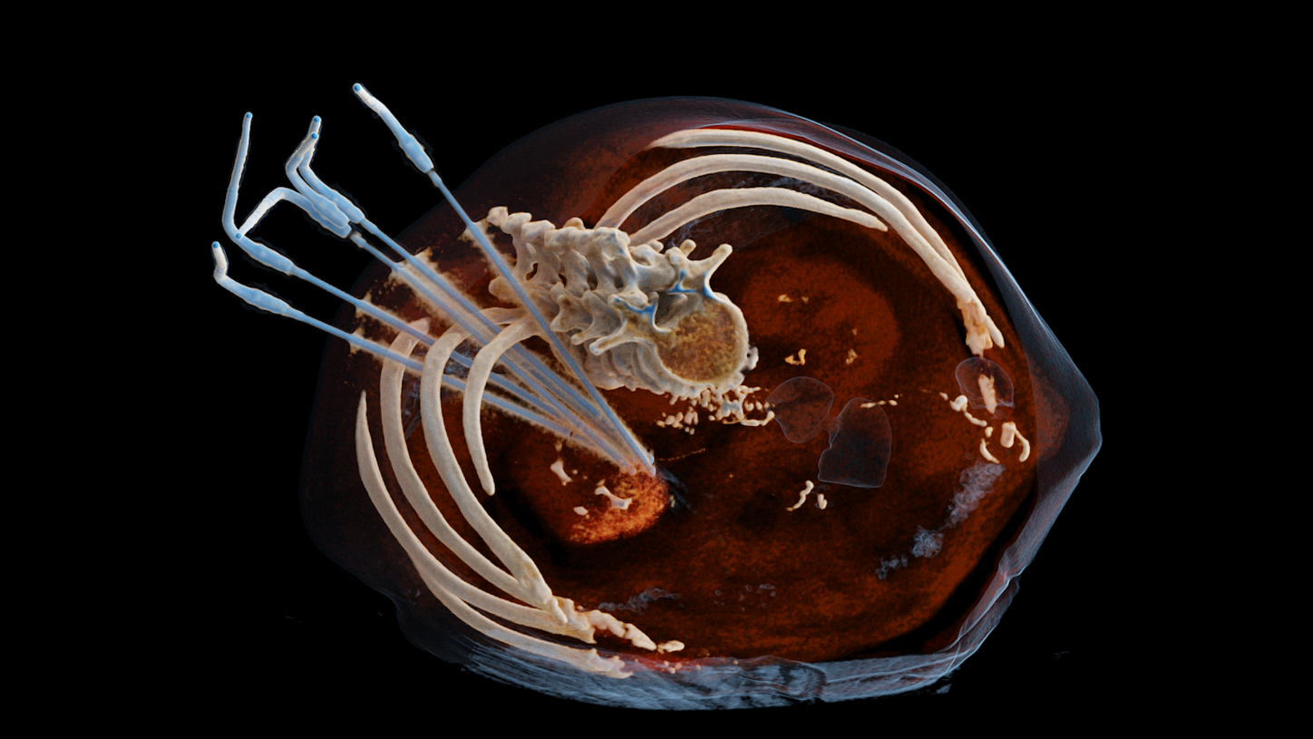
Cryoablation of an adrenal metastasis
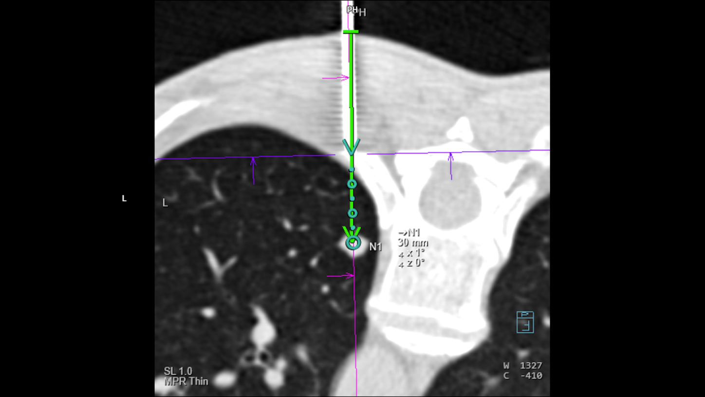
Percutaneous lung biopsy of a small metastatic nodule under CT guidance
Did this information help you?
Thank you.
Professor Frank Wacker, MD and Professor Ralf Bauer, MD receive financial support from Siemens Healthineers for collaborations.