Clinical Image Gallery
03

Courtesy: Prof. Wan-You Guo, MD, Taipei Veterans General Hospital, Taiwan
Post-treatment: A syngo Neuro PBV Neuro map depicted the recovery of CBV values (circle) in part of the hypoperfused parenchyma after revascularization.
syngo DynaPBV Neuro depicted a large area of hypoperfusion in the corresponding left MCA.
Related Study Toward the Era of a One-Stop Imaging Service Using an Angiography Suite for Neurovascular Disorders

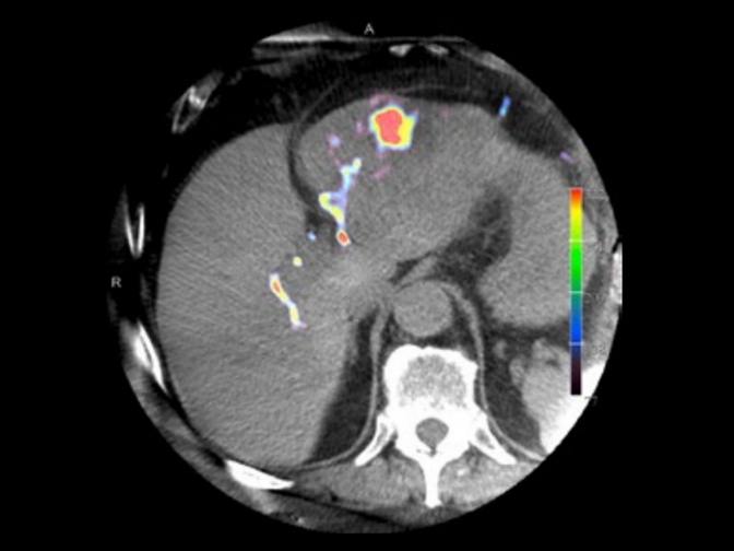
Courtesy: Prof. Gerd Groezinger, Interventional Radiology, University Hospital Tuebingen, Germany
syngo DynaPBV Body blood volume map only

Courtesy: Prof. Vogl, Dept. of Diagnostic and Interventional Radiology, University of Frankfurt, Germany
Blood volume map overlaid on native syngo DynaCT images provided by syngo DynaPBV Body.

Courtesy: Prof. Vogl, Dept. of Diagnostic and Interventional Radiology, University of Frankfurt, Germany
Blood volume map overlaid on native syngo DynaCT images provided by syngo DynaPBV Body.

Courtesy: Prof. Vogl, Dept. of Diagnostic and Interventional Radiology, University of Frankfurt, Germany
Blood volume map overlaid on native syngo DynaCT images.

Courtesy: Prof. Vogl, Dept. of Diagnostic and Interventional Radiology, University of Frankfurt, Germany
Blood volume map overlaid on native syngo DynaCT images provided by syngo DynaPBV Body.

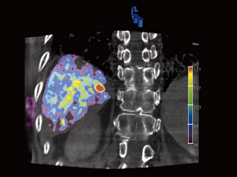
Prof. Vogl, Dept. of Diagnostic and Interventional Radiology, University of Frankfurt, Germany
High-resolution contrast-enhanced syngo DynaCT images provided by syngo DynaPBV Body.

Courtesy: Prof. Martin Skalej MD, University Hospital Magdeburg, Germany
syngo DynaPBV Neuro helps to monitor the functional state during endovascular stroke treatment.

Courtesy: Prof. Wan-You Guo, MD, Taipei Veterans General Hospital, Taiwan
Pre-treatment: A parenchymal cerebral blood volume (syngo Neuro PBV Neuro) map depicted a large area of hypoperfusion in the corresponding left MCA territory.
Related Study Toward the Era of a One-Stop Imaging Service Using an Angiography Suite for Neurovascular Disorders
Courtesy: Prof. Wan-You Guo, MD, Taipei Veterans General Hospital, Taiwan
Post-treatment: A syngo Neuro PBV Neuro map depicted the recovery of CBV values (circle) in part of the hypoperfused parenchyma after revascularization.
syngo DynaPBV Neuro depicted a large area of hypoperfusion in the corresponding left MCA.
Related Study Toward the Era of a One-Stop Imaging Service Using an Angiography Suite for Neurovascular Disorders
Courtesy: Prof. Gerd Groezinger, Interventional Radiology, University Hospital Tuebingen, Germany
syngo DynaPBV Body blood volume map only
Courtesy: Prof. Vogl, Dept. of Diagnostic and Interventional Radiology, University of Frankfurt, Germany
Blood volume map overlaid on native syngo DynaCT images provided by syngo DynaPBV Body.
Courtesy: Prof. Vogl, Dept. of Diagnostic and Interventional Radiology, University of Frankfurt, Germany
Blood volume map overlaid on native syngo DynaCT images provided by syngo DynaPBV Body.
Courtesy: Prof. Vogl, Dept. of Diagnostic and Interventional Radiology, University of Frankfurt, Germany
Blood volume map overlaid on native syngo DynaCT images.
Courtesy: Prof. Vogl, Dept. of Diagnostic and Interventional Radiology, University of Frankfurt, Germany
Blood volume map overlaid on native syngo DynaCT images provided by syngo DynaPBV Body.
Prof. Vogl, Dept. of Diagnostic and Interventional Radiology, University of Frankfurt, Germany
High-resolution contrast-enhanced syngo DynaCT images provided by syngo DynaPBV Body.
Courtesy: Prof. Martin Skalej MD, University Hospital Magdeburg, Germany
syngo DynaPBV Neuro helps to monitor the functional state during endovascular stroke treatment.
Courtesy: Prof. Wan-You Guo, MD, Taipei Veterans General Hospital, Taiwan
Pre-treatment: A parenchymal cerebral blood volume (syngo Neuro PBV Neuro) map depicted a large area of hypoperfusion in the corresponding left MCA territory.
Related Study Toward the Era of a One-Stop Imaging Service Using an Angiography Suite for Neurovascular Disorders
Courtesy: Prof. Wan-You Guo, MD, Taipei Veterans General Hospital, Taiwan
Post-treatment: A syngo Neuro PBV Neuro map depicted the recovery of CBV values (circle) in part of the hypoperfused parenchyma after revascularization.
syngo DynaPBV Neuro depicted a large area of hypoperfusion in the corresponding left MCA.
Related Study Toward the Era of a One-Stop Imaging Service Using an Angiography Suite for Neurovascular Disorders