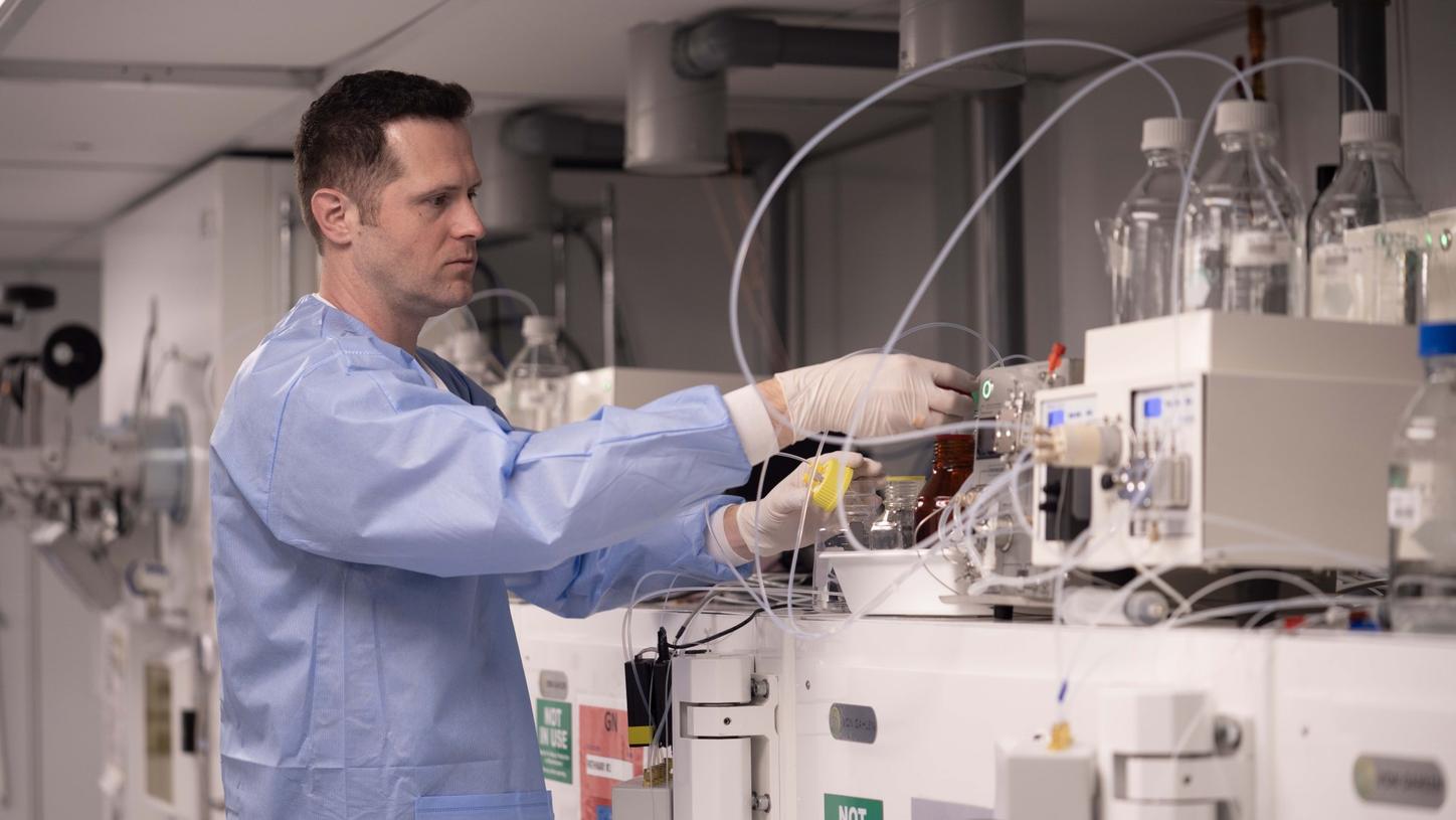
Due to their high sensitivity, modern PET/CT scanners provide unprecedented insights for patients with chronic conditions. This makes them an essential tool in combatting non-communicable diseases.
Photography by Simon Lau and Annika Daria Neubauer.
According to the World Health Organization’s Global Burden of Disease study, half of the world’s top 10 leading causes of early death and disability are non-communicable diseases (NCD).1 Non-communicable diseases, such as cancer, cardiovascular disease, and neuropsychiatric diseases, may largely define healthcare in the decades to come. Fittingly, a session on diagnostic precision spanning oncology, neurology, and cardiology clinical pathways launched the Molecular Imaging World Summit 2026 in Copenhagen, Denmark. Global leaders in the molecular imaging community presented research, insight, and case studies focused on the expanding role of PET/CT and its impact on diagnostic precision.
High-sensitivity PET/CT for improving cancer care
High-sensitivity PET/CT imaging can play an important role in improving cancer care. During his presentation, a nuclear medicine physician from Munich, Germany, was asked why PSMA imaging and PSMA-based theranostics in prostate cancer are so successful. His answer: “PSMA imaging enables therapies, not only radionuclide therapies, but also surgical therapies. Too often, oncologic imaging is only about staging. And staging usually does not expand but limit the therapeutic options.” The presentation shared how the PSMA success story could potentially be copied to other oncologic entities.
ImmunoPET: Getting a grip on tumor biology and treatment response
Dr. Erik Aarntzen, nuclear medicine physician from the University Medical Center Groningen, Netherlands, envisions a future in which PET/CT will be used much more widely in cancer care―not only for diagnostics and staging, but also for personalized treatment decisions. This, he argued, can be reached with novel antibodybased or even nanobody-based tracers. The goal would be to not only target tumor antigens, but also immune cells to get a more complete picture of what is going on in the tissue. Imaging immune cells is tricky, though. “It adds another layer of complexity, especially since target expression is usually low and many of the potential targets are shared across different immune cell populations.” What is needed to address this complexity, said Aarntzen, is high sensitivity of the kind that is provided by large axial field-of-view (LAFOV) scanners. “LAFOV scanners unlock unprecedented opportunities for immunoPET.”
Progress in the field of immunoPET is imperative. Immunotherapies have taken centerstage in oncology in recent years―but much remains to be understood in terms of how they work and who will or will not respond. LAFOV PET/CT scanners also unlock opportunities in “traditional” molecular cancer cell imaging explained Aarntzen. “They help us detect lesions with only moderate expression levels. We can reduce the radiation dose massively, and we can do multiple-time imaging with scans up to 40 days after injection.” The latter can be helpful, for example, for image-driven early phase clinical trials.

Aarntzen believes LAFOV PET/CT scanners unlock unprecedented opportunities for immunoPET.
Mapping perfusion and myocardial flow reserve
Beyond cancer, PET/CT scanners that provide high-sensitivity total-body imaging open up new care pathways in cardiovascular care. Dr. Frederico Caobelli is a leading physician in the Department of Nuclear Medicine at Inselspital, Bern University Hospital, Bern, Switzerland. He has specialized in nuclear cardiovascular imaging, including myocardial blood flow quantification in perfusion imaging. Caobelli is currently working on several projects that look at myocardial perfusion imaging. This can be done with different tracers, depending on local needs and availabilities.
“Using modern high-sensitivity PET/CT scanners, we can rule out hemodynamically obstructive coronary artery disease with a high degree of certainty,” said Caobelli. In this regard, estimating myocardial flow reserve and coronary flow capacity gives additional information and improves the prognostic value. “PET/CT gives us a probability score and allows us to reclassify patient risk. This is the kind of information clinicians need to know.” Most importantly, LAFOV PET enables the quantification of blood flow in various organs of interest, reflecting the systemic nature of atherosclerosis.

Caobelli takes the stage to discuss flow quantification.
Effective non-invasive method for microvascular dysfunction
PET/CT is showing yet another remarkable capability with its detection of cardiac microvascular dysfunction.2 This is something that can be caused by various underlying conditions, among them diabetes. It can mimic epicardial coronary artery disease symptom-wise, but there are no stenoses to be dilated― something cardiologists need to know ideally early-on.
Caobelli showed a meta-analysis of clinical studies that compared different diagnostic modalities, with a clear result.2 It is his opinion that PET is the best non-invasive method to detect microvascular dysfunction. Thanks to highly sensitive LAFOV PET/CT scanners, these types of measurements can be done in nearly every type of patient. “We scanned a 165 kg patient with a total radiation exposure of 0.7 mSv.a,b That’s quite impressive I would say! Now we are performing all our myocardial perfusion imaging on LAFOV PET/CT scanners”3
How do heart problems affect the rest of the body?
Another exciting perfusion imaging opportunity that LAFOV PET/CT scanners provide is total-body perfusion imaging. This was the topic of Prof. Juhani Knuuti, director of the Turku PET Centre, Turku, Finland. Knuuti is using 15O-H2O for his whole-body perfusion scans on Biograph Quadra Vision PET/CT, and this, he said required an activity of only 350 MBq (9.46 Mci) per examination.a,b
A very interesting topic, according to Knuuti, and one that could not have been addressed without a LAFOV PET/CT scanner, was how the rest of the body, including the brain, reacts to cardiovascular problems. To research this, Knuuti and his team have created a total-body standard space for 15O-H2O perfusion that takes into account parameters like sex, bodyweight, age, and hemoglobin levels. “Based on that, we now can observe what happens in other parts of the body when we stimulate, for example, the heart.” In a very recent publication, the Finnish experts did exactly that for adenosine stimulation of the heart. They could show that women have higher brain perfusion during adenosine-induced stress than men.4 “We also saw a trend towards lower brain perfusion in ischemic heart disease, but that needs to be confirmed.”

Knuuti discusses total-body perfusion imaging.
A new era in dementia care
More than 55 million people worldwide suffer from dementia, around two-thirds of which are Alzheimer’s disease (AD). By 2050, it is expected that around 150 million patients will be living with AD globally.5 With the advent of anti-amyloid therapies, AD may transition into a manageable condition. But there is no anti-amyloid therapy without imaging, said Prof. Alexander Drzezga, head of the Department of Nuclear Medicine at Cologne University. “We need to select patients carefully. Treatments are expensive and can have severe side effects.” PET/CT, according to Drzezga, is well positioned to be a cornerstone of AD care. “We have amyloid and tau imaging to depict the major abnormalities in AD, and we have FDG PET as the Swiss Army Knife for measuring neuronal injury.”
Unlike fluid biomarkers, amyloid and tau imaging were strictly validated against brain pathology, said Drzezga. This is a huge advantage in terms of diagnostic precision. While laboratory biomarkers are good at ruling in and ruling out clear-cut cases, there is a large intermediate group of patients in which diagnosing AD with high diagnostic certainty can be difficult. This is where PET/CT can deliver much more precise answers. “Fluid and imaging biomarkers are complementary, not interchangeable. But Amyloid-PET is the clear gold-standard for patient inclusion, staging, and therapy response monitoring.”
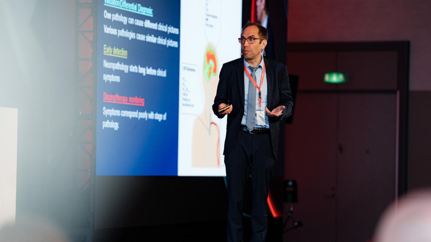
PET/CT, according to Drzezga, is well-positioned to be a cornerstone of AD care.
PET/MR as a powerful molecular neuroimaging platform
Prof. Maria Rosano Ponisio of the Mallinckrodt Institute of Radiology at Washington University in St. Louis concurs. The value of PET/CT in the context of anti-amyloid therapies, she said, was clearly illustrated by usage statistics in the USA. “Biomarker usage changed after the approval of the new therapies: PET/CT is going up, fluid biomarkers are going down.”
For Ponisio, molecular brain imaging is about more than PET/CT, though. She is a huge supporter of PET/MRI diagnostics, which, she said, could be extremely valuable not only in neurodegenerative diseases, but also in neuro-oncology and in epileptic seizure management. Her department has recently acquired a BIOGRAPH One PET/MR from Siemens Healthineers that features full 3T MRI performance, a 35-cm axial field of view and ultra-fast time-of-flight.6
In epileptic seizure management, the new machine was a huge step forward, she said, since it allowed to identify critical epileptogenic zones for neurosurgical therapy more reliably. Ponisio is convinced that there is more to come. “We are really seeing a paradigm shift here. PET/MR is the future of neuromolecular imaging, it will become the platform of choice.”

Ponisio discusses epileptic seizure management and other neurological conditions.
Shaping the future of personalized care
Advancements in PET/CT technology are revolutionizing the fight against non-communicable diseases by enabling earlier and more precise diagnoses. As these innovations continue to shape personalized care, they hold the potential to significantly improve patient outcomes worldwide.
About the author
Philipp Grätzel is a medical doctor by training, who has been working as a healthcare and technology journalist in Berlin for many years.