
The Cardiovascular Center at the Sugimura Hospital, Japan, was looking to offer advanced imaging and diagnosis to aid physicians in treating acute and chronic conditions. The solution? A SOMATOM Definition AS+ upgrade with a Stellar detector.
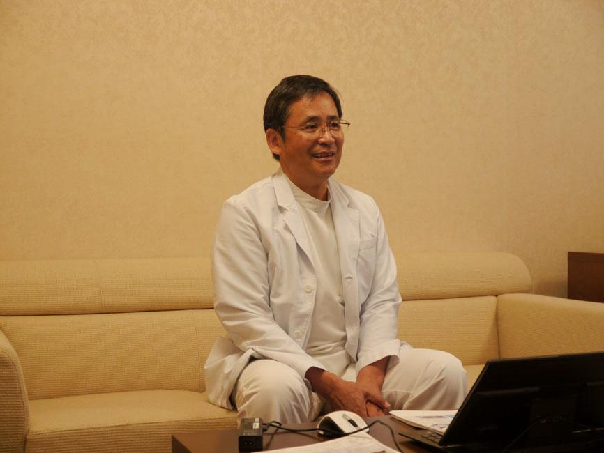
The Cardiovascular Center at Sugimura Hospital, in Kumamoto city, Japan, is a specialist unit that provides rehabilitation, and the treatment of acute and chronic conditions. In its quest to deliver advanced and reliable therapy and diagnosis, an investment decision was made in May 2014 to upgrade the SOMATOM Definition AS+ with a Stellar detector for the newly established Department of Cardiovascular Therapy. This was the first such upgraded system available in the whole of Japan. In this interview, head of the hospital and cardiovascular center, Kenji Horiuchi, MD, together with radiology department director, Yasuhiro Yano, and Koichi Hamakawa, a technologist at the Department of Cardiovascular Therapy explain the background to their decision and report on their experiences so far.
What made you decide to upgrade the detector on your system?
Horiuchi: Our guiding mission at the Cardiovascular Center of Sugimura Hospital is to handle the vascular pathology of the entire body. This involves maintaining consistent quality in treating cardiovascular diseases, including acute and chronic conditions, and rehabilitation, and offering tailor-made and timely medical care.
"Ever since I learned that the next generation X-ray detector, the Stellar detector, could be retrofitted on our scanner I became very interested in this solution."
"In the end, we decided in favor of the version upgrade due to its clinical and financial benefits. Now we can use our existing equipment for at least another 5 years."


One-third of patients with acute coronary syndromes, especially myocardial infarction, do not experience any warning symptoms, and more than half of patients pass away on the way to hospital while experiencing their first heart attack. By contrast, two-thirds do have warning symptoms, and if they come to the hospital on time, we can perform proper treatment.
We have been thinking about how to provide tailored medical care in a short period of time for those who are very busy and are not able to come to hospital. This demands fast, accurate diagnosis performed in a non-invasive manner. A 128-slice CT was the most advanced scanner at the time we purchased our SOMATOM Definition AS+, but there are CTs nowadays with more than 128 slices. I was fairly satisfied with its temporal resolution and image quality; however, I felt there was room for improvement in terms of spatial resolution for stent lumen assessment. In recent years, there has also been increasing awareness of patient radiation exposure as an issue.
Our radiation technologists have been actively trying to reduce such exposure, currently down from approximately 60 mGy to 15 mGy of the dose we had with the initial cardiac CT scans. We need to further reduce exposure without affecting image quality, because radiation dose accumulates after a CT examination, catheter intervention, or CT confirmation scan.

Ever since I learned that the next generation X-ray detector, the Stellar detector, could be retrofitted on our scanner I became very interested in this solution. After the upgrade, I believe that we’ve achieved our goals and we have an equipment configuration capable of competing with other hospitals that have newer CT scanners. With this version upgrade,* the existing equipment was “reborn“ and while it would normally have been hugely expensive to replace the scanner itself, the upgrade solution also delivered considerable financial advantages for us.
Yano: To be honest, we had reached our performance limits with the existing system. So I felt we needed to reinforce our configuration both in terms of hardware and software by obtaining the latest functionalities available in the version upgrade.
We were also driven by the idea of being the first hospital in Japan to have the latest functionalities on our system. Unlike replacing the entire system, another significant financial advantage for hospitals operating just one CT is that the upgrade process can be completed in a short period of time. In the end, we decided in favor of the version upgrade due to its clinical and financial benefits. Now we can use our existing equipment for at least another 5 years.
Please tell us a little about the improvements you experienced after the upgrade and what you hope for in the future
Horiuchi: We saw improvements in stent image quality; especially in challenging cases and smaller stents. Previously, we pondered long over the diagnosis in these cases and so now we’re also saving time. In the future, I expect to lower patient dose even further.
Yano: We experienced benefits in domains other than cardiovascular, as well – due to the improved spatial resolution. For example, we received very positive evaluations from other hospitals who asked us to perform scans on the temporal bone because our image quality is so much more improved. Our mission is to contribute to community medicine, and now we are able to respond better to requests from general practitioners.
Hamakawa: I’m not merely interested in low dose. We want to acquire images that can support better diagnostic ability – even at a low dose. One particular focus we have, among others, is on stent assessment after PCI. Even though stent blooming was already low, the image quality is sharper thanks to the iterative reconstruction algorithm, SAFIRE, which is included in the upgrade with Edge technology. This gives us the ability to reconstruct images with 0.5 mm resolution thus improving spatial resolution in the body axis, in addition to the plane view. The continuity of temporal bone coronal images is now better, and we can provide much smoother images with lower noise. Presently, we scan with the same amount of dose, and we have heard such wonderful feedback on the dramatically improved image quality. We are therefore hoping to receive more order requests in the future.
"I’m not merely interested in low dose. We want to acquire images that can support better diagnostic ability – even at a low dose."


In this version, we also upgraded to FAST IRS (a high-speed image reconstruction computer). I am very satisfied with the outcome because the processing speed is considerably faster, which means I can work on iterative reconstruction without becoming frustrated. In the future, I would like to provide more valuable image information by making use of low dose applications, such as CARE kV from the Optimized CARE CT program.
What do you think about the version upgrade as an investment?
Horiuchi: We are a specialized hospital and we are constantly driven to become a leading hospital in our field. To stay at the cutting edge, it is important for us to remain up-to-date. I am sure that this approach will become the preferred investment method for equipment upgrades due to the excellent cost performance.
About the Author
Ryo Mitsuhashi, Siemens Healthcare, Japan