
Two European hospitals are combining the human approach and radiology technology in symbiosis. Their personal touch together with high-end 3D measurement cameras1 helps provide better image quality and an optimized radiation dose.
Photos: Philipp Frowein
The Swiss University Hospital Zurich (USZ) and the Dutch academic hospital Erasmus MC in Rotterdam are leading European hospitals that provide fundamental medical care and cutting-edge medicine for national and international patients. Erasmus MC ranks as the top European institution in clinical medicine according to the Times Higher Education rankings. It is the largest and one of the most authoritative scientific university medical centers in Europe. USZ treats around 542,000 patients in 43 specialist departments and institutes each year. It is one of the largest hospitals in Switzerland, and the percentage of patients with complex diseases is particularly high there.
Ronald Booij is Coordinator of Research and Innovation at the CT Unit at Erasmus MC, which is situated in a brand-new hospital building in Rotterdam. The transparent architecture, made up of various layers, appears inviting. “With its fitting design and its ‘healing environment’, it claims to reduce the time a patient has to spend in hospital, while at the same time providing an enhanced quality of care,” says Booij.
Revolution in radiology
“The clear structure of the building is beneficial to stress reduction and patient well-being,” he says. “During the last few years of my 20-year career here, I have witnessed a rapid evolution in patient care and technology. As Innovation Coordinator and PhD candidate in Radiology and Research, I have always been driven by innovative solutions for healthcare. “I have always wanted to know how these solutions really perform in a clinical context,” comments Booij. “I am convinced that the advent of computed tomography has revolutionized radiology.”
3D camera with great potential
“We have six scanners in our radiology department. Two are already equipped with a 3D measurement camera,” says Ronald Booij. According to Booij, the increasing awareness of the risks associated with radiation exposure has always been one of the department’s main concerns. “We can only enhance the patient experience and care with a clear operating protocol and accurate patient positioning. And the 3D camera has great potential when it comes to positioning.”
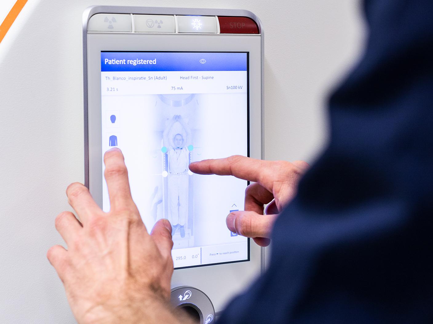
“With 3D camera technology, the patient can be accurately positioned to within a few millimeters.”

“In the past, this patient positioning technology simply did not exist,” states Natalia Saltybaeva. As a medical physicist and scientific researcher at USZ, she says radiation dose optimization algorithms used to be a kind of black box. “It kept its secrets,” she says. ”But nowadays we can benefit from technologies for very precise patient positioning. This allows us to avoid mistakes. For example, when our technicians position the patient manually, they are typically about three centimeters off-center. With 3D camera technology, the patient can be accurately positioned to within a few millimeters using infrared images and 3D data in combination with deep learning algorithms.”
Significant improvement in patient centering
Natalia Saltybaeva is the co-author of a study performed in Zurich that evaluated the accuracy of a 3D camera algorithm for automatic and individualized patient positioning based on body surface detection.[1] Together with Professor Hatem Alkadhi, MD, Head of the Institute of Diagnostic and Interventional Radiology at USZ, she compared the results of the 3D camera workflow with manual positioning carried out by radiology technologists in chest and abdomen CT examinations.
The study included the data of 120 patients undergoing consecutive CT examinations with and without the help of a 3D camera. The team found a significant improvement in patient centering (offset 5 ± 3 mm) when using the automatic positioning algorithm with the 3D camera compared with manual positioning (offset 19 ± 10 mm) performed by technologists (P < 0.005). Automatic patient positioning based on the 3D camera reduced the average offset in vertical table position from 19 mm to 7 mm for chest and from 18 mm to 4 mm for abdominal CT scans. The absolute maximal offset was 39 mm and 43 mm for chest and abdominal CT scans, respectively, when patients were positioned manually, whereas with automatic positioning using the 3D camera the offset never exceeded 15 mm.
“The 3D camera is my personal backup. It feels like we’re working together.”

Determining the optimal position
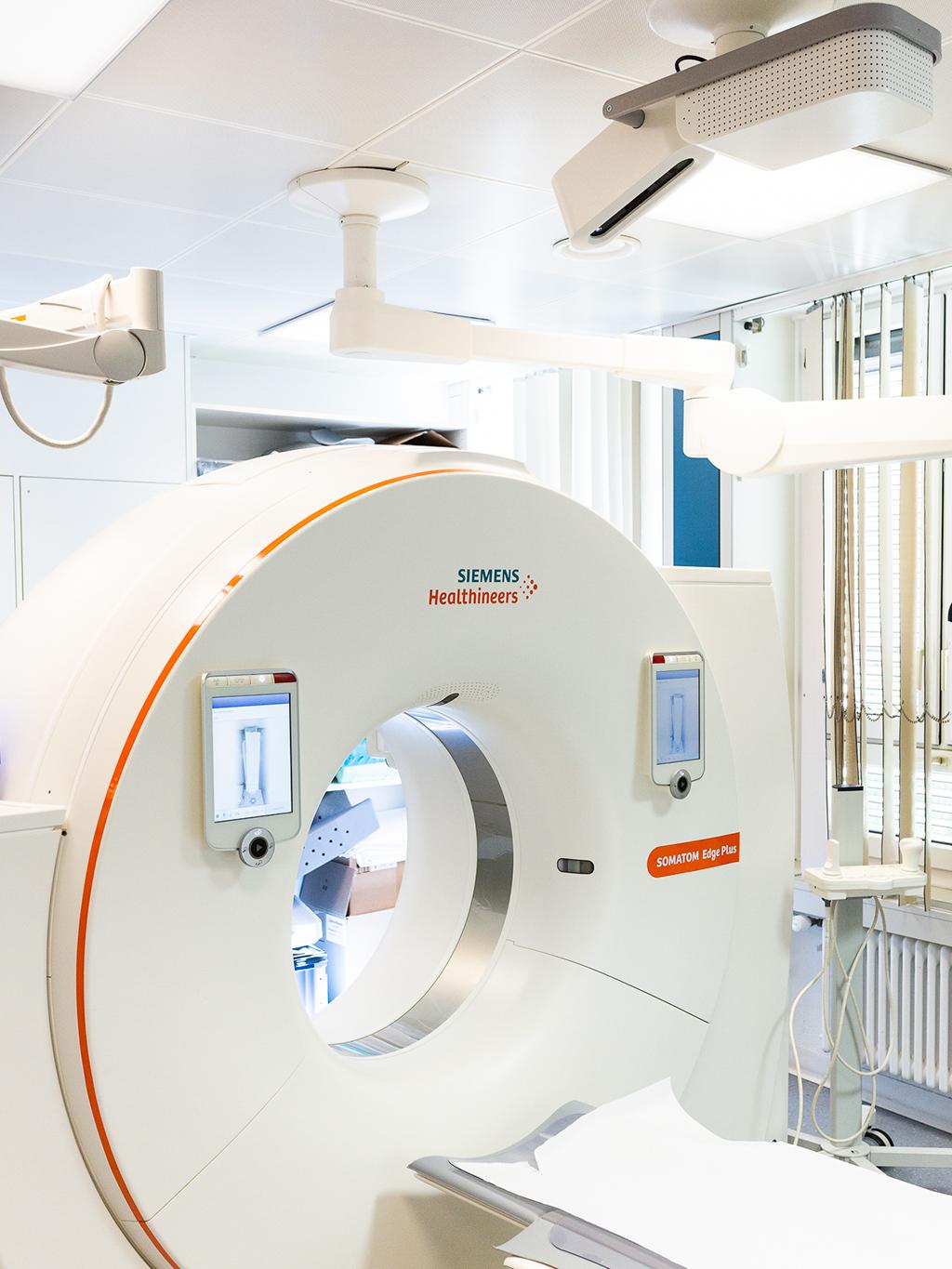
In the radiology department in Rotterdam, the 3D camera has been used in connection with the SOMATOM Drive and the SOMATOM Edge Plus for more than a year. “At first, I was not convinced about the added value of the 3D camera,” says Ronald Booij. “In fact, I was rather skeptical. We are well-trained, professional radiographers and we prefer to position the patient ourselves.” But he does admit that it is sometimes difficult to interpret the optimal table position for the body part to be examined. “Every patient is unique, from their body shape to the clothing they are wearing. And the isocentering itself, which is done on the spot by the radiographer with the aid of a laser beam, is not always easy to accomplish.” He points out that the curve in the middle of the scanner gantry is also one of the reasons why it can be more difficult for a radiographer to accurately estimate patient positioning and define the right scan protocol: “We tend to position the patient lower than needed, which can lead to serious deviations from the ideal isocentering.”
“Believe it or not,” says Professor Alkadhi, “ positioning is indeed an issue. We know from a study by J. Li and his team, which was published eleven years ago already, that approximately 95 percent of the patients undergoing a CT scan were not positioned accurately in the gantry isocenter.[2] So it is obviously a big issue. It is not a rare occurrence.”
Alkhadi also states that from a dose optimization study by Schmidt and his team they learned that the dose from localization radiology may contribute significantly to the total effective dose of low-dose CT examinations such as lung cancer screenings.[3] Optimal settings can reduce the localizer radiography dose substantially but adaptations have to consider scanner characteristics, detector technology, and patient size.
Image:
In Zurich, the SOMATOM Edge Plus together with its 3D camera allows for improved patient positioning in comparison to manual positioning as study results show.
Image quality and radiation dose

Booij, Alkadhi, and Saltybaeva are all convinced that there is still room for improvement and that the 3D camera can be of great help for the accuracy of patient positioning. Says Ronald Booij: “You will never hear me say that we no longer need radiographers. On the contrary, I believe we need a symbiosis. The human factor has to go hand-in-hand with the technology to offer more accurate patient positioning, resulting in an optimized result. At the end of the day, a better patient outcome is always our goal. Optimization of image quality and radiation dose leads to a better diagnosis and consequently to better healthcare. The 3D camera is my personal backup. It feels like we are working together. One cannot work without the other.” In the upcoming years, artificial intelligence (AI) is likely to fundamentally transform diagnostic imaging. But AI will not by any means replace radiologists and radiographers – rather it will provide them with tools to meet the rising demand for diagnostic imaging and actively shape the transformation of radiology into a data-driven research discipline.
About the Author
Erika Claessens works as an independent journalist from Antwerp, Belgium.