
Ευχαριστούμε για τη συμμετοχή σας!
Θα θέλαμε να ευχαριστήσουμε θερμά όλους τους επαγγελματίες υγείας, συνεργάτες και συμμετέχοντες που παρευρέθηκαν και στήριξαν με την παρουσία τους.

Συνέδριο
|Ωδείο Αθηνών
|11 – 14 Δεκ 2025
Θα θέλαμε να ευχαριστήσουμε θερμά όλους τους επαγγελματίες υγείας, συνεργάτες και συμμετέχοντες που παρευρέθηκαν και στήριξαν με την παρουσία τους.

11–14 Δεκεμβρίου 2025 · Ωδείο Αθηνών
Σας περιμένουμε στο περίπτερό μας για να ενημερωθείτε για τις τεχνολογίες αιχμής στον τομέα της Ακτινολογίας και να παρακολουθήσετε παρουσιάσεις κλινικών περιστατικών από τους θεματικούς σταθμούς μας.
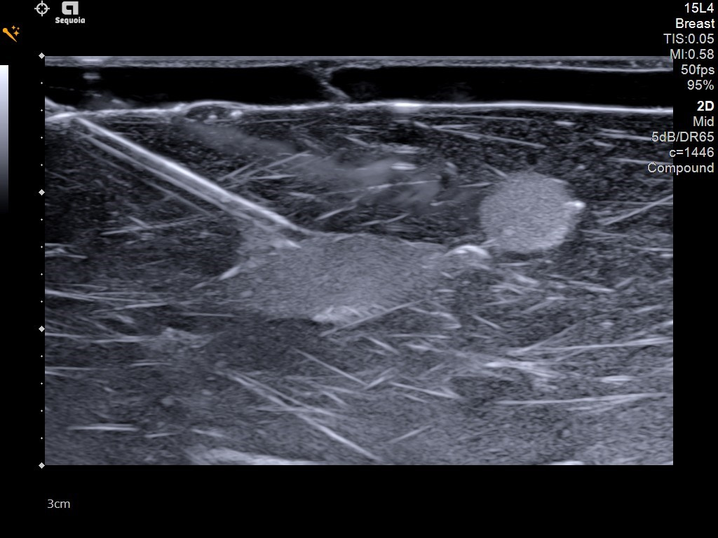
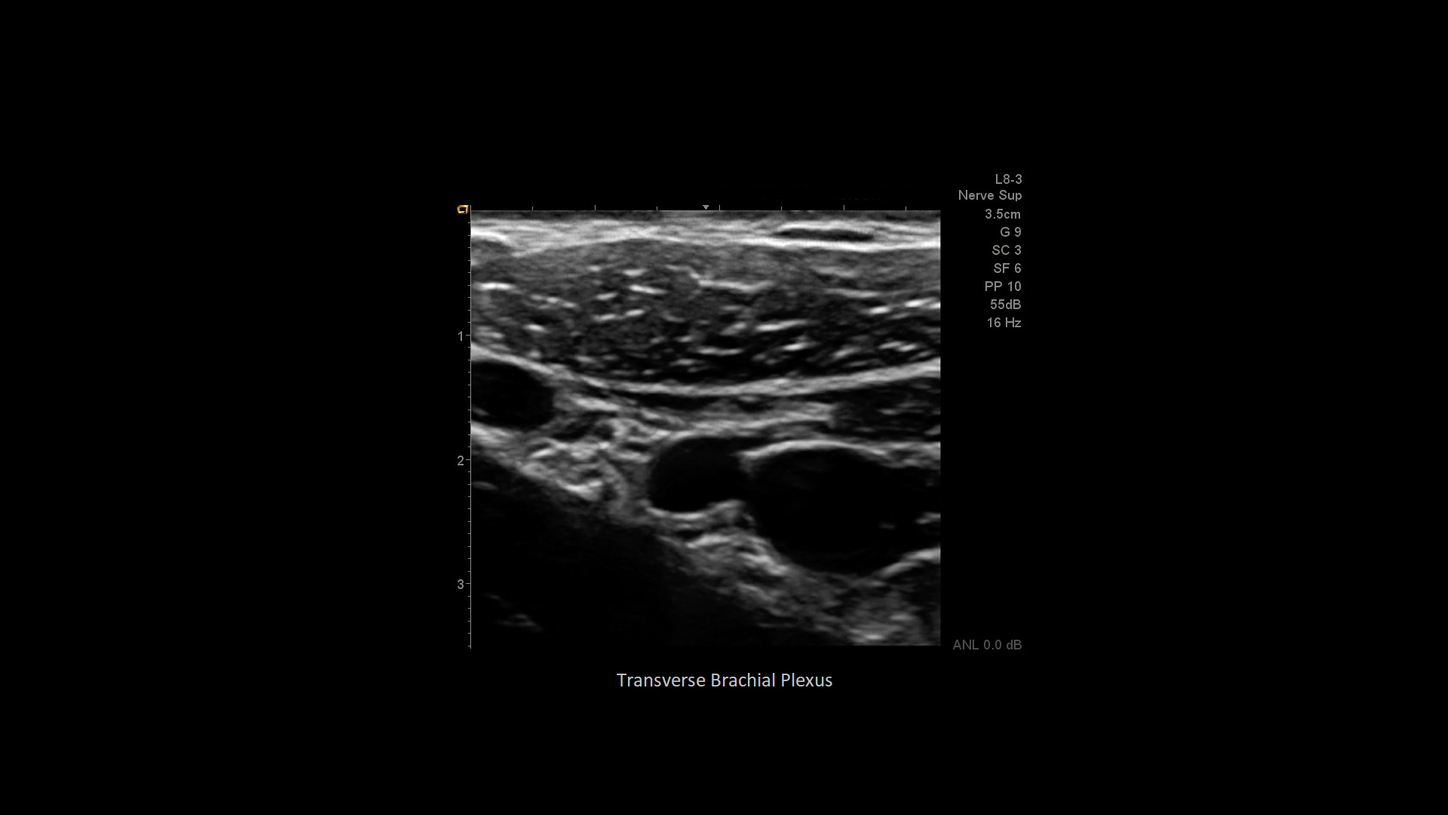
Ζήστε από κοντά την πρακτική εμπειρία των Yπερηχοτομογράφων μας στο περίπτερό μας, με τη καθοδήγηση των εξειδικευμένων συνεργατών μας.
Faster labeling. Accurate measurements. Empower faster, smarter exams, exclusively onboard ACUSON Sequoia. Change the way you ultrasound and make the most of the precious time and talent of your clinical teams.
Επισκεφθείτε το εκθεσιακό μας περίπτερο και ενημερωθείτε από τους εκπροσώπους μας για τις τελευταίες καινοτόμες λύσεις της Siemens Healthineers.

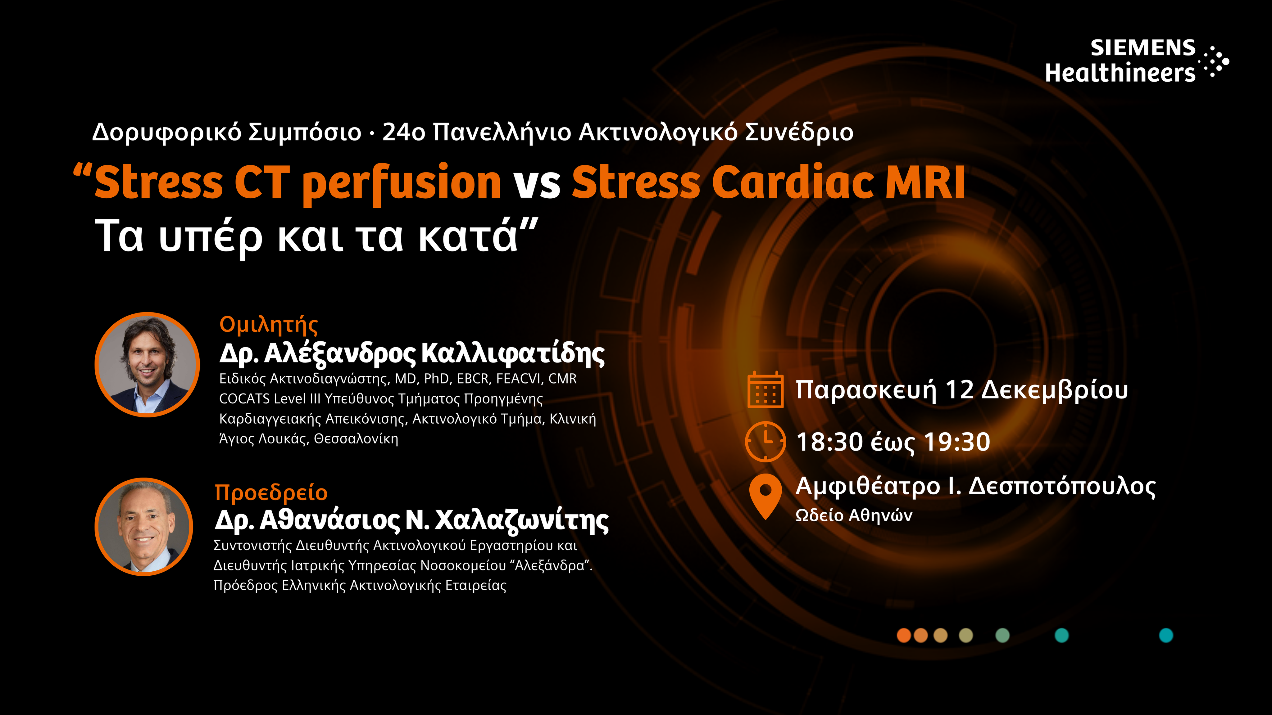
Με ιδιαίτερη χαρά σας προσκαλούμε να παρακολουθήσετε το δορυφορικό μας Συμπόσιο, το οποίο θα πραγματοποιηθεί την Παρασκευή, 12 Δεκεμβρίου 2025, από 18:30 έως 19:30, στο Αμφιθέατρο Ι. Δεσποτόπουλος.
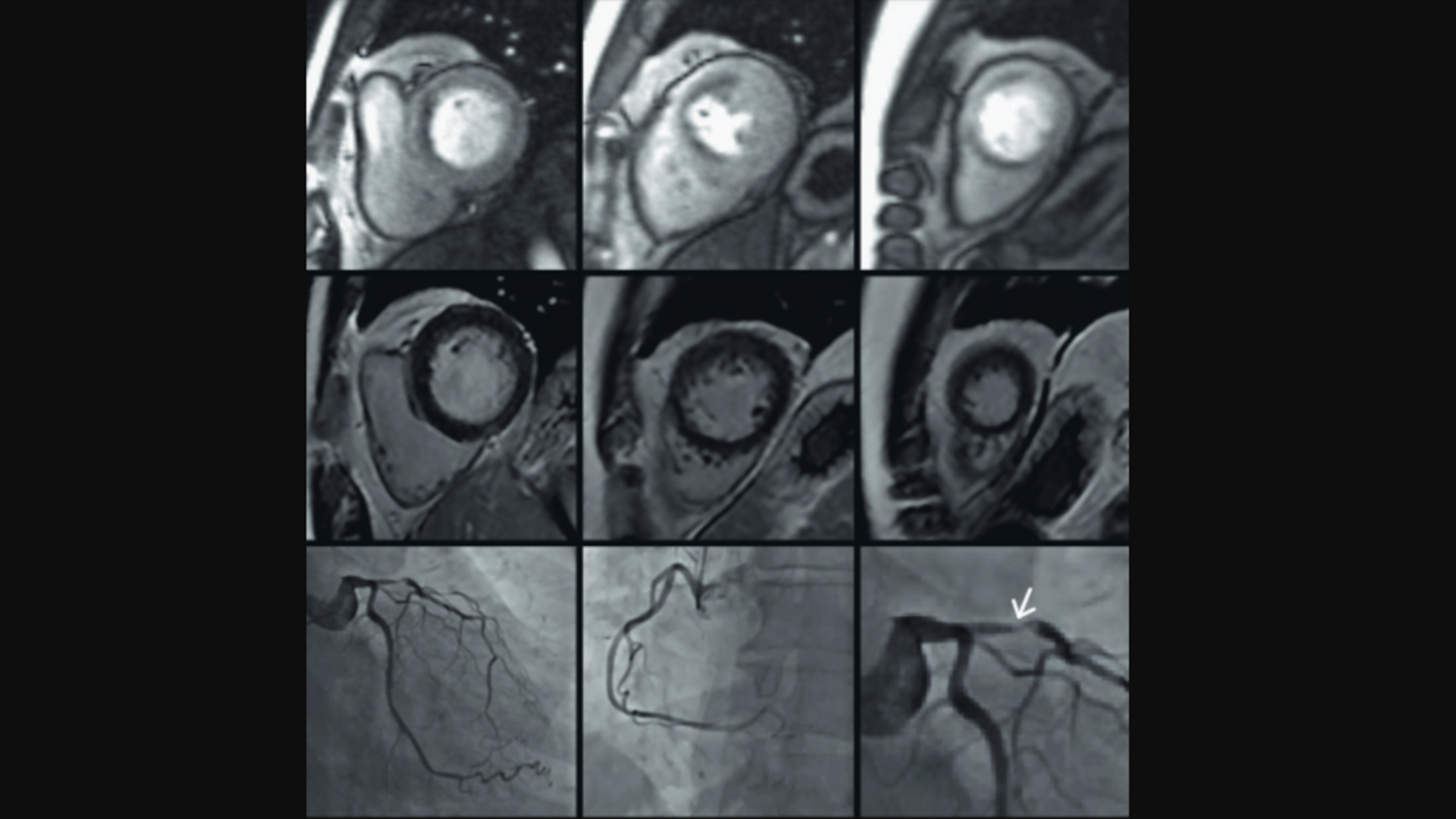
Θεματολογία: “Stress CT perfusion vs Stress Cardiac MRI: Τα υπέρ και τα κατά”

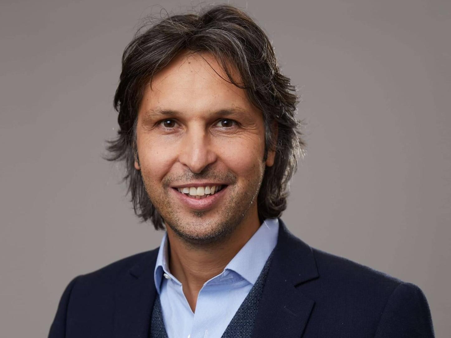
Ομιλητής θα είναι ο Δρ. Αλέξανδρος Καλλιφατίδης, Ειδικός Ακτινοδιαγνώστης, ενώ το προεδρείο θα τιμήσει με την παρουσία του ο Δρ Αθανάσιος Ν. Χαλαζωνίτης, Πρόεδρος Ελληνικής Ακτινολογικής Εταιρείας.
Η παρουσία σας θα αποτελέσει ιδιαίτερη τιμή για εμάς και θα δώσει την ευκαιρία για ενημέρωση και συζήτηση πάνω στις τελευταίες εξελίξεις της καρδιακής απεικόνισης.

Σάββατο 13 Δεκεμβρίου 2025
Ώρα 16:15- 18:15
Αίθουσα 4

Παρασκευή 12 Δεκεμβρίου 2025
Ώρα 12:00- 14:00
Αίθουσα 3

Παρασκευή 12 Δεκεμβρίου 2025
Ώρα 12:00- 14:00
Αίθουσα 4

Παρασκευή 12 Δεκεμβρίου 2025
Ώρα 14:00- 16:00
Αίθουσα 3

Παρασκευή 12 Δεκεμβρίου 2025
Ώρα 14:00- 16:00
Αίθουσα 4

Σάββατο 13 Δεκεμβρίου 2025
Ώρα 13:00- 15:00
Αίθουσα 4

Σάββατο 13 Δεκεμβρίου 2025
Ώρα 16:15- 18:15
Αίθουσα 4
*Για δηλώσεις συμμετοχής και λεπτομέρειες, επικοινωνήστε με τη γραμματεία του συνεδρίου.
Transverse thyroid image utilizing the 18L6 transducer. Notice the noise-free image, and the fine pixel and contrast resolution.
AI Abdomen labels and measures anatomical structures in milliseconds, helping to reduce repetitive movements and exam times.
AI Abdomen continuously identifies and labels anatomy during the abdominal scan.

An accessible and quantitative ultrasound index to aid in the management of patients with hepatic steatosis. Complimentary to liver elastography.

Rapidly reduce liver elastography acquisition time up to 75%1 by acquiring up to 15 valid pSWE measurements in less than 5 seconds.
1. Data on file. Many variables exist in the customer environment including sonographer techniques, which may affect individual customer experience.
In contrast imaging, the ACUSON Sequoia ultrasound system has twice the sensitivity than previous systems for improved diagnostic confidence1.
1. Compared to ACUSON Sequoia 512 ultrasound system
The DAX, Deep Abdominal Transducer, is designed to help enhance diagnostic confidence in adult and pediatric patients with high BMI >40 for general, gyn, and OB imaging, deep lesions, deep vessels or advanced disease, helping radiologists keep patients in their care stream. The DAX images at a depth of 55 cm for difficult abdominal exams.
Color Doppler image demonstrating clear delineation and absence of flow in the common bile duct utilizing the single crystal technology of the 9C2 transducer.

Sagittal 5C1 image of the liver, gallbladder and inferior vena cava demonstrates excellent detail and contrast resolution utilizing the harmonic high setting and an UltraArt setting of 3. UltraArt can be used on both live and frozen images with a choice of off, 1 to 5.

With 2D-SWE on the ACUSON Sequoia, you can confidently downgrade many BI-RADS 4A – 4C lesions to BI-RADS 3 lesions leading to a decrease in the number of benign biopsies.

Needle Enhancement provides clear, real-time needle visualization at steep angles to support precise targeting during complex procedures, helping improve diagnostic accuracy and patient satisfaction.
18L6 high resolution image of a breast hamartoma utilizing a high fundamental frequency and UltraArt setting of 3.

Breast image utilizing the 15L4 transducer demonstrating excellent resolution of tissue behind the nipple.
4D HeartAI uses AI to process complex cardiac data instantaneously for real-time quantification of key parameters across all four heart chambers. Average up to five beats with or without physio.
The 2D HeartAI feature provides single click analysis of size, function, and bull's-eye analysis with segmental strain curves. Analysis can be performed on contrast enhanced images and does not require an ECG.

Assess myocardial motion and mechanics with global longitudinal strain (GLS), global circumferential strain (GCS), and global radial strain (GRS) using AI-powered 2D Heart.

AI-powered measurements alleviate tedious, time consuming and variable Routine Echo calculations.
Semi-automated measurements for 2D, M-Mode, and PW measurements.
Reduces variability and standardizes exams across users to improve diagnostic accuracy on follow-up exams.
Auto Doppler can reduce the number of exam keystrokes by greater than 25%.1 Auto Spectral Doppler automatically optimizes for gain, baseline, scale, and wall filter keeping operator adjustments to a minimum.
1. Data on file. Keystroke is defined as any interaction with the ultrasound machine including touchscreen taps and swipes, mouse movement, flat key presses, soft key twists, and soft key presses.

Bi-Plane+ image of mitral valve prolapse with color Doppler demonstrating severe regurgitation utilizing the Z6T transesophageal transducer.
A sagittal image of the internal jugular vein utilizing the 10L4 transducer and the new Vascular Access preset.

The 14L5 transducer demonstrates the exceptionally clear and detailed spectral Doppler waveform obtained from the superficial femoral vein.

Reduce strain and pressure during scans with the lightweight HLX transducer, designed with an ergonomic grip and narrow shape allowing for constant light pressure and tight access.

Utilizing the 15L4 transducer, structures can be visualized in greater detail resolution as shown in this image of the supraspinatus tendon.

This image demonstrates a 9VE4 endovaginal 3D surface rendering image of an early OB.

The new 9C2 high frequency curved single crystal transducer provides superior contrast resolution in obstetric imaging.

The 10EV3 transducer demonstrates excellent image quality in the gynecology segment as seen in this fully-focused sagittal image of the uterus.

Neonatal imaging of the pylorus utilizing the 18L6 transducer demonstrating exceptional detail and contrast resolution throughout the field of view.
Experience single crystal technology with the microconvex 11M2 transducer. Notice the excellent spatial and temporal resolution in all major modes as demonstrated in this image of the celiac axis in a pediatric patient.
Spectacular image resolution can be seen in this sagittal 9C2 pediatric image of the liver and inferior vena cava, utilizing an UltraArt setting of 2.
Use of slow flow color Doppler offers high sensitivity and exceptional detail resolution of small vessels as shown in this Live Dual transverse pediatric kidney image in color only mode.
18L6 transverse image of the testis utilizing trapezoidal imaging which is available on all linear transducers providing a larger imaging area without sacrificing image resolution.
Use of slow flow color Doppler offers high sensitivity and exceptional detail resolution of small vessels as shown in this transverse image of the testis.
Experience a larger field of view utilizing trapezoid mode on the HLX as demonstrated in this transverse image of the thyroid.
Appreciate the detail of small vessels and increased sensitivity with Slow Flow microvascular color Doppler as demonstrated in this Live Dual – Color Only mode image of a thyroid mass.
Transverse thyroid image utilizing the 18L6 transducer. Notice the noise-free image, and the fine pixel and contrast resolution.


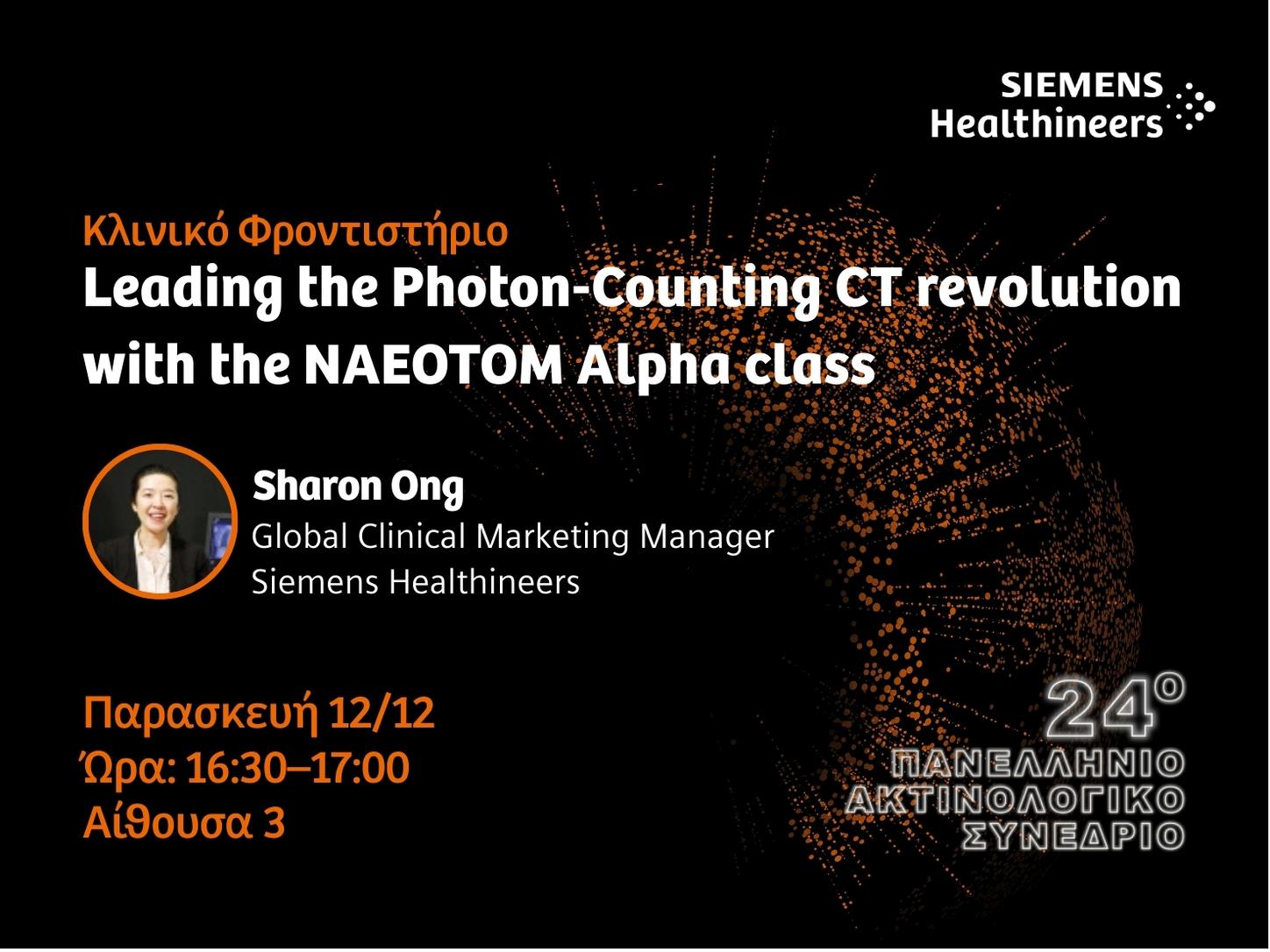
Με ιδιαίτερη χαρά σας προσκαλούμε να παρακολουθήσετε το Κλινικό Φροντιστήριο με τίτλο:
“Leading the Photon-Counting CT revolution with the NAEOTOM Alpha class”
Ομιλήτρια:
Sharon Ong (Global Clinical Marketing Manager, Siemens Healthineers)
Το Δορυφορικό Κλινικό Φροντιστήριο θα πραγματοποιηθεί την Παρασκευή, 12 Δεκεμβρίου 2025, από 16:30 έως 17:00, στην Αίθουσα 3, μετά το πέρας των MRI Hands-On Courses.