History
A patient with prostate cancer, elevated prostate-specific anti-gen (PSA) values and known bone metastases was enrolled in treatment initialization with an androgen synthesis inhibitor. Prior to the first treatment cycle, a bone scan using SPECT/CT xSPECT Bone™ and xSPECT Quant™ acquisition and reconstruction was performed on a Symbia Intevo™ scanner. To monitor treatment effects, the patient was referred for follow-up scans three months and six months after treatment onset.

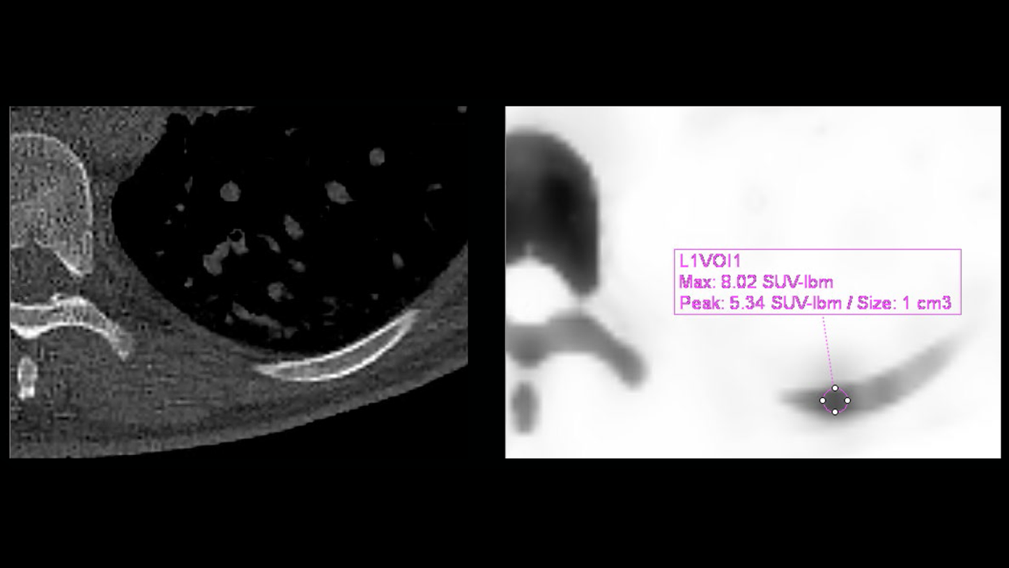
Findings
Initial lesions in the rib and the contralateral scapula were identified with highly active bone metabolism. Three months after treatment onset with the androgen synthesis inhibitor, the rib lesion remained strongly avid while the scapula lesion nearly disappeared. CT demonstrated the beginning of sclerosis in both lesions.
Six months after treatment onset, the tracer uptake was significantly reduced and CT demonstrated further progression of sclerosis in both lesions.
xSPECT Quant quantified the decrease of metabolic activity in the bone. In addition, the uptake measured at three time-points correlated with a decline of PSA, suggesting the therapeutic approach was effective. This shows xSPECT Quant may be a tool for early detection, as well as a better estimate of therapeutic effect.

![Figure 2: This series of xSPECT MIPs demonstrates the lesion’s visual intensity decline over time. Prior treatment [A] shows two lesions: on rib and one contralateral scapula lesion. Three months after treatment [B] displays a significant decline in tracer uptake for the scapula but only barely reduced uptake in the rib, considering visual analysis only [B]. Six months after treatment [C] shows that both lesions nearly disappeared.](https://marketing.webassets.siemens-healthineers.com/1800000006219451/d3b984e0ce95/v/be5226dc16d4/xspect-quant-treatment-monitoring_Figure2_1800000006219451.png)
Comments
Currently, anti-androgen-based treatment is a common therapy for metastasized prostate cancer. A recent trial, however, is raising concerns about the usage of anti-androgen treatment due to treatment-related increased morbidities and mortalities.1
Image-based treatment evaluation can be difficult. Although different efforts have been evaluated, none have reached clinical rou-tine.2 Particularly in patients with multiple metastases, pure visual assessment becomes difficult and unprecise or delayed, due to unspecific changes. This might lead to unreported treatment effects because of a lack of the necessary granularity in decrease or increase of tracer uptake, or in the example of bone scans, bone metabolism. Quantification in molecular imaging enables reading and reporting physicians worldwide to identify subtle changes compared to purely visual approaches. For example, a recently published study found that a quantitative approach in SPECT/CT was leading to different results when compared to visual interpretation alone.3
To our knowledge, this is the first case that demonstrates a three-time-point follow-up of a prostate cancer patient with bone metastases under treatment. Considering the rib lesion as an example, a clear classification as a responder was nearly impossible after the first three months. However, quantification was able to demonstrate a decline of nearly 50% SUV, compared to the baseline scan.
Although these are preliminary results, quantification in SPECT/CT may finally lead clinicians to earlier and more precise treatment response evaluation.


Conclusion
xSPECT Bone and xSPECT Quant helped to visualize early treatment effects in prostate cancer patients with bone metastases under androgen synthesis inhibitor.