iMARIterative metal artifact reduction
The iterative algorithm for metal artifact reduction, iMAR,1 allows artifacts—often caused by implants, artificial joints, or pacemakers—to be significantly reduced, thus improving the image quality of your CT and attenuation-corrected PET and SPECT images. The ability to see more detail in these types of challenging exams leads to efficient image interpretation and accurate quantification.
Features & Benefits
- Enhance PET and SPECT image quality when metal is present by improving the attenuation correction (AC), as compared to non-iMAR CT images used for AC
- Improve process efficiency with an algorithm that can handle different metal implants in a smooth workflow
- Leverage your ability to address more challenging cases (e.g. dental fillings, pacemakers, and extremity implants) and better satisfy your referral base by delivering outstanding image quality
- Use iMAR with conventional reconstruction methods or iterative reconstruction, such as SAFIRE
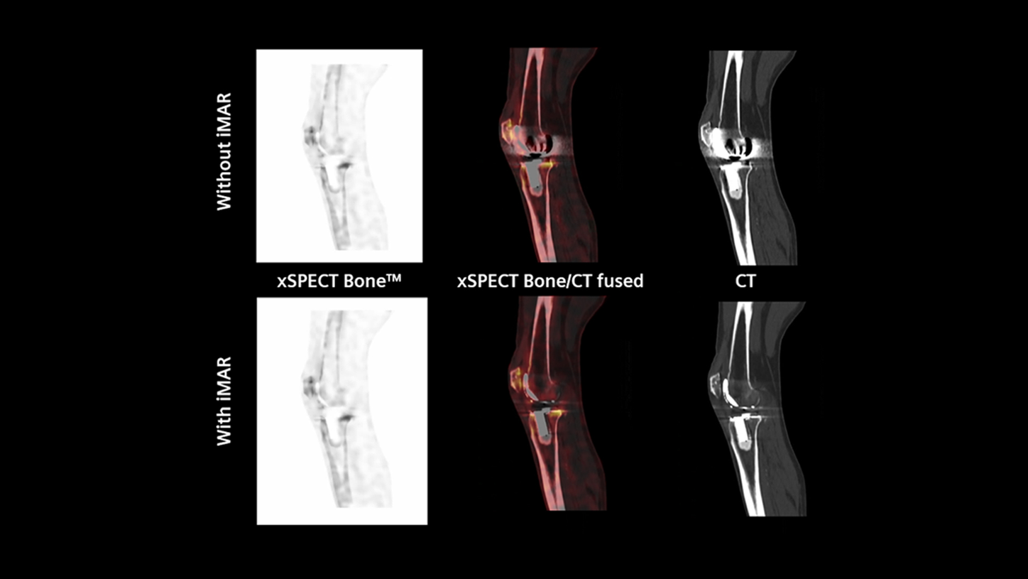
- Enhance xSPECT Bone™ image quality by improving the bone zone maps for advanced reconstruction

More accurate quantification
In this study, iMAR reduced the ipsilateral and contralateral metal artifacts and diminished artifacts impacting SPECT AC and xSPECT Quant™ values, which resulted in accurate quantification. Additionally, iMAR xSPECT Bone images clearly demonstrated focal uptake in the mandible, showing suspected osteomyelitis.

Rule out additional findings
Whether PET/CT or SPECT/CT, iMAR helps rule out additional findings in metal artifact-impacted areas as seen on these Symbia Intevo Bold™ SPECT/CT images. The reduction of metal artifacts in the hip enabled thorough inspection of all bony structures of the pelvis.
Decrease metal artifacts in staging
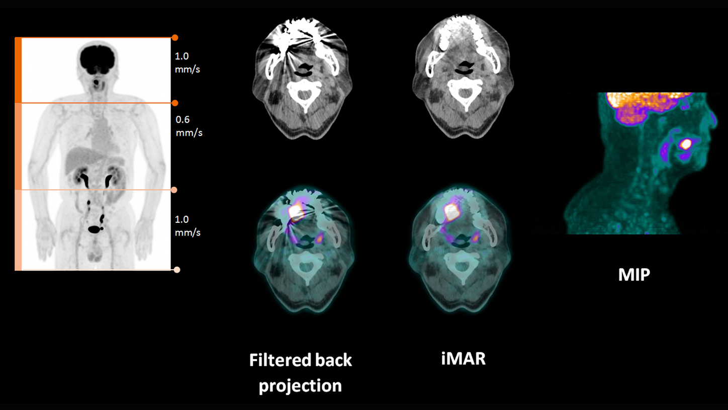
In this example Biograph™ Horizon PET/CT with iMAR decreased metal artifacts in staging head and neck disease. iMAR allowed for the correction of obscuring artifacts from metal implants and was used for the reconstruction of AC PET images.
Technical Specifications
How it works
 Without iMAR
 With iMAR
Courtesy of Universitaetsklinikum Heidelberg, Heidelberg, Germany
Without iMARWith iMAR Without iMAR
 With iMAR
 Without iMAR
 With iMAR
 Without iMAR
 With iMAR
General Requirements
System
- Biograph Vision™2 PET/CT
- Biograph mCT PET/CT
- Biograph Horizon PET/CT
- Symbia Intevo Bold™ SPECT/CT
Minimum Software Version
- Symbia Intevo Bold: MI SPECT VB20
- Biograph Horizon: PETsyngo VJ10
- Biograph mCT: PETsyngo VG60
- Biograph mCT Flow: PETsyngo VG60
- Biograph Vision: PETsyngo VG75
Other
Please Note: Additional technical pre-requisites may apply. Upon receiving your request, your local Siemens Healthineers representative will clarify whether your system meets the requirements.
Header images data courtesy of University Radiology Associates, Syracuse, NY, USA