By Klaas Pieter Koopmans, MD, UMCG Groningen and Partha Ghosh, MD, Siemens Healthineers
Data and images courtesy of EMC Grha Kedoya Hospital, Jakarta, Indonesia
History
65-year-old woman with history of uterine carcinoma treated total hysterectomy with bilateral salpingo-oophorectomy, followed by chemoradiation presented with suspected recurrence. Patient underwent Fludeoxyglucose F 18 Injection (18F-FDG) PET/CT evaluation for metastatic spread and local recurrence.
Study was acquired 60 minutes following intravenous injection of 3.3 mCi F-FDG (0.05 mCi/kg)** on the Biograph Vision Quadra long axial field of view (aFOV) PET/CT scanner (106 cm aFOV). Total scan time was approximately 4 minutes (229 seconds) for head-to-toe acquisition. Low-dose, noncontrast CT was performed for attenuation correction. PET image reconstruction parameters were 4i5s, 440x440 matrix, all-pass.
* Measured value, based on phantom studies performed on a single system. Biograph Vision Quadra has ≤249 ps based on acceptance values. Data on file.
** Based on bench testing (e.g. improved sensitivity and ToF per NEMA NU-2:2018). Please refer to the approved PET drug prescribing information for dosing and administration instructions.
Findings
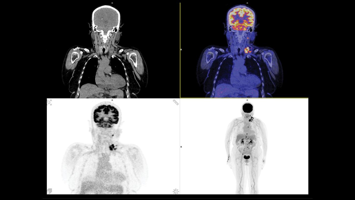
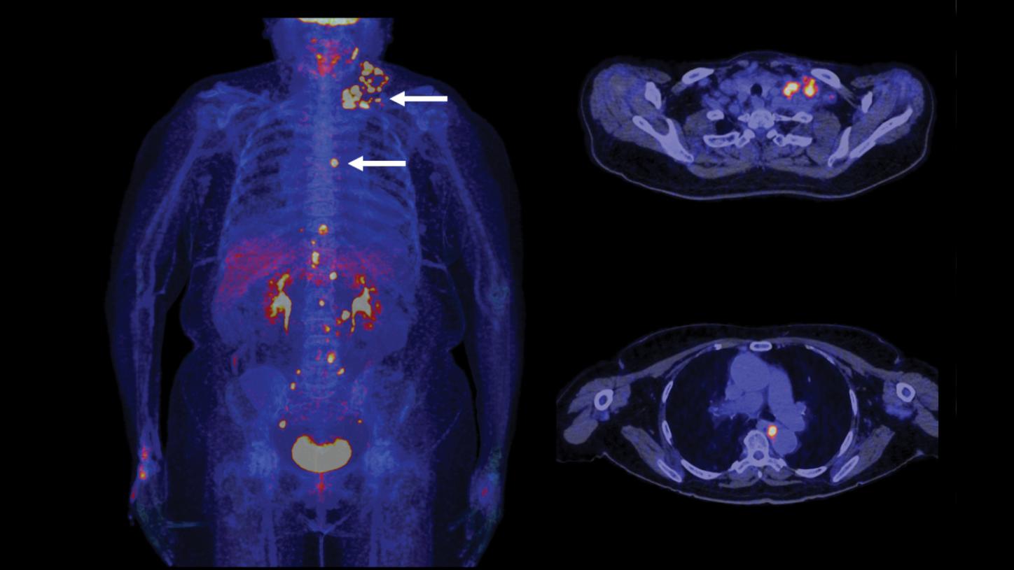
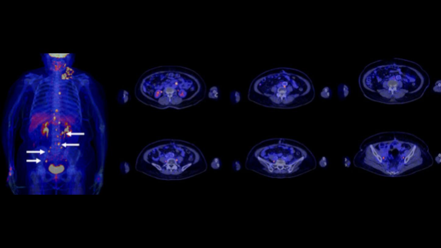
MIP image (Figure 1) shows high 18F-FDG uptake in hypermetabolic lymph nodal mass in the left supraclavicualar and upper jugular region. Multiple 18F-FDG-avid lymph nodes (SUV 5.1) are visualized in the left upper jugular region (level II, measuring approximately 1.7 × 0.6 cm), left lower jugular region (level IV, largest measuring approximately 1.5 × 1.3 cm), and most prominently, in the left posterior triangle (level V, largest measuring approximately 1.5 × 1.3 cm) (Figures 2 and 3).
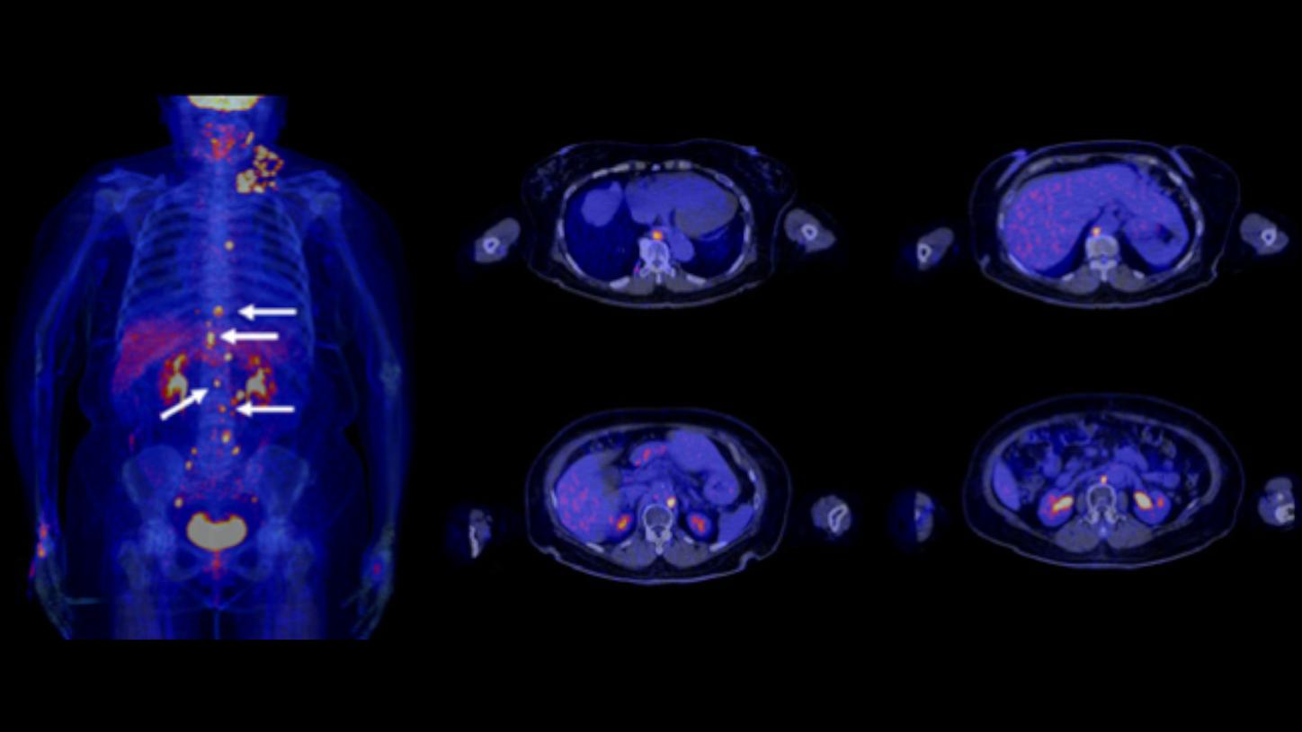
MIP images also show focal hypermetabolic metastatic lymph nodes in the mediastinum, including the subcarinal (station 7, measuring approximately 1.5 × 1.2 cm) and paraesophageal region (station 8, largest measuring approximately 2.2 × 1.2 cm) (Figure 3). Multiple hypermetabolic nodal metastases are also visualized in the para-aortic region extending to bilateral common iliac arteries. Enlarged 18F-FDG-avid lymph nodes (SUV 4.6) are defined in the retrocrural, para-aortic, aortocaval, and bilateral para-iliac regions (Figures 4 and 5), the largest node measuring approximately 1.3 × 1.1 cm in the para-aortic region.
No focal 18F-FDG-avid lesion is visualized in the pelvis in the hysterectomy bed or pelvic wall or obturator nodes.
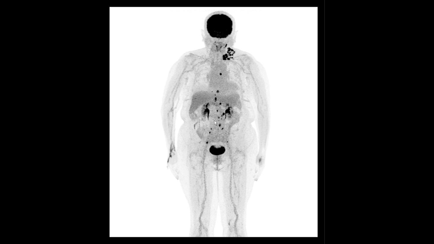
Figure 1: MIP image of 18F-FDG PET/CT scanned on Biograph Vision Quadra with head-to-toe acquisition of only 4 minutes shows multiple lymph node metastases from bilateral common iliac to para-aortic, mediastinal to left supraclavicular lymph nodes. These findings reflect nodal spread of metastases from uterine cancer from pelvis to thorax and cervical nodes via the thoracic duct.
Discussion
The 18F-FDG PET/CT images clearly show multiple lymph node metastases involving bilateral internal iliac nodes; para-aortic nodes in the abdomen and lower thorax; subcarinal and para-esophageal nodes in the thorax; and finally, in the metastatic lymph nodal mass in left supraclavicular and upper and mid jugular nodes in the neck. This reflects the pattern of lymph nodal metastatic spread from primary uterine malignancy to para-aortic, mediastinal, and left supraclavicular node (historically known as Virchow’s nodes) via the thoracic duct.
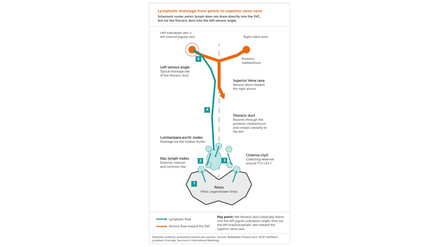
As depicted in Figure 6, uterine tumor malignant spread initially via lymphatic channels to the iliac lymph nodes and subsequently, to the para-aortic and lumbar nodes and also the cisterna chyli. Subsequently, the malignant cells spread via the thoracic lymphatics which predominantly involve the thoracic duct. The thoracic duct connects to the systemic venous system in the neck at the junction of the left subclavian and internal jugular veins. Because of this connection, remote cancer cells often deposit in the left supraclavicular lymph nodes, as apparent from the PET images in this example. The high uptake of 18F-FDG within the enlarged matted lymph nodal mass in the left supraclavicular region reflects the enlarged Virchow’s node. Palpable left supraclavicular mass is often associated with advanced abdominal and pelvic malignancies like uterine and gastric carcinoma typically associated with Virchow’s nodal metastasis presentation.
This clinical example shows hypermetabolic nodal lesions at every stage of the lymphatic metastatic spread. This includes lesions in bilateral common iliac, para-aortic, para-esophageal, left supraclavicular, and left upper jugular nodes, defining the spread from the uterine primary to the thorax and neck via thoracic duct.
18F-FDG PET/CT shows high lesion contrast in hypermetabolic lymph nodal metastases despite ultra-fast acquisition time of just 4 minutes for a head-to-toe PET study (FlowMotion AI enables continuous movement and region-specific acquisition speeds; in this case, two zones with 3-minute acquisition for head-to-mid-thigh and 1 minute for lower extremities). The low injected dose (3.3 mCi)* achieved high lesion contrast, low background, and sharp vascular definition, reflecting the high sensitivity and resolution of the Biograph Vision Quadra.
* Based on bench testing (e.g. improved sensitivity and ToF per NEMA NU-2:2018). Please refer to the approved PET drug prescribing information for dosing and administration instructions.
Examination protocol
Scanner: Biograph Vision Quadra
Prescribing information
Please see Indications and Important Safety Information.