
Three nuclear medicine professionals reflect on the history of the field and how technology, over the course of 60 years, has helped shape nuclear medicine. Through their experiences, they offer insights into where they think the latest innovations will take nuclear medicine.
Download your print version here.
Photos: Site photography courtesy of Siemens Healthineers, CHUV, and UMCG; xSPECT Quant photography by Alex Teuscher
Hindsight is always 20/20, or so goes the saying. Looking back we are able to see past events far more clearly, and the history of nuclear medicine is no exception. We can look back and reflect on the milestones and achievements that comprise the rich history of the science. Yet, in hindsight, one thing that was less clear were the images that came from early efforts in the technology.
“Although the function of the organs was exquisitely measured by nuclear medicine, you had to have a little bit of faith about the morphology when you looked at an image and wondered, ‘well, is that the shape of a lung, is that the shape of a myocardium, is that the shape of a liver?’” reminisces John Prior, PhD, MD, FEBNM, head of nuclear medicine at Centre Hospitalier Universitaire Vaudois (CHUV) in Lausanne, Switzerland. To summarize his experiences in the early 1990s, Prof. Prior states, “what we were doing back then was sometimes more of an art than science. You had to have faith in the images you were seeing.”
“Today you can see a huge difference from years ago in image quality, lesion detectability, and the impacts on diagnosis of patients.”
Over the years nuclear medicine progressed, and what continues to fascinate Prof. Prior is, “the ability to really understand the disease and to be able to bring, if you will, the latest technological advances forward to take care of patients.” For Ronald Boellaard, PhD, medical physicist at the University Medical Center Groningen (UMCG) in the Netherlands—and an 18-year veteran in the field—the effects of technological advancements within nuclear medicine are also notable. “Today you can see a huge difference from years ago in image quality, lesion detectability, and the impacts on diagnosis of patients,” he states. Such progress has a profound effect on the quest to deliver the best patient care.
The culmination of innovation
Technological advances reside at the core of nuclear medicine, spanning a time from when systems first detected tracers administered to patients to today’s multi-functional tracers used for quantitative analysis. Technology’s benefits to nuclear medicine continue to evolve, even now as the field celebrates 60 years of the gamma camera and 20 years since the introduction of PET/CT.
As physicians strive to enhance patient care, manufacturers continue to work and produce technologies that define innovations within nuclear medicine. For physicians at CHUV and UMCG, the utilization of such innovations impacts their clinical decisions today.

Quantifying nuclear medicine
“SPECT quantification has been around for a long time in nuclear medicine,” Prof. Prior says. “In the beginning, we were basically trying to perform quantification with regular nuclear medicine every time we did dosimetry studies, but that was very cumbersome.”
Needless to say, implementing a cumbersome quantitative approach to SPECT/CT in a busy, daily workflow was a challenge. With xSPECT Quant, Prof. Prior and his team now have access to a technology that enables them to incorporate automated quantification in their daily workflow. “When we could see that a manufacturer had the possibility to deliver something that could be intrinsically calibrated, this gave us a lot of hope for quantitative SPECT/CT. The more quantitative we can be, the higher resolution and sensitivity we can have, the better,” summarizes Prof. Prior.
“In SPECT/CT, we can now precisely quantitate how much of the radiopharmaceutical we administer to the patient ends up in a given organ. Siemens Healthineers was the first one with this tool.”

“We can now precisely quantitate how much of the radiopharmaceutical we administer to the patient ends up in a given organ. Siemens Healthineers was the first one with this tool. We are currently working with a range of isotopes—Iodine-123, Lutetium-177, Indium-111, and Technetium-99m—and we can quantify things we could only see before.”
He further explains that the technology enables physicians to obtain an absolute quantitative value. “We do not need to make a ratio. And because of that, we thought, ‘this is interesting and maybe we can catch disease earlier with this.’ With these radiotracers we can follow the pathology; we can detect a disease in a more efficient way since we have an absolute, quantifiable value,” Prof. Prior concludes.


Delivering precision
Automated quantification in SPECT/CT is more than a clinical breakthrough: it enables precision. Prof. Prior emphasizes the benefit that quantification brings to SPECT/CT: “It’s bringing a little bit more precision in what we call precision medicine.”
While SPECT/CT looks to quantification as its gateway to precision, PET/CT looks to the latest advancements in scanner technology to bring precision to molecular imaging.
UMCG, an institution that wants to harness the latest advancements in PET/CT imaging capabilities, recently installed the world’s very first Biograph Vision. Discussing the initial appeal, Prof. Boellaard explains, “we were interested in working with new technology and new innovations, not only because we could see more patients a day, but also because of the expected improvement in image quality as a result of the excellent time-of-flight performance. We envisioned that we could get more accurate image quantification and an improved image quality.”1
“There is a potential in better discriminating lesions from physiological background activity. And maybe upstaging your patients from, let’s say, uncertain up to the presence of a disease.”

Installed in May 2018, Biograph Vision was able to quickly meet expectations at UMCG. Drawing on Prof. Boellaard’s recount on the appeal of the new scanner, Walter Noordzij, PhD, MD, a nuclear physician at UMCG, reveals his first clinical impressions: “There is a potential in better discriminating lesions from physiological background activity. And maybe upstaging your patients from, let’s say, uncertain up to the presence of a disease.”
Dr. Noordzij further stresses the importance of technological advancements as physicians move toward personalized medicine. “It’s becoming very important for us—even more nowadays since we know more about the disease and the heterogeneity of tumors and their metastases.”


Making a difference
The ability to be precise, as the field strives to contribute to personalized medicine, is paramount.
At CHUV, while Prof. Prior works to bring quantification and precision to SPECT/CT, he can also bring the most current innovations in PET/CT to his patients, as CHUV recently installed the world’s second Biograph Vision. “I’m really lucky to be at a center where our oncologists are so interested in molecular imaging. With xSPECT Quant and Biograph Vision, it’s really helping us push the technology that best serves our patients,” Prof. Prior emphasizes.
At UMCG Biograph Vision will be the third PET/CT scanner, enabling them to significantly increase the number of scans they can perform—the organization topped 4,000 scans in 2017. Dr. Noordzij discusses how the additional capacity will enable the implementation of more clinical, as well as research-focused, PET/CT scans. “On the one hand, we’ve seen an increase in the demand for clinical PET/CT scans over the past five to six years. But on the other hand, there’s an enormous demand for research PET/CT and it’s pretty easy to say the addition of the Biograph Vision scanner will help us meet this need." As institutions expand their clinical offerings and work to improve patient care, using technology that enables confident and precise decisions makes a difference.
The robust history and technological innovations of the past 60 years are a significant testament to nuclear medicine’s progress. Given the recent development of groundbreaking technologies, it’s apparent that the future looks a lot clearer than the past.

PET: 7 beds, 3 minutes/bed, 440 x 440 matrix.
Data courtesy of UMCG, Groningen, The Netherlands.
Precision with Biograph Vision
The images showed sharp delineation of small hypermetabolic metastasis in the right lobe of liver near the dome of diaphragm (Segment VII). Intense uptake in the abdominal wall is suggestive of metastatic deposit. Mildly increased uptake in mediastinal lymph nodes are possibly secondary to inflammation. High contrast of small liver metastases without significant motion-related blurring even without respiratory gating reflects high overall image quality and increased lesion contrast to background in the Biograph Vision images.
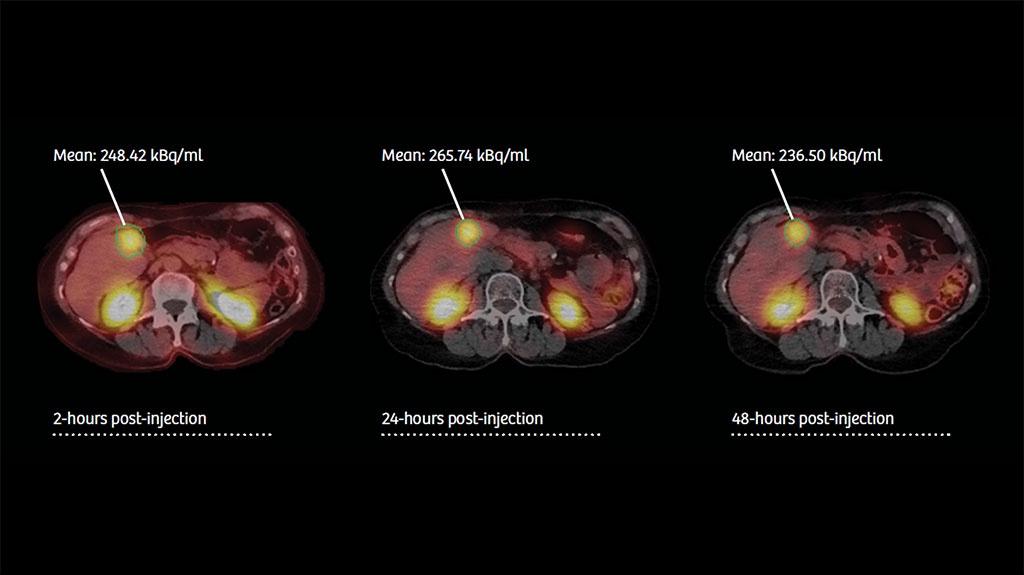
DOTATATE; 2-, 24-, and 48-hour post-injection delay. CT: 130 kV, 30 mAs.
Data courtesy of CHUV, Lausanne, Switzerland.
Quantifying nuclear medicine with xSPECT Quant
Following peptide receptor radionuclide therapy (PRRT) for metastatic neuroendocrine tumor with 177Lu DOTATATE, sequential SPECT/CT images were acquired. With xSPECT Quant, total lesional concentration of the therapy dose can be calculated and tracked over time. This enables dosimetric evaluation of tumor dose (as in this case) or assessment of therapeutic response following multiple treatment cycles.
About the Author
Joseph J. Diorio is a freelance writer based in Nashville, Tennessee, USA, specializing in the human and personal aspects of technology.