Artis Freestyle Access - a unique solution for ultrasound imaging in the interventional suite - Clinical experience from Göttingen University Hospital
Clinical applications for your everyday procedures
Our dedicated clinical solutions such as Low-dose Acquisition, Roadmap and DSA Roadmap, Overlay Reference, syngo iFlow, 3D Wizard and syngo Dyna3D fit into your workflow when treating everyday procedures.

Low-dose Acquisition
- Dedicated low-dose acquisition protocols saving up to 67% of the dose*
- Outstanding DSA image quality utilizing 0.3 micro-focus and 2K resolution
- Up to five magnification formats
- Selectable frame rates independent of organ program, changeable at tableside
- CLEARmatch: next generation real-time pixel shift for movement compensation
*Compared to standard system settings

Roadmap and DSA Roadmap
- DSA Roadmap: Increase image quality and save contrast and
X-ray dose by using a DSA image as vessel map - Individual windowing of vessel map and devices
- CLEARmatch: next generation real-time pixel shift for movement compensation
- CLEARmap: a set of features to support an easy and efficient Roadmap workflow including: starting Roadmap DSA with the press of a button, zoom and pan during Roadmap mode, and more

Overlay Reference
- Save dose and contrast agent by overlaying existing DSA images to guide your intervention
- See the vessel map in relation to anatomical landmarks
- Fade in and out the overlay image according to personal preference
- Easily select your preferred overlay mode at tableside: Roadmap, Roadmap DSA or Overlay Reference
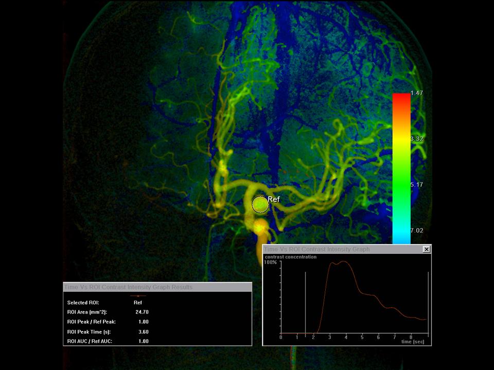
syngo iFlow
- Easily visualize flow characteristics and identify the regions with blood flow anomalies
- Analyze and understand flow at specific points of interest along the vessel tree
- Demonstrate changes in flow post-intervention and quantify level of post-procedural improvement
- Get additional information to support decision-making during the intervention
- syngo iFlow also works with previously acquired DSA scenes – no need for a dedicated acquisition

3D Wizard
- Choose the desired image result from a pool of possible cases and let the system guide you through the 3D scan
- Provides all required parameters for a 3D scan at the time you need them
- Supports definition and establishment of clinical and departmental standards (e.g. for clinical studies, quality assurance, etc.)

syngo Dyna3D
- Automatic reconstruction with user defined presets
- Full 3D control from tableside
- Real-time update of 3D view with C-arm movements
- Subtracted scans for optimal visualization of the stent, graft or coils in relation to the contrast-filled vessels – with syngo DualVolume
- Simply select your desired 3D imaging result using the 3D Wizard and let the system guide you through the acquisition step by step, including recommended injection parameters and acquisition delays
Clinical applications for your most challenging interventions
Our dedicated clinical solutions such as syngo Fusion Package, syngo DynaCT, syngo Dyna3D HighSpeed, syngo DualVolume, syngo DynaPBV Body, syngo Embolization Guidance fit into your workflow when treating most challenging interventions.

syngo Toolbox and syngo 3D Roadmap
- Overlay of marked anatomical landmarks or points of interest from the 3D information onto live fluoroscopy
- One-click generation of anatomical outlines
- Allows for better orientation during catheter navigation through complex vessel anatomy
- Changes in C-arm angulation, SID, zoom and table movement are automatically updated in real time
- Efficient tools to support a high patient throughput: Bookmarks allow to plan the procedure in advance, save and later recall the procedure planning data easily when needed. Parallel patient processing allows the independent use of the post-processing applications in the control room, while maintaining full workplace functionality in the exam room, even for datasets of different patients

syngo Fusion Package
- Integrate the unique information of MRI, CT or PET·CT into your angio image using syngo Fusion Package
- Make the most of available pre-procedural information and avoid radiation dose and contrast injections of repeating 3D scans
- Overlay information from other modalities using syngo 3D Roadmap or utilize applications like syngo Toolbox with existing three-dimensional datasets
- Select between syngo 3D/3D Fusion, or syngo 2D/3D Fusion for easy multimodality integration, which does not require an intra-procedural 3D scan
- syngo Fusion Package enables multimodality integration without leaving the tableside

syngo DynaCT
- Provides most updated 3D soft-tissue information directly in the angio suite for planning, monitoring of interventions, and confirmation of treatment results
- Differentiation of 10 HU in 5 mm slice thickness or 5 HU in 10 mm slice thickness
- 200 degree rotational angiography for reconstruction of the complete volume
- Reconstructed volume of 24 cm x 18.5 cm (9.4” x 7.3”)
- Visualization as MPR, MIP, or VRT
- Simply select your desired 3D imaging result using the 3D Wizard and let the system guide you through the acquisition step by step, including recommended injection parameters and acquisition delays

syngo DynaCT with low-dose
- Perform syngo 3D/3D Fusion at lower dose using a dedicated Low-dose syngo DynaCT protocol, with up to 72% less dose
- Confirm catheter position in dose-sensitive regions with syngo DynaCT Body CARE - a dedicated soft-tissue imaging protocol with 30% less dose and shorter acquisition time

syngo DynaCT SMART
- Reduce artifacts from dense objects using the iterative syngo DynaCT SMART volume reconstruction
- Make relevant aspects in soft tissue visible even close to e.g. coil packages or glue for sounder decision-making during interventions

syngo DynaCT Micro
- syngo DynaCT Micro provides 40% more resolution to enhance the smallest details and up to 68% less dose due to reduced field of view
- Improved spatial resolution compared to CT to better visualize small structures like stents, cochlear implants or cartilage structures
- Expand the scope of your lab by accommodating additional procedures and diagnostic studies in e.g. musculoskeletal or ENT field

syngo DynaCT 3601 and syngo DynaCT Large Volume1
- Get extended syngo DynaCT coverage with these ARTIS pheno unique acquisition protocols:
- a 32 cm x 23.5 cm (12.6'' x 9.3'') volume in only 6 seconds using syngo DynaCT 360
- a 43 cm x 17.4 cm (16.9'' x 6.9'') volume using syngo DynaCT Large Volume
- Obtain the complete axial view of the entire abdomen, also on obese patients
- Demonstrate the accurate skin entry point for needle procedures
1 Available exclusively with ARTIS pheno
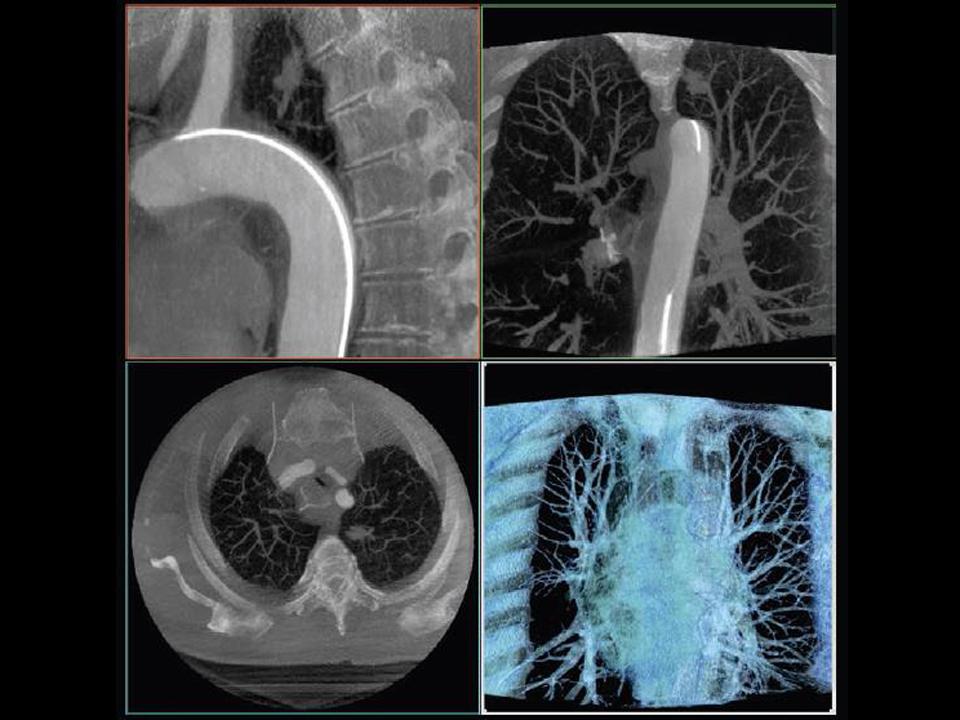
syngo Dyna3D HighSpeed1
- This ARTIS pheno exclusive protocol enables the shortest acquisition time available – 3 seconds only – for high contrast 3D images
- This means:
- less contrast material used
- fewer motion artifacts with moving organs
- shorter breath-hold for your patient
- Most useful for lung imaging, for example: demonstrating lung tumors, stenoses or AVMs
1 Available exclusively with ARTIS pheno

syngo DualVolume
- Clearly differentiate between different types of objects by using distinctive visualization presets
- Visualize two high-contrast datasets to demonstrate the contrast-filled vessels in relation to implanted devices such as stents, grafts or coils
- Visualize one high- and one low-contrast dataset in one volume to carefully study vessels in relation to surrounding soft tissue
- Individual visualization parameters can be applied to each volume
- “DSA Layout”: intelligently preprocessed visualization combining three volumes: fill run visualized as cross-sectional MPR images, as well as mask run and subtracted run visualized as a DualVolume in VRT

syngo DynaPBV Body
- Check distribution of blood in lesions and surrounding tissue by obtaining cross-sectional blood volume information in your lab
- Perform qualitative measurement of blood volume in order to assess changes in perfusion caused by e.g. tumor embolization
- Presets available for:
- pre-/post-therapy comparison dual-volume representation of blood volume map together with anatomic information from the mask run
- Supports identification of the optimal endpoint during embolization
- Potential to identify non-responders directly in the angio suite
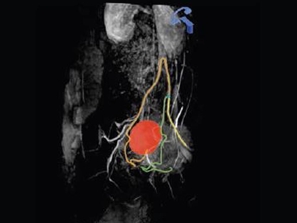
syngo Embolization Guidance
- Simple planning and guidance for catheter-based interventions
- Automatic path computation from the tip of a diagnostic catheter to the tiny vessels feeding a lesion
- Easy lesion segmentation incl. automatic tumor volume calculation
- Planning can be done on syngo DynaCT as well as CT, PET·CT or MR volumes
- Overlay of planning data onto live fluoroscopy with syngo 3D Roadmap

syngo Needle Guidance
- Increase safety, confidence and accuracy of needle procedures using a dedicated planning and guidance application
- Planning can be done on syngo DynaCT as well as CT, PET·CT or MR volumes
- Control scans are automatically registered with the planning scan
- Free up your CT by facilitating needle procedures in the interventional suite
- Get better access in obese patients and complex needle procedures
- Unique integrated cross-hair laser light visualizes skin-entry point and allows for radiation-free needle advancement
- Allows for simultaneous planning and overlay of multiple puncture paths
Artis Family of Imaging Systems

With our Artis family we offer a broad portfolio of systems to cover all clinical needs to support image-guided therapy for interventional radiology. The product lines include a floor-, a ceiling-mounted, a biplane, a multi-axis system with robotic technology, and a seamlessly combined Angio-CT suite.