SOMATOM go.SimSimülasyon yeniden icat edildi
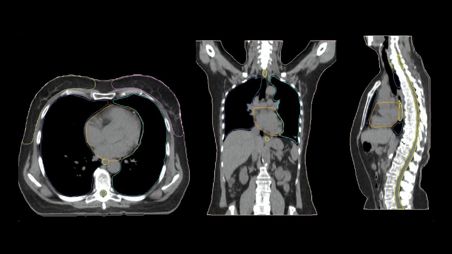
SOMATOM go.Sim, oldukça esnek, sezgisel bir CT simülatörüdür. Gereksinimlerinize özel olarak uyarlanmış tam entegre donanım ve yazılımla, kesinliği artırmak ve hata olasılığını azaltmak için tasarlanmıştır. Ayrıca gelişmiş algoritmalar, AI destekli risk altındaki organ (OAR) otomatik şekillendirme ve hedef marjlarını potansiyel olarak azaltan mükemmel yumuşak doku kontrastı içerir. Dahası, basit bir işletim konsepti ve tek bir satıcı hizmet sözleşmesi ile hem hastalara hem de kullanıcılara özen göstermek üzere tasarlanmıştır.
SOMATOM go.Sim, CT simülasyonunu yönetmek için daha az, hastalara odaklanmak için daha fazla zaman harcayabilmeniz için resmin tamamını daha hızlı elde etmenize yardımcı olur.
Özellikler ve Faydalar

Klinik Kullanım
CT simülasyonunu SOMATOM go.Sim ile yeniden icat ediyoruz – radyasyon onkologları, tıbbi fizikçiler, dozimetristler, RTT'ler ve finansal karar vericiler gibi 300'den fazla RT uzmanıyla birlikte tasarladığımız, son derece esnek, sezgisel bir CT simülatörü ve bize dünyanız hakkında bilgi vermek için. Sonuç, CT simülasyonuna çığır açan bir yaklaşımdır. Optimum RT planlaması için nihai başlangıç noktasına ulaşmak için ihtiyacınız olan her şeyi size verecektir.
Be certain in simulation
Kontürleme için sürüş hassasiyeti
Hastalar ve kullanıcılar için bakım
Testimonials
Müşterilerimizin ne dediğini dinleyin:
Teknik Özellikler
Optional
As shown by measurements with a Gammex 467 Tissue Characterization Phantom comparing [T]standard reconstruction and DirectDensity reconstruction. Image value to relative electron/mass density conversion for the standard reconstruction was based on a two-linear-equations approach with individual calibration for each tube voltage. For DirectDensity images, a single tube-voltage-independent linear conversion was used.
DirectDensity reconstruction is designed for use in Radiation Therapy Planning (RTP) only. DirectDensity reconstruction is not intended to be used for diagnostic imaging.
The DirectBrachy positioning board is currently under development; it is not for sale in the U.S.A.
The DirectBrachy positioning board is not commercially available in all countries. Its future availability cannot be guaranteed.
The urology stirrups (leg support) seen here are optional. The information contained here refers to products from third party manufacturers and are therefore in their regulatory responsibility.
The CT simulator and the CT table seen here are not part of the DirectBrachy positioning board.
DirectBrachy positioning board is only compatible with SOMATOM go.Sim and SOMATOM go.Open Pro with Multi-index RTP Overlay. Afterloader needed for brachytherapy. Shielding of the CT room is required when it its used for brachytherapy.
Verification of mutual compatibility of medical devices combined in a system in accordance with Article 22 (MDR Regulation (EU) 2017/745) on Medical Devices is pending. The combination is not commercially available in the European Union.
The DirectBrachy positioning board does not yet fulfill all applicable General Safety and Performance Requirements according to the European Medical Device Regulation 2017/745. The product is not commercially available. Its future availability cannot be guaranteed.
Jameson MG et al. A review of methods of analysis in contouring studies for radiation oncology. J Med Imaging Radiation Oncol. 2010; 54(5): 401–10.
Wu X, Udupa JK, Odhner D, et al. Knowledge-Based Auto Contouring for Radiation Therapy: Challenges in Standardizing Object Definitions, Ground Truth Delineations, Object Quality, and Image Quality. Int J Radiat Oncol Biol Phys. 2017; 99(2): E740.
Cheung CW, Leung KY, Lam WW, et al. Application of Model-based Iterative Reconstruction in Auto-contouring of Head and Neck Cases. Scientific Informal (Poster) Presentation at: LL-ROS-TH Radiation Oncology and Radiobiology Lunch Hour CME Posters; 2012 Nov 29; Chicago, IL.
Requires Anzai or Varian RGSC
Kelly E et al, Reduced patient anxiety as a result of radiation therapist‐led psychosocial support: a systematic review, J Med Radiat Sci Sep; 64(3): 220–231 2017.
Use of cone-beam imaging to correct for catheter displacement in high dose-rate prostate brachytherapy, Holly R.et al., Brachytherapy (2011) 10:4 (299-305), Doi: 10.1016/j.brachy.2010.11.007