A quarter of a century ago, the world’s first commercial PET/CT scanner was developed. Although initially met with uncertainty, the medical community vigorously embraced the modality as an indispensable diagnostic imaging tool and one that can provide life-saving information for countless patients, even for one of the scanner’s inventors.
How it all began
Time flies. Looking back, David Townsend, PhD, can hardly believe it has been twenty-five years since he and his team, based at the University of Pittsburgh, developed the device in collaboration with Ronald Nutt, PhD, CEO of CTI PET Systems, in Knoxville, Tennessee, USA. Since then, numerous innovations have propelled PET/CT to even higher and wider applications.
The story of PET/CT begins in the early 1970s in Geneva, Switzerland, almost 30 years before the first PET/CT scanner was developed. Back then, Townsend joined the European Organization for Nuclear Research (CERN), the European particle physics laboratory. At the time, PET was far from being a clinical tool—it was a research project with uncertain applications. Townsend started looking at potential detector applications for PET in 1975 along with Alan Jeavons, PhD.
Click on each year to explore the timeline
Four years later, Townsend moved to University of Geneva Hospital to focus entirely on PET scanner development. In 1988, while working at University of Geneva Hospital, Townsend collaborated with CTI PET Systems and Terry Jones, DSc, from Hammersmith Hospital, London, UK, to design and build a cost-effective, rotating PET scanner based on the BGO (bismuth germanium oxide) block detectors developed by CTI PET Systems. Townsend and Jones‘s rotating prototype scanner imaged patients at the University of Geneva Hospital and was later commercialized by CTI PET Systems as the Advanced Rotating Scanner (ART) that represented a cost-effective PET scanner.
2000
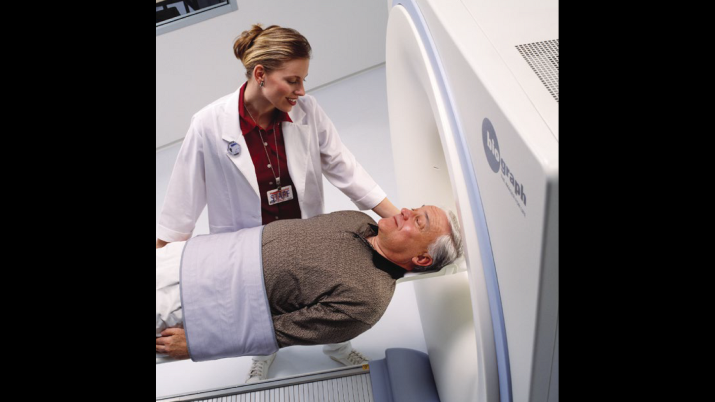
Biograph is the first commercial PET/CT system installed at the University of Pittsburgh Medical Center, Pittsburgh, Pennsylvania, USA. This system is revolutionary in that it combines PET and CT in a single diagnostic system. TIME magazine hails the PET/CT as “Medical Invention of the Year.”1
The birth of PET/CT
How did the idea of combining PET and CT come about? As with many groundbreaking innovations, it started with a simple question—and a bit of chance.
According to Townsend, an oncology
surgeon at the University of Geneva
Hospital walked by the PET scanner
one day and wondered if it would be
possible to add CT to the scanner
design. His reasoning was practical:
PET, at the time, was mostly confined
to research centers, while every
physician was familiar with CT.
Townsend considered that idea.
“So, I called Nutt at CTI and asked
him, ‘How about adding CT to PET?’” Townsend recalls. “For us, as scientists
and engineers, it made sense.
You’re combining complementary
information—the high-resolution
anatomical detail of CT with the
functional imaging of PET—and
offering physicians both in a single
device. While some degree of
alignment of two separate CT and PET
scans could be achieved using software,
this approach was really only effective
and reliable for brain imaging.”
Fast forward to 1995 at the
University of Pittsburgh. Townsend,
along with physicist Thomas Beyer,
PhD, MBA, and bioengineer Paul
Kinahan, PhD, teamed up with Nutt
to turn their concept into reality,
funded by the National Cancer
Institute (NCI). In close partnership,
CTI contributed the PET technology,
while Siemens provided the CT
components. The team was also
backed by the Department of
Radiology at the University of
Pittsburgh, where Townsend worked.
The prototype PET/CT was built at
the CTI factory in Knoxville and then
moved to the PET facility at the
University of Pittsburgh to start
clinical scanning. Within three years,
they had developed a prototype and
began acquiring the first-ever clinical
PET/CT images of patients.
2008
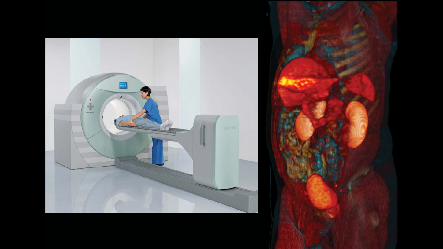
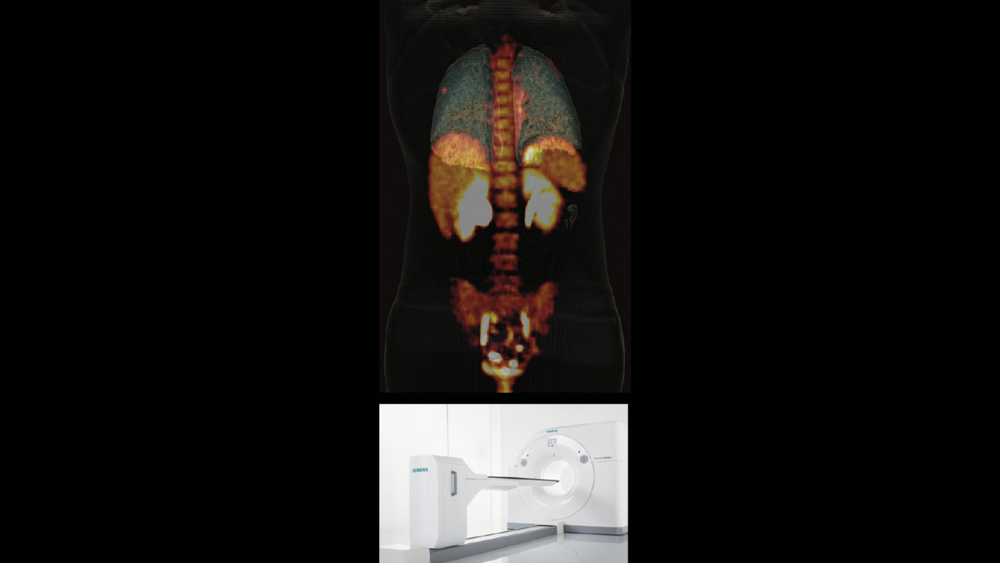
Biograph mCT is announced as the world’s first molecular computed tomography system and represents the evolution of integration in imaging. Biograph mCT is a combination of a state-of-the-art CT scanner with a high-performance PET system.
A modality looking for a home
At the 1998 Society of Nuclear Medicine (SNM) annual meeting in Toronto, Canada, Townsend shared the first clinical PET/CT images with the world. Townsend looks back, “It was a very exciting time to be to be involved in nuclear medicine.” CTI and Siemens did not begin looking at the commercialization of PET/CT until about 1999, after enough clinical image data was acquired to convince physicians that it was a useful clinical concept.
While surgeons and oncologists were generally more receptive to the idea, the wider medical community’s initial reaction to the new PET/CT scanner was, at best, uncertain. Why? Because it was hybrid technology that did not quite fit. Was it nuclear medicine? Was it radiology? No one knew exactly where it belonged. And would it require two technologists to operate it? Two physicians to read the scans? Many questions were asked.
The co-inventors, Townsend and Nutt, and members of the team remained involved in PET/CT’s development. In November 2000, the new scanner was unveiled to the world at the Radiological Society of North America’s (RSNA) annual meeting in Chicago, Illinois, USA. In December of the same year, the PET/ CT concept and prototype design was honored by TIME magazine as “Medical Invention of the Year.”1 By mid-2001, the first commercially available Siemens Biograph PET/CT system was installed at the University of Pittsburgh.
2013
Biograph mCT Flow is the first PET/CT system to move the patient through the gantry while continuously acquiring PET data.
“’How about adding CT to PET?’…For us, as scientists and engineers, it made sense. You’re combining complementary information—the high-resolution anatomical detail of CT with the functional imaging of PET—and offering physicians both in a single device.”

2015
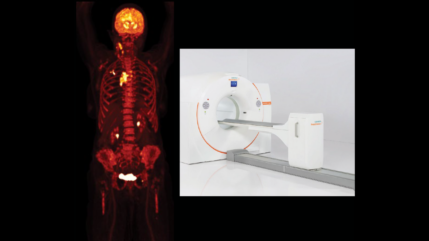
Biograph Horizon offers high-resolution imaging, advanced PET and CT technologies, and AI-powered workflows. Its compact design and simplified operations enable more healthcare providers to offer a higher standard of care to more patients.
Pushing PET/CT forward with ground-breaking innovation
Since PET/CT’s unveiling at RSNA, the modality’s capabilities continue to expand. Maurizio Conti, PhD, joined the Siemens molecular imaging team in 2000. Today, he serves as director of PET Physics and Reconstruction at Siemens Healthineers in Knoxville. When asked about the biggest technological developments in PET/CT over the past 25 years, he quickly points to two key milestones: replacing BGO detector blocks with lutetium oxyorthosilicate (LSO) scintillators, which enabled time-of-flight (TOF) imaging, and the later introduction of silicon photomultipliers (SiPMs), which dramatically improved time resolution. These two breakthroughs significantly elevated image quality and greatly accelerated the scanning time, which in turn helped increase patient comfort.
Conti also speaks with particular pride about two projects: the first TOF PET/CT scanner from Siemens Healthineers, Biograph mCT, which was “a revolution in those years—a truly outstanding machine,” he says— and Biograph Vision Quadra total-body PET/CT. “It was an unusually fast project. Can you believe that it was done in about 1.5 years?”
In the last 25 years, software
development has also played a critical
role. Advances in reconstruction and
data correction have greatly improved
image quality, especially for how
attenuation correction and image noise
are handled in obese patients. TOF
reconstruction now helps compensate for patient size and even motion—from
breathing to heartbeats.
“Using different tracers, and more specific tracers, we can understand not only what a disease is, but if it’s aggressive or not. And that correlates with the organs and the anatomy, which only PET/CT can do.”

2017
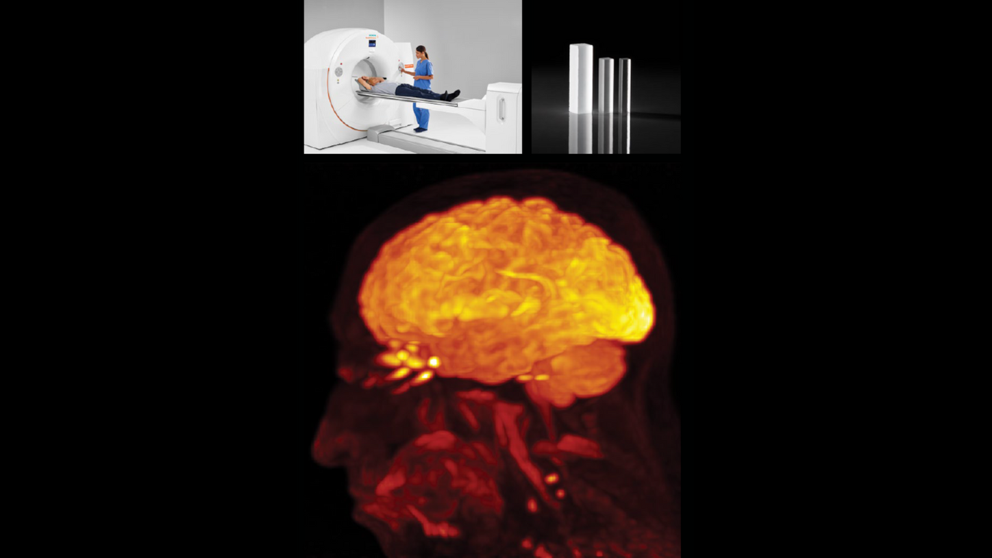
Biograph Vision PET/CT is launched with an innovative design that reduces the size of the detector’s crystal elements from 4 x 4 mm to 3.2 x 3.2 mm and generates 214-picosecondtrue TOF performance that leverages the full potential of SiPM technology.
Reflecting on the progress over
the past 25 years, Townsend says
“Nowadays, you have very powerful
instruments compared to what we
had in 2000. And they provide superb,
unbelievable imaging capabilities in
times we didn’t even dream of back in
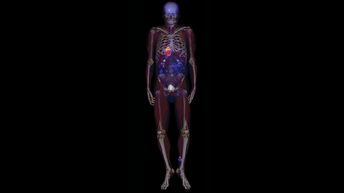
those days. The development of total body
imaging with large field-of-view
systems is now a reality. We used to
work with 15-cm axial extent, and now
you have a system like Biograph Vision
Quadra, which has 106-cm axial field
of view (aFOV). You can look at
dynamics between different organs,
which we could never do, and that’s
been a tremendous step forward.
Because you know we would image
either the brain or the heart or the liver,
maybe the lungs, lower abdomen—
but the body is a total system.”
Ability to understand disease and improve treatment
The defining strength of PET/CT remains its ability to combine functionality and anatomy in a single scan. But there is even more to it today. “Using different tracers, and more specific tracers, we can understand not only what a disease is, but if it’s aggressive or not. And that correlates with the organs and the anatomy, which only PET/CT can do,” Conti explains.
It all started with the generic tracer fludeoxyglucose injection F 18 (FDG) to measure metabolism. But now, a growing number of novel tracers are available, providing deeper insight into disease function that comes with PET. “This is a great future,” says Conti. “Today, we have access to highly specific tracers such as PSMA for prostate cancer, tau and amyloid for dementia and Alzheimer’s disease evaluation, and rubidium for cardiac imaging. As the spectrum tracers broadens, so does the potential for earlier, more accurate disease detection and improved treatment monitoring.”
Full circle: from pioneer to patient
Last year, Townsend himself
underwent a PET/CT scan following
the discovery of a suspicious nodule
in his left lung. The PET/CT was
consistent with an adenocarcinoma
that was confirmed by biopsy.
Fortunately, it was an early stage 1
tumor that was removed surgically
and a 6-month follow-up scan showed
no evidence of disease. Reflecting on
his own patient experience, Townsend
recalled that the imaging team who
performed the scan at the University
of British Columbia Cancer Centre,
Vancouver, Canada, were aware of
Townsend‘s background. The
technologist who gave him the
injection of FDG quipped, “‘I guess I
don’t need to explain the imaging
procedure to you, do I?‘” remembers
Townsend, with a smile.
2020
Biograph Vision Quadra takes its place as the first 106-cm aFOV whole-body scanner with ultra-high sensitivity, for advanced research, clinical flexibility, and improved patient outcomes. The scanner fits traditional PET/CT spaces while enabling dynamic, multi-organ, and low-dose imaging.
What may the next 25 years bring?
When asked about the next developments in PET/CT, Conti envisions a future full of potential—from widespread use of artificial intelligence to more advanced motion correction, improved speed and image quality, and increased patient comfort. Theranostics and opportunities for more personalized therapy are also on the horizon.
And what are his personal hopes for the next 25 years of PET/CT? Conti pauses, then offers two wishes: “More tracers that unlock the full potential of our amazing technology. And that PET/CT becomes more accessible to patients earlier in the care pathway. Right now, a patient needs to have a cancer diagnosis to maybe get a PET/CT scan. I would love a world where PET/CT is routine, where patients receive a PET/CT when there is suspicion of cancer.” As for Townsend? His wish echoes a similar sentiment: that PET/CT technology is widely available to all patients no matter where they live.
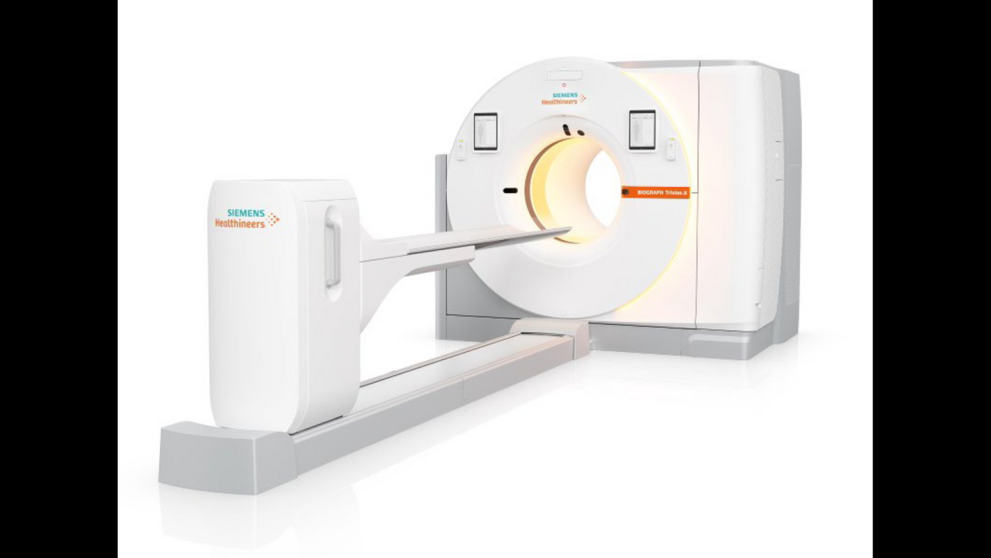
2025
Biograph Trinion.X meets evolving clinical demands with greater speed, sensitivity, and a patient-focused design. The system’s ultra-fast 197-picosecond time-of-flighta capability is a key advancement, along with an extended aFOV of up to 48 cm.
The defining strength of PET/CT remains its ability to combine functionality and anatomy in a single scan. With the use of more specific tracers, PET/CT can provide deeper insights into diseases, their aggressiveness, and their correlation with organs and anatomy. Paving the way for earlier, more accurate detection and improved treatment monitoring and working toward making PET/CT accessible to all patients is an ambition shared by clinicians and researchers alike.
PET/CT served as catalyst for other hybrids of nuclear medicine with anatomical measures, such as in PET/MR. The success of PET/CT commercially encouraged the industry to develop and market other hybrids. The audacity of PET/CT was daunting; its success equally so. The next 25 years will undoubtedly hold more exciting developments.
Fludeoxyglucose F 18
Please see Indications and Important Safety Information for Fludeoxyglucose F 18 (18F FDG) Injection.
