

Early Detection is Critical – Get Checked!
Breast cancer does not always present symptoms. Many physicians recommend an annual screening to help find cancer early, when the tumor may still be small and the risk of it spreading is lower.
The American Cancer Society encourages women over 45 with average cancer risk to get a mammogram every year. At age 55, you can transition to every other year. Even at 40, women can ask for a screening and be confident in the results.
Early detection is helping women realize better outcomes. More than 3.1 million women are breast cancer survivors. Advances in early detection technology like 3D mammography are fueling the drive.
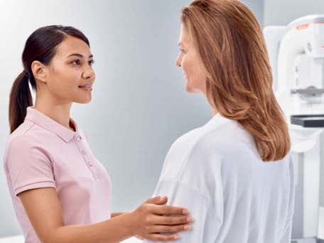
Your best chance to be a survivor is early detection. One of your best tools is the 3D mammogram delivered on MAMMOMAT Revelation with Wide-angle Breast Tomosynthesis.

What Is a Wide-Angle 3D Mammogram?
A mammogram is an X-ray image of your breast. The breast is compressed to spread out the tissue inside so X-rays can capture black-and-white images. These images are displayed on a monitor and examined by your doctor for cancer.
For years, the typical mammogram would provide doctors with 2D images. They’d search these images for tumors or lesions. But 2D images can present challenges like overlapping tissue layers. Often, the results were inconclusive and required more testing. Worse, they sometimes missed cancer.
A wide-angle 3D exam is different. Images of your breast are taken over a 50o angle and gathered into a 3D view, layer by layer. This way, doctors can separate layers of breast tissue for a deeper view. The wide-angle 3D exam is wider than any mammogram, so your doctor gets more information for a confident diagnosis.
The result is an image that looks inside your breast - layer by layer - to find tumors that may not be defined in 2D. It results in fewer follow-up exams. And it can save lives because it leaves breast cancer with no place to hide.

Why Choose a Wide-Angle 3D Mammogram?
Reason 1: Better and earlier detection of breast cancer
The studies back it up – a wide-angle 3D mammogram is a much better way to screen for breast cancer than a traditional 2D exam.
The 50o wide-angle sweep is the widest on the market. It acquires more images of your breast than any other mammogram. That means more information for your doctors than other mammograms so they can make a more confident diagnosis.
Reason 2: It’s more comfortable
Why do mammograms have to hurt? We asked ourselves the same question, and we answered by building a system that personalizes the squeeze. MAMMOMAT Revelation’s personalized soft compression technology prevents unnecessary pain by adjusting to the anatomy of each woman’s breast. OpComp calculates and applies optimal compression: Your breasts are compressed based entirely on the structure of your breast tissue – so you get the least amount of pressure needed for the best impression possible of your breast. SoftSpeed slows the compression paddle down as soon as it reaches the breast: The paddle that applies pressure to your breast is built with soft edges that contour to your shape. The result? Greater comfort! No pinching. No pulling. More time for proper positioning and less operator variation. And a better experience for you.

Category B: There are scattered areas of dense tissue seen as white areas on the mammogram.
Category C: More of the breast is made of dense tissue and is described as heterogeneously dense. This can make it harder to see small abnormalities around the dense tissue, which also appear as white areas.
Category D: Breasts are extremely dense, which makes it harder to see abnormal findings which also appear as white areas on the mammogram.
What You Need to Know About Breast Density
What is dense breast tissue?
Inside each breast is a complicated network of fibrous tissue. Each woman is different, and the density of her breast tissue will be different from others. When your breast tissue is denser, it presents diagnostic challenges to your doctors – and health dangers to you.
The challenge for doctors
In a traditional mammogram, suspicious lesions typically present themselves as white masses. Unfortunately, breast tissue also appears as a white network of fibers. Sometimes suspicious lesions can be hidden under the layers or misrepresented by the white fibers of breast tissue.
The dangers for you
Physicians monitor breast density because dense breasts can be connected to a heightened risk of breast cancer. Screenings become even more important because of the challenges described above, where dense tissue can hide tumors – particularly in a standard 2D mammogram.
You are more likely to have dense breasts if you have a high degree of fibrous or glandular tissue in the breast and lower levels of fatty tissue. Every woman has a different level of breast density, and very dense breasts are common. This density tends to decrease as women age, though it persists for some women without much change.
How does Wide-Angle 3D mammography tackle dense breasts?
3D mammography may be able to detect more cancers1 thanks to the increased imaging range and quality. The Wide-Angle 3D mammogram can deliver the confidence you need. You’ll know you’re doing everything you can to detect breast cancer early, in a way that’s comfortable, safe and accurate.
What to Know Before Your Wide-Angle 3D Exam
Ready to schedule an exam, or have a mammogram coming up? Here’s what you’ll need to know.
When Scheduling Your Mammogram:
- Let your doctor know about any history of breast cancer in your family, or if you’ve had pain, discomfort or any other changes related to your breasts.
- Some women’s breasts may be tender in the week leading up to their period. To avoid discomfort, we recommend scheduling for the week after your period if possible.
- If there’s a possibility you might be pregnant, please let your doctor know prior to scheduling, as they may elect to skip the exam or provide special instructions in order to ensure safety.
How to Prep:
- Avoid deodorant, talcum powder or lotion under your arms or on your breasts, as these can appear similar to calcium spots when viewed.
- Wear comfortable clothing that you can easily change out of.
- Obtain the results of any prior mammograms and provide these to your medical team prior to or upon arrival.