The facts about photon-counting CTGet to know the technology and the benefits
Photon-counting CT marks the beginning of a new era in computed tomography. This radically new detector technology directly converts X-rays into electrical signals, helping the NAEOTOM Alpha class overcome limitations once considered unavoidable in conventional CT. But how does photon-counting CT actually work? What are the facts, and what is just marketing hype?
FACT: Photon-counting is more than just spectral imaging
Computed tomography has traditionally required trade-offs between dose, resolution, speed, and spectral information. Conventional CT users must compromise between high spatial resolution and low dose, and between spectral information and high-speed acquisition.
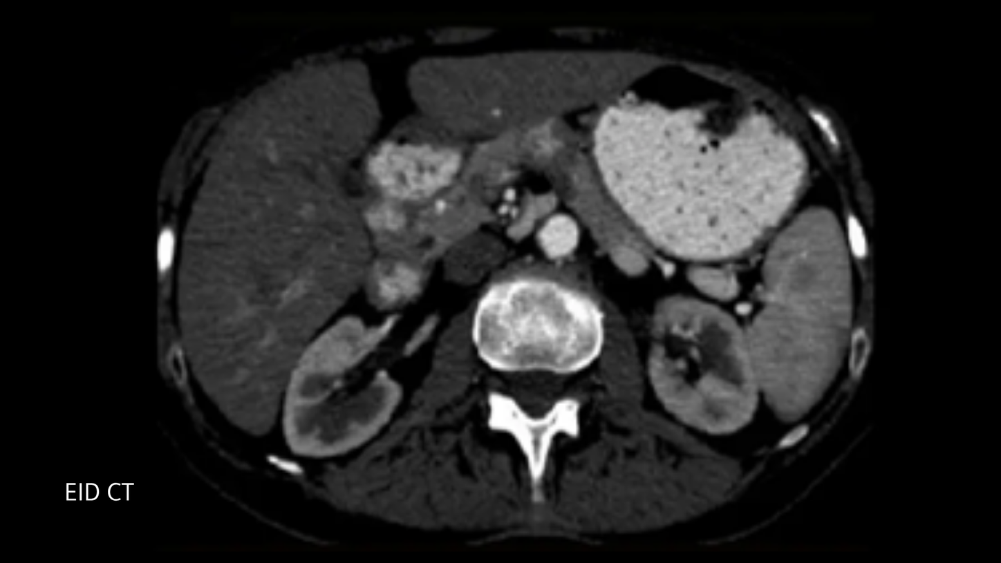
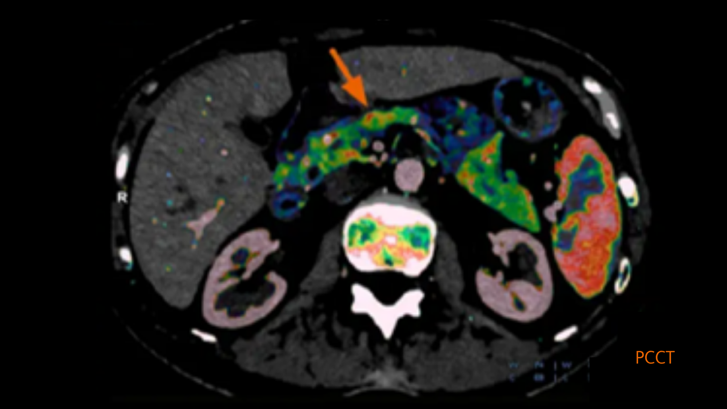
Photon-counting CT enables low dose, high resolution, fast acquisition, and spectral information in a single scan—without compromise. Use the slider on the images below to see the difference the world’s first photon-counting CT can make.
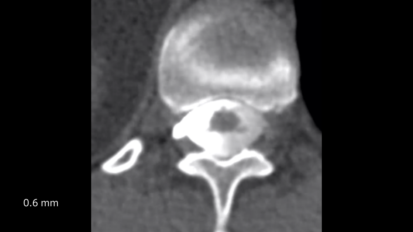
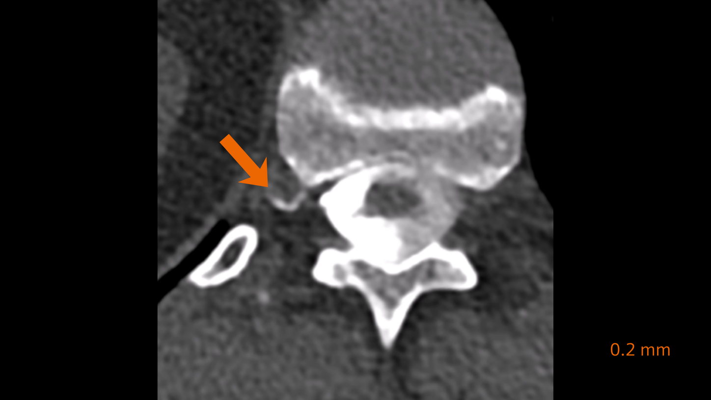
Courtesy of Duke University Medical Center, Durham, NC, USA
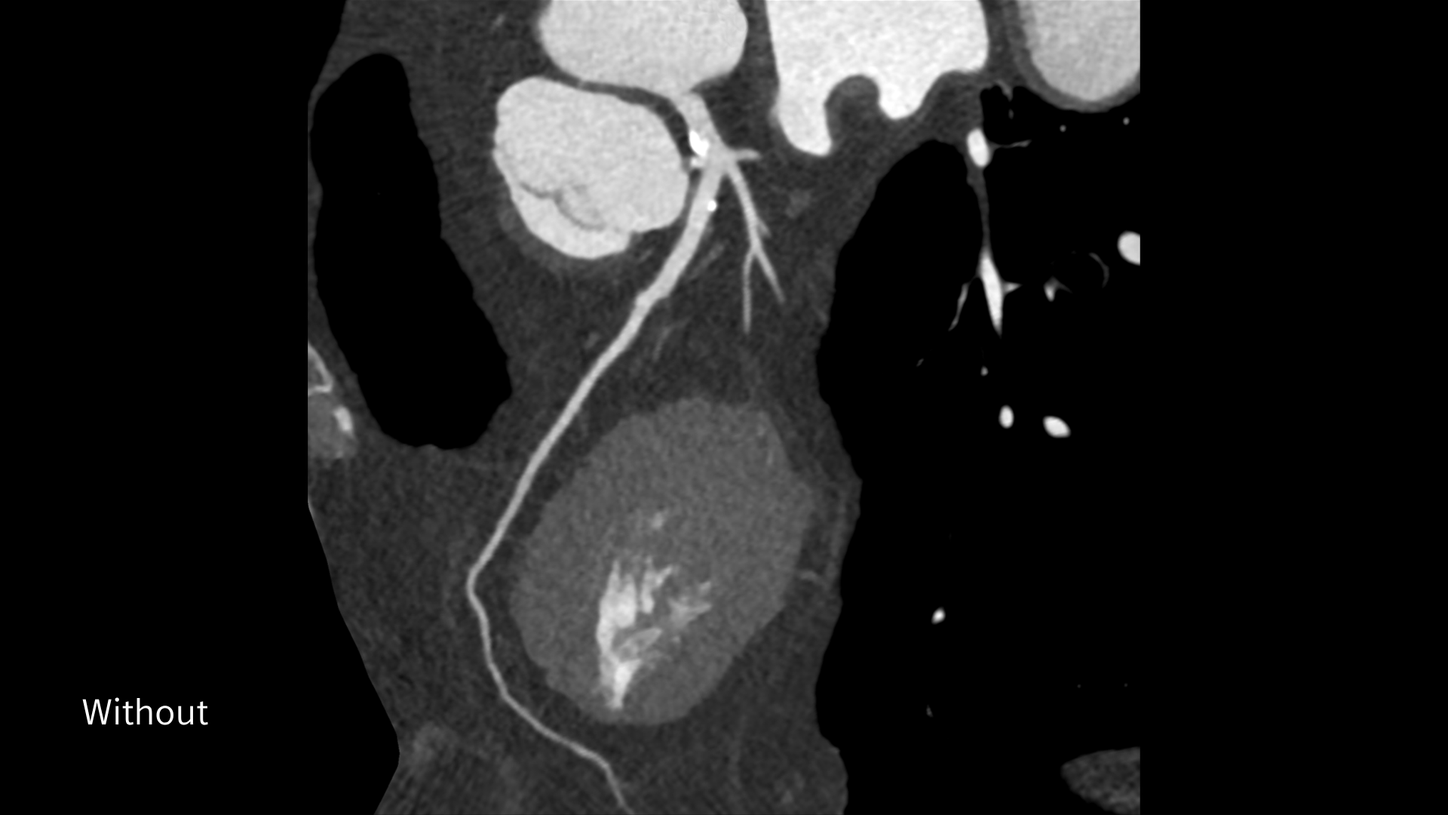
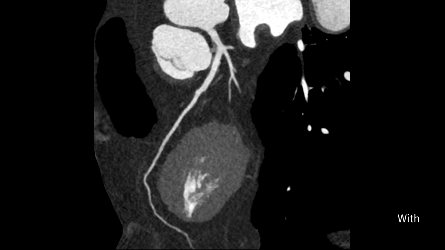
Visualize fine anatomical details with Quantum HD
With 0.2 mm ultra-high resolution, PCCT can "improve the detection of fistulas that might otherwise be missed".1
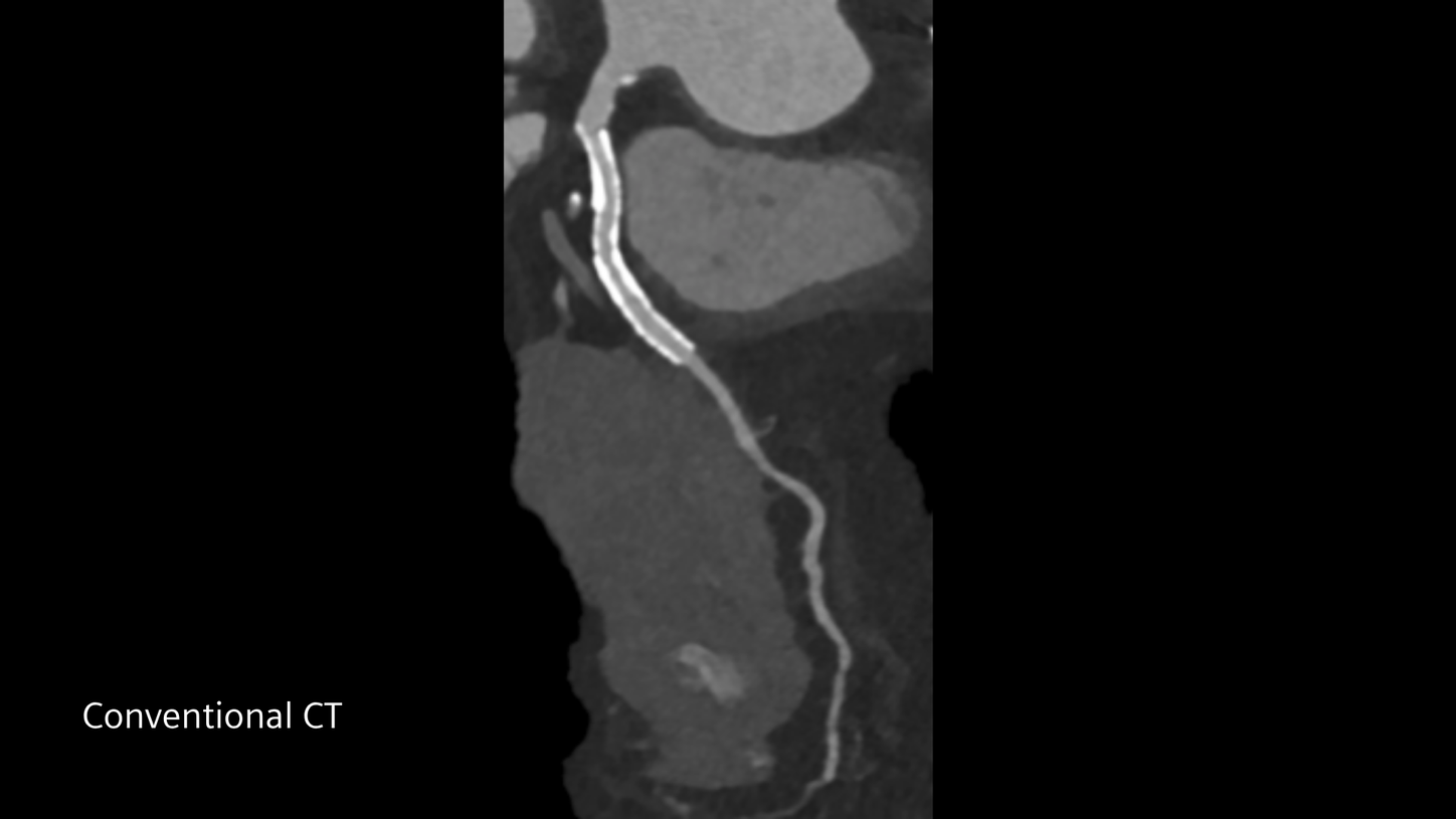
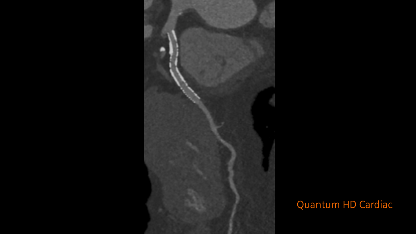


Four fundamental advantages
Photon-counting CT provides intrinsic spectral sensitivity in every scan by measuring each X-ray photon against multiple energy thresholds. But that's just one piece of the puzzle. Explore more key advantages of photon-counting CT.
FACT: Photon-counting is a radically different detector technology
How does photon-counting work?
Resolution starts at acquisition
Spatial resolution can be influenced by many factors, including reconstruction kernels, smaller fields of view, and deep learning. While these tools can improve image appearance or reduce noise, true native spatial resolution starts with the data captured at the detector.
That is why detector design matters. The CdTe photon-counting detector enables native 0.2 mm ultra-high resolution, capturing fine anatomical detail directly at the point of acquisition. AI and postprocessing can support image quality, but they cannot replace the physical resolution acquired by the detector.
Detector material matters
Photon-counting detectors can be based on different semiconductor materials, including cadmium telluride or silicon. After decades of detector material research, cadmium telluride, or CdTe, was chosen for the NAEOTOM Alpha class because of what it helps make possible.
CdTe’s high atomic number and density support efficient X-ray absorption and a compact detector geometry designed for small pixels. These material properties help the NAEOTOM Alpha class combine high resolution, low-dose imaging, fast acquisition, and spectral information in a single scan—without compromise.
More than dual layer
Dual layer detectors are not equivalent to photon-counting detectors. Dual layer detectors are still scintillator-based and require light as an intermediate step in converting X-rays to signals. They have the same physical limitations as all conventional energy integrating detectors.
In addition, dual layer detectors have the same challenge as single layer detectors when it comes to integrating electronic noise. The problem is amplified with a dual layer detector because of the interference with more layered components. Photon-counting, on the other hand, allows for the complete elimination of electronic noise.
FACT: NAEOTOM Alpha class comes with simple, proven usability
The technology runs deep. The workflow stays simple.
Advanced technology should not mean added complexity. With AI-powered workflow support across the imaging process, the NAEOTOM Alpha class helps care teams move through patient preparation, acquisition, reconstruction, and reading. Intelligent guidance, a mobile, tablet-based workflow, automated reconstruction, and advanced visualization tools help make photon-counting CT simple to incorporate into broad clinical use.

Personalized procedures
AI-powered imaging guidance helps tailor exam parameters and dose to the patient and clinical question.

Consistent results
Zero-click reconstructions support help consistency across users, patients, and procedures.

Streamlined reading
Interactive visualization and postprocessing tools support efficient review and interpretation.

Guided scanning
myExam Companion supports technologists through patient preparation, scan setup, and acquisition.
Accelerate scanning workflows and empower technologists with myExam Companion
myExam Companion guides technologists through CT scan procedures using clinical language and intuitive visuals, helping them move more confidently through exam preparation, scan setup, and parameter selection. By reducing guesswork and decision overload, it helps accelerate scanning workflows while supporting high-quality, consistent results for each patient.
Unleashing the full power of photon-counting
Our proven usability features support and guide users throughout the scanning workflow, while Al-enabled solutions help radiologists when reading cases.

High-Resolution Auto Mode
Balance the desire for details with the increased processing time required. Auto Mode selects the appropriate matrix size based on the FoV and Kernel.
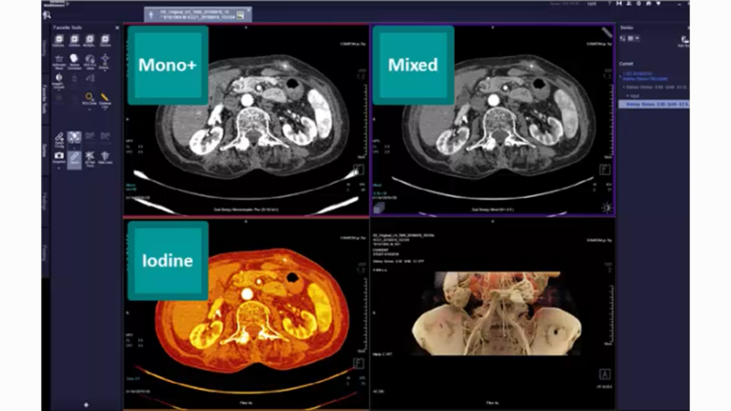
Interactive Spectral Imaging
Interactive Spectral Imaging (ISI) datasets enabling radiologists to switch between spectral imaging views on the fly.

SPP Data Format
Always archive intrinsic spectral imaging source data, while minimizing the number of series sent to PACS thus reducing transfer time, image storage size and overall PACS clutter.



A peer community for NAEOTOM Alpha class users
With more than 2,000 NAEOTOM Alpha class users worldwide, NAEOTOM Connect offers a space to exchange best practices, engage with clinical and technical experts, and continue building confidence with photon-counting CT in daily clinical use.
FACT: NAEOTOM Alpha class delivers profound clinical impact

Abundant clinical evidence after a decades-long journey
Siemens Healthineers started basic research on photon-counting more than 20 years ago. The CdTe semiconductor material is at the core of this innovative technology— both the quality and the fabrication of the material. In 2012, Acrorad was brought into the Siemens Healthineers family, adding specialized CdTe crystal production expertise directly into the detector development process.
The next step in the journey was working with global collaboration partners to evaluate the technology from both technical and clinical perspectives. Beginning in 2014, two generations of clinical prototypes provided invaluable feedback that helped advance the technology from research to clinical use. This journey culminated in November 2021 with the launch of NAEOTOM Alpha: the world’s first clinical photon-counting CT.
Today, the NAEOTOM Alpha class continues to advance photon-counting CT with a growing portfolio of scanners and an expanding body of clinical evidence. With more than 4 million patients scanned on the NAEOTOM Alpha class and over 1,300 peer-reviewed publications, the evidence is clear. As the pioneer of clinical photon-counting CT, Siemens Healthineers continues to build on more than 20 years of research and clinical experience. Explore the research, symposia, and webinars that demonstrate the full potential of photon-counting CT.
Photon-counting CT publication summaries
Explore summaries of peer-reviewed research on the clinical applications of photon-counting CT.

CT webinars, symposia, and scientific presentations
Watch recordings of webinars and scientific presentations from past events.
User experience
Read what some of our early adopters have to say.
“This will redefine our clinical decision-making right from scan one.”2

University Hospital Augsburg, Germany


What photon-counting means for the future of your department
The NAEOTOM Alpha class with Quantum Technology is redefining CT imaging. Based on the revolutionary direct signal-conversion of its QuantaMax detector, NAEOTOM Alpha class offers high-resolution images at minimal dose, improved contrast at lower noise, and breakthrough consistency you’ve never seen before.

Scan previously excluded patients
The NAEOTOM Alpha class may redefine which patient populations can be addressed with CT. It unlocks patient groups, like those with heavy calcifications, stents, pacemakers, or artificial valves, by offering intrinsic spectral imaging independent of scan speed, and of temporal or spatial resolution.
Impact treatment outcomes
Benefit from confident clinical decision-making in cardiology, oncology, and pulmonology, with answers that are meaningful, precise, and reproducible.
Did this information help you?
The statements by Siemens Healthineers’ customers described herein are based on results that were achieved in the customer's unique setting. Because there is no "typical" hospital or laboratory and many variables exist (e.g., hospital size, samples mix, case mix, level of IT and/or automation adoption) there can be no guarantee that other customers will achieve the same results.